

Introduction
Old World species in the family Gesneriaceae (Lamiales, Eudicots) are well known for their unorthodox cotyledon development, termed anisocotyly (Jong & Burtt, Reference Jong and Burtt1975). During germination just after cotyledon unfolding, the two cotyledons are equal in size, as in most other dicotyledonous species. However, soon afterwards the two cotyledons develop at different rates, with one cotyledon eventually being significantly larger than the other (Nishii et al., Reference Nishii, Huang, Wang and Möller2017 and references therein). This phenomenon was first reported in the mid-nineteenth century in Streptocarpus Lindl., an African genus (Caspary, Reference Caspary1858; Crocker, Reference Crocker1860).
In previous studies, the mechanism behind anisocotyly was investigated mainly in Monophyllaea R.Br. and Streptocarpus species (Oehlkers, Reference Oehlkers1923; Jong, Reference Jong1970; Jong & Burtt, Reference Jong and Burtt1975; Tsukaya, Reference Tsukaya1997; Imaichi et al., Reference Imaichi, Nagumo and Kato2000, Reference Imaichi, Inokuchi and Kato2001; Nishii et al., Reference Nishii, Kuwabara and Nagata2004). It has been found that unequal cotyledon development is caused by differential meristem activity in the proximal part of the lamina of the two cotyledons, the so-called basal meristem. Just after germination, both cotyledons are equal in size and both show cell division in the proximal region of the lamina. Soon afterwards, the basal meristem activity in one cotyledon ceases and it becomes the microcotyledon, while in the other the basal meristem activity continues to form the macrocotyledon. It has been suggested that cotyledon fate is determined after germination (Oehlkers, Reference Oehlkers1923; Tsukaya, Reference Tsukaya1997; Nishii et al., Reference Nishii, Kuwabara and Nagata2004; Saueregger & Weber, Reference Saueregger and Weber2004).
Burtt (Reference Burtt1963) suggested that anisocotyly is characteristic of Old World Gesneriaceae, but not many species had been studied at that time. In addition, it seems there are large variations in the expression of anisocotyly between species. In the unifoliate Streptocarpus (so-called one-leaf plants), the macrocotyledon can sometimes grow up to 1 m in length (Hilliard & Burtt, Reference Hilliard and Burtt1971). In contrast, some species in other genera, such as Aeschynanthus Jack or the former Chirita Buch.-Ham. (now split into five different genera; see Weber et al., Reference Weber, Middleton, Forrest, Kiew, Lim, Rafidah, Sontag, Triboun, Wei, Yao and Möller2011) show only moderate anisocotyly (Hill, Reference Hill1938; Burtt & Woods, Reference Burtt and Woods1958).
Although mostly tropical and subtropical Gesneriaceae genera have been previously been studied for anisocotyly, only brief reports on the European Haberlea Friv. and Ramonda Rich. have been published. Fritsch (Reference Fritsch1904) reported simply that the cotyledons of Ramonda myconi (L.) Rchb. were unequal. Hill (Reference Hill1938) reported that Ramonda serbica Pančić and Haberlea rhodopensis Friv. “have seedlings with unequal cotyledons, though less markedly so than in Streptocarpus”. In Haberlea, apparently, the larger one is 1.00–1.25 mm in length, whereas the smaller one is 0.85–0.95 mm long. No further observations or images were presented and no detailed study of the involvement of meristem activity was undertaken. Therefore, whether the same meristem activity observed in Streptocarpus and Monophyllaea underlies this mild anisocotyly is unknown.
The underlying mechanisms of weak anisocotyly have received little attention thus far, and this study presents quantitative data relevant to the subject. The cotyledon development of the two European Gesneriaceae species Haberlea rhodopensis and Ramonda myconi was studied in detail, assessing basal meristem activity and growth of the cotyledons in particular. The results were compared with those for typical anisocotylous Old World species and typical isocotylous New World species.
Materials and Methods
Plant material
Besides the European Haberlea rhodopensis (RBGE accession no. 19281002) and Ramonda myconi (19710970), the plant material used in this study included the typical anisocotylous Old World Streptocarpus rexii (Bowie ex Hook.) Lindl. (20080299) and the isocotylous New World Corytoplectus speciosus (Poepp.) Wiehler (19540131). Seeds were sown on soil (vermiculite to peat moss, 3:1) in pots or on filter paper in 9-cm Petri dishes and cultivated in a growth room at the National Taiwan University (NTU), Taipei, Taiwan, at 23°C, with 16 h of light and 8 h of darkness.
Morphological observations
To observe the process of germination, seeds sown on filter paper were observed under a stereomicroscope, SMZ-10 (Nikon, Tokyo, Japan). To be able to comparatively observe the consecutive seedling development, the first day of cotyledon expansion was defined as 1 day after cotyledon unfolding (1 DAU). Cotyledon area was measured from images obtained using ImageJ (Schneider et al., Reference Schneider, Rasband and Eliceiri2012). Student’s paired t-tests were performed with Microsoft Excel (Microsoft Corporation, Washington, USA) to statistically detect significant size differences between larger and smaller cotyledons in a seedling.
For scanning electron microscopy (SEM), seedlings were fixed in FAA (50% ethanol, 10% acetic acid, 3.7% formaldehyde) under vacuum. The samples were dehydrated in an ethanol series, followed by treatment with acetone–ethanol (1:1) and then 100% acetone. Following critical-point drying with an HCP-2 (Hitachi, Tokyo, Japan), the samples were coated with gold palladium in a IB-2 ion sputter coater (Eiko Engineering, Tokyo, Japan) and observed under a Quanta 200 scanning electron microscope (FEI, Hillsboro, Oregon, USA) at either the Institute of Plant and Microbial Biology, Academia Sinica (Taipei, Taiwan) or the TC5-Bio-Image group, Tech-comm, College of Life Science, NTU (Taipei, Taiwan).
To observe the epidermal cell size, seedlings were fixed in ethanol–acetic acid (3:1) and hydrated in an ethanol series. The hydrated seedlings were cleared with chloral hydrate (Nishii et al., Reference Nishii, Kuwabara and Nagata2004) and observed under Nomarski optics on a BX51 microscope (Olympus, Tokyo, Japan). Epidermal cell size was measured from SEM or light microscopy images of cleared samples using ImageJ.
Observation of meristem activity
Cell division activity was detected by aniline blue staining (Nishii et al., Reference Nishii, Kuwabara and Nagata2004). Briefly, aniline blue stains callose (β-1,3-glucans), which exists mainly in the cell plane of freshly divided cells but rapidly degrades after division. Seedlings were fixed in ethanol–acetic acid (4:1) and further treated with 100% ethanol then ethanol plus 100 mM pH 9.0 phosphate buffer (1:1) before finally being transferred to pH 9.0 phosphate buffer. The seedlings were stained with 0.02% aniline blue in 100 mM phosphate buffer (v/v) and observed under ultraviolet excitation with a fluorescence microscope BX51 (Olympus); images were taken with a DP71 camera (Olympus). The images were analysed in ImageJ and XY coordinates of the cotyledon contour and aniline blue–stained cell planes obtained. The XY data were transferred into Microsoft Excel to produce graphs by using the scatter graph function.
Results
Seedling development of European Gesneriaceae
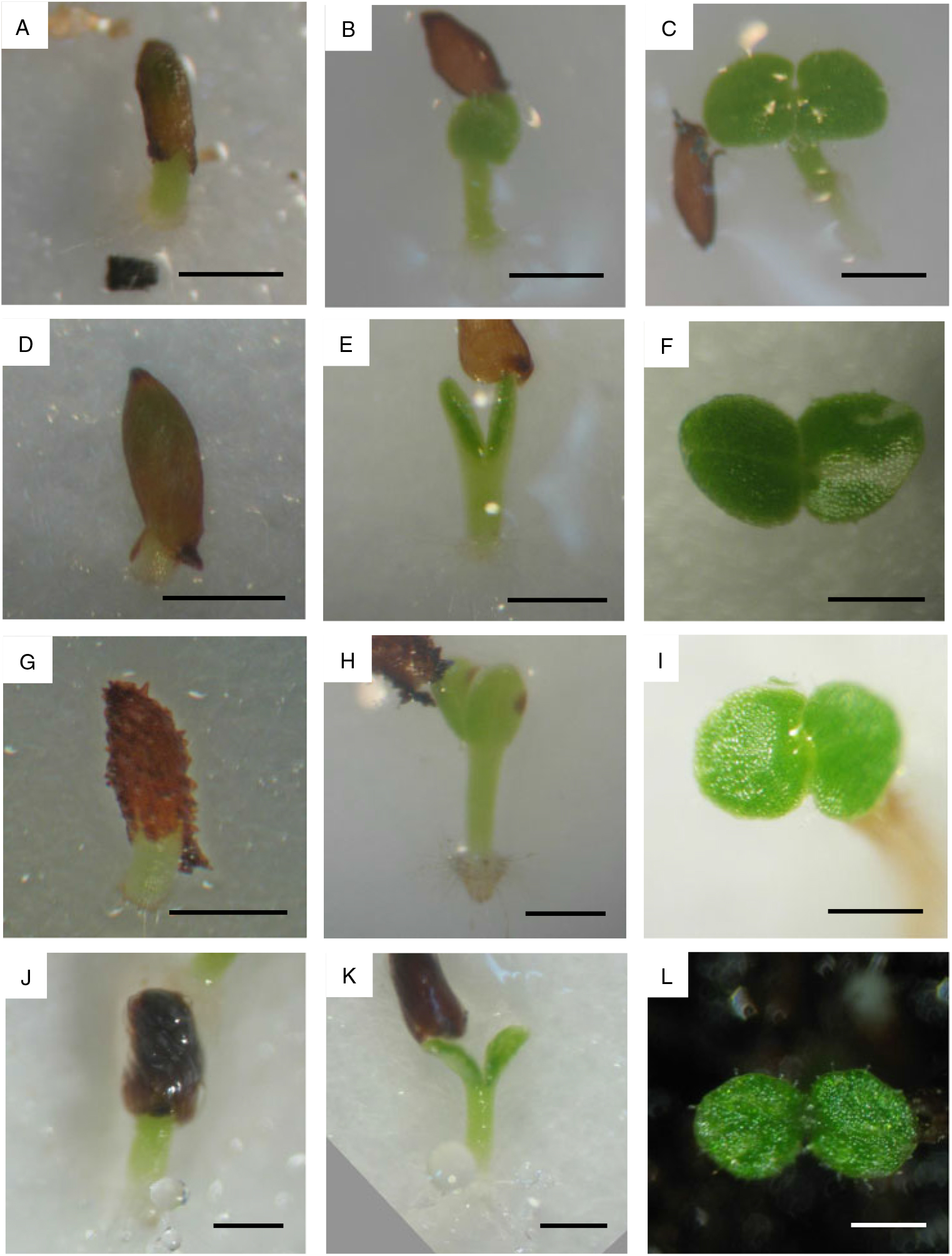
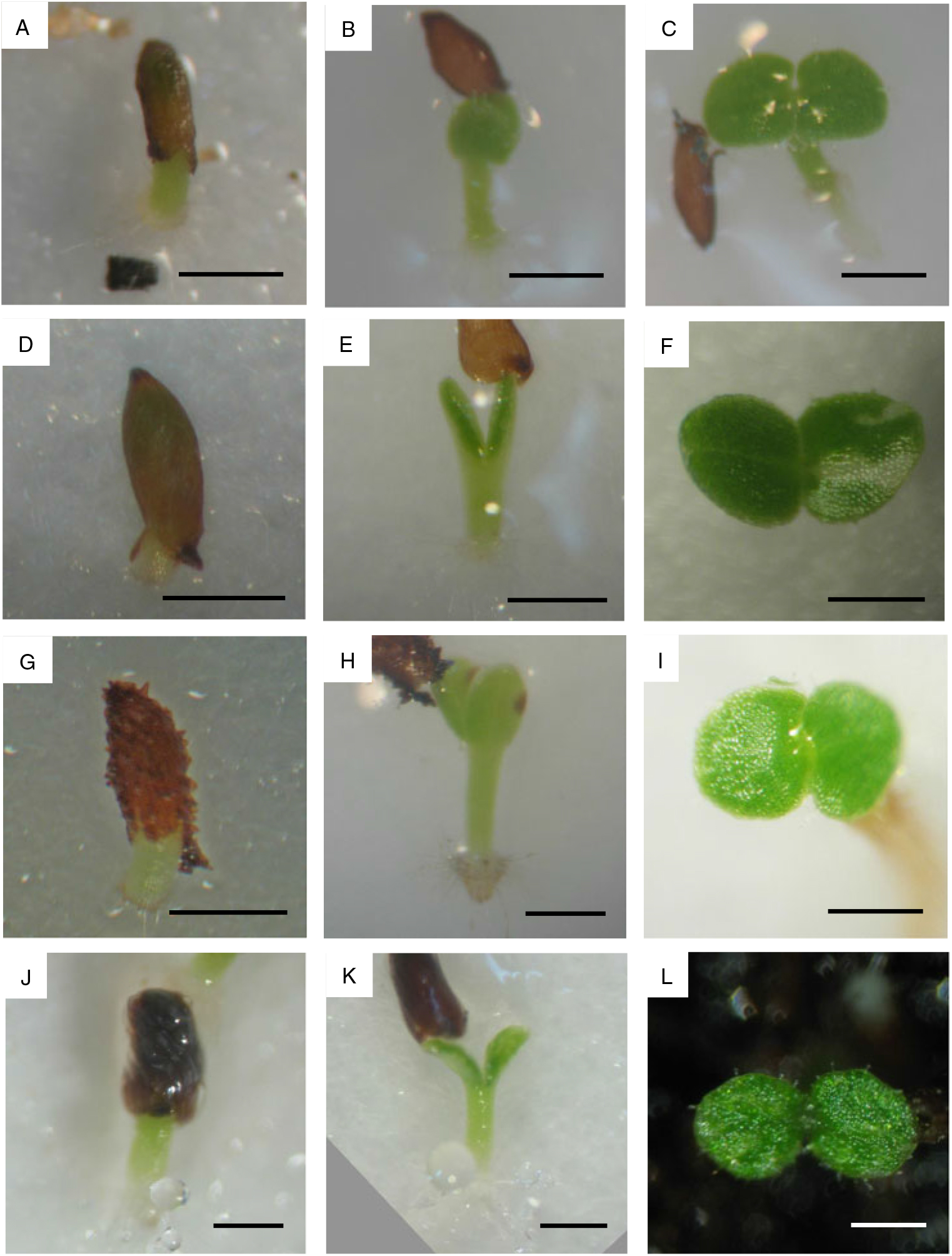
The initial stages of seed germination were similar in the four species examined (Fig. 1). Between 10 and 15 days after sowing, the seed coat was ruptured at one end by the hypocotyl, pushed out by the expanding cotyledons and hypocotyl. The radicle emerged earlier in New World Corytoplectus speciosus than in Old World species. Shortly afterwards, the cotyledons unfolded (1 DAU). At this point both cotyledons were equal in size (see Fig. 1).
Germination in Streptocarpus rexii (A–C), Haberlea rhodopensis (D–F), Ramonda myconi (G–I) and Corytoplectus speciosus (J–L). A, D, G and J, Germinated seeds; B, E, H and K, seedlings with folded cotyledons; C, F, I and L, seedlings with unfolded cotyledons. Scale bars, 500 μm.
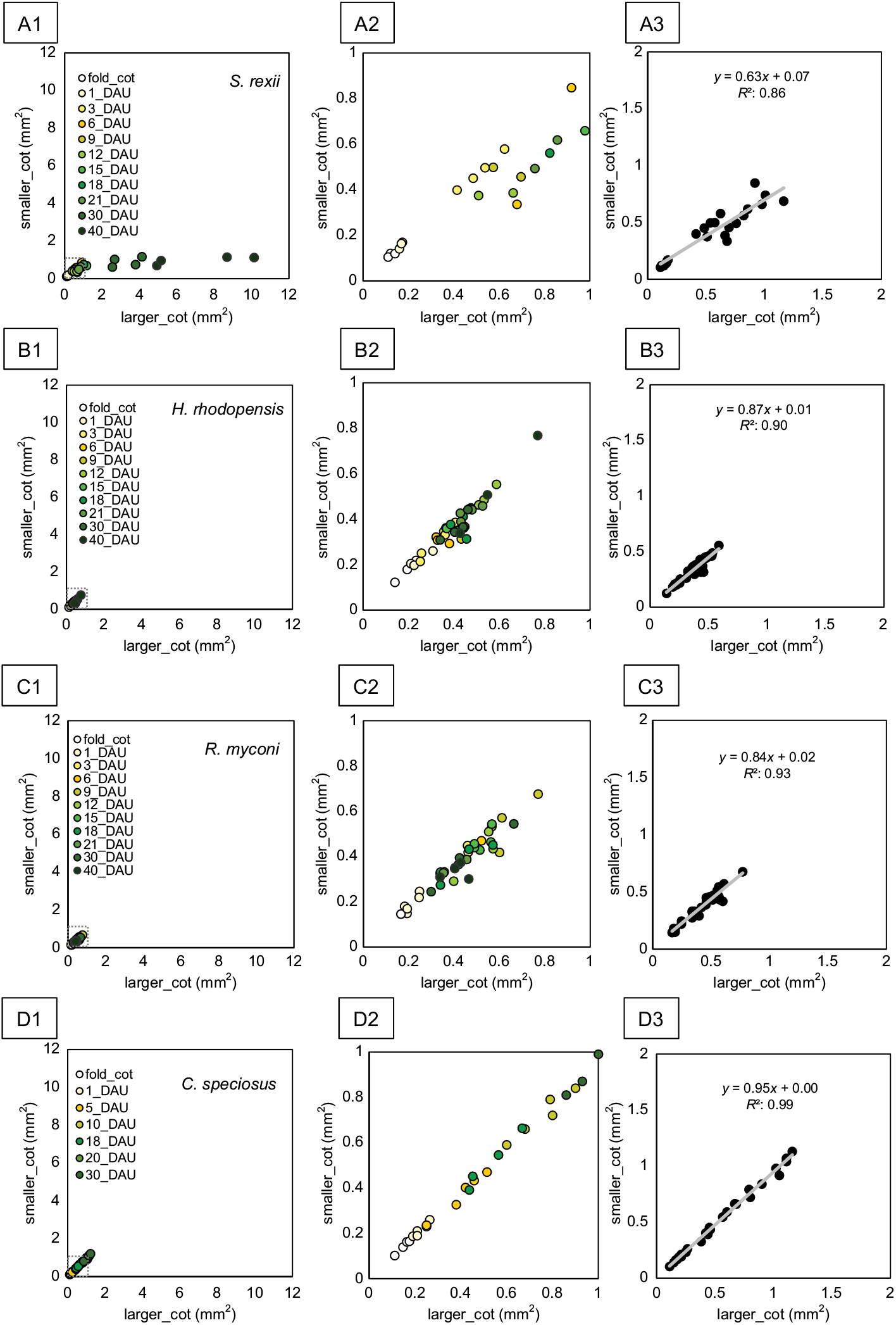
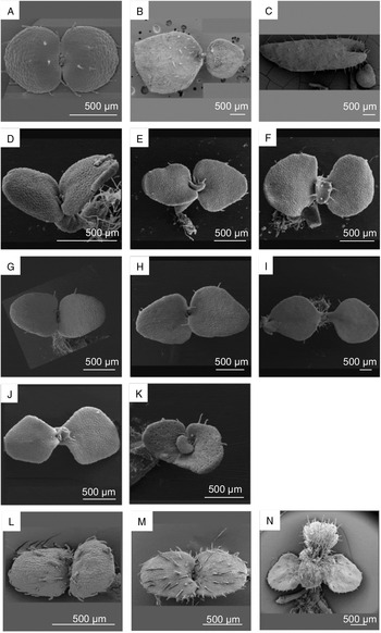
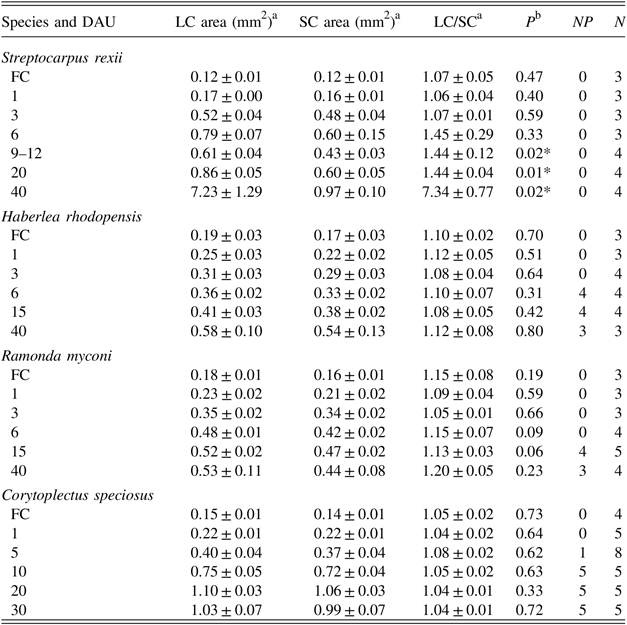
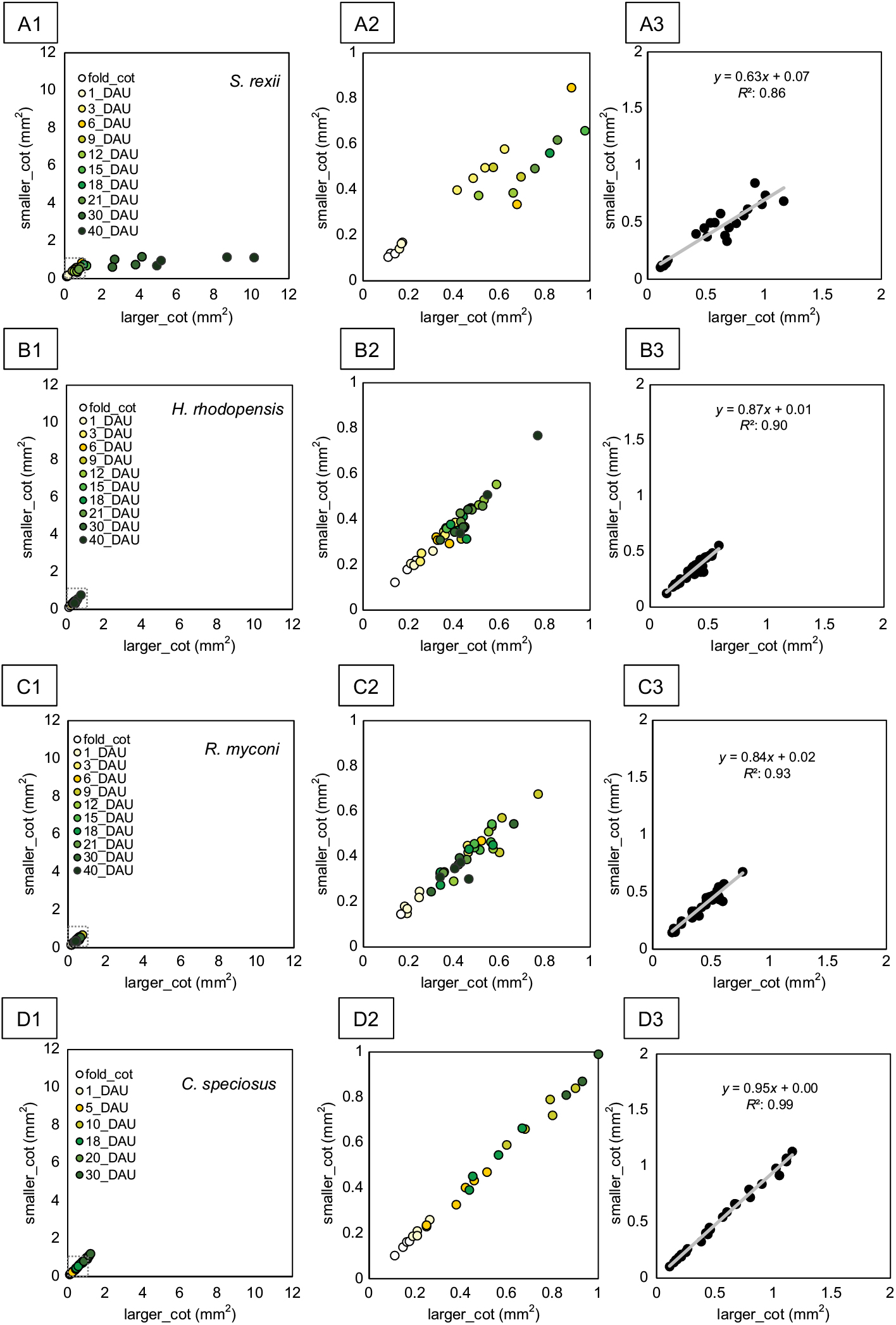
Anisocotyly became statistically established in Streptocarpus rexii at 9–12 DAU (Table 1) and visually apparent between 21 and 30 DAU (Figs 2A, 3B). In contrast, Haberlea rhodopensis and Ramonda myconi seedlings did not show clear anisocotyly (see Table 1; Figs 2B,C, 3D–I). Most seedlings remained isocotylous, with a few seedlings showing weak anisocotyly (Figs 2B2,C2, 3J,K) but with no statistical significance (see Table 1). In Corytoplectus speciosus, all seedlings remained isocotylous, with very little variation between the two cotyledons (see Table 1; Fig. 2D).
Growth patterns of cotyledon area. A1–3, Streptocarpus rexii with anisocotylous growth; B1–3, Haberlea rhodopensis; C1–3, Ramonda myconi; D1–3, Corytoplectus speciosus with isocotylous growth. A1, B1, C1 and D1, Scatter plots of cotyledon area from folded cotyledon stage to 40 days after cotyledon unfolding (DAU); A2, B2, C2, and D2, magnified views of 1 × 1 mm2 plots of A1, B1, C1 and D1, respectively, indicated by stippled boxes; A3, B3, C3 and D3, scatter plots of cotyledon area from folded cotyledon stage to 20 DAU and linear trend lines.
Scanning electron micrographs of seedlings. A–C, Streptocarpus rexii: A, 5 days after cotyledon unfolding (DAU); B, 25 DAU; C, 40 DAU. D–F, J, Haberlea rhodopensis: D, 5 DAU; E, 15 DAU; F, 25-DAU seedling with equal cotyledons; J, 25-DAU seedling with slightly unequal cotyledons. G–I, K, Ramonda myconi: G, 5 DAU; H, 15 DAU; I, 25-DAU seedling with equal cotyledons; K, 25-DAU seedling with slightly unequal cotyledons. L–N, Corytoplectus speciosus: L, 1 DAU; M, 5 DAU; N, 20 DAU.

Descriptive statistics for cotyledons during seedling development

DAU, days after cotyledon unfolding; FC, folded cotyledon stage; LC, larger cotyledon in a seedling; LC/SC, ratio of area of larger cotyledon to area of smaller cotyledon in a seedling; N, total number of seedlings examined; NP, number of seedlings forming a plumule; SC, smaller cotyledon in a seedling.
a Values represent means ± standard errors.
b For size differences between larger and smaller cotyledons (determined by Student’s paired t-test).
* P< 0.05.
To illustrate the differences between the studied species, a linear trend line was fitted to the scatter plots of seedling cotyledon sizes in relation to time, from the folded cotyledon stage to 20 DAU (Fig. 2A3–D3). Corytoplectus speciosus showed a very high level of linear fit (R 2, 0.99; Fig. 2D3), whereas in Streptocarpus rexii the correlation was not strong (R 2, 0.86; Fig. 2A3); the results for the European species were somewhat in-between (Haberlea rhodopensis; R 2, 0.90; Fig. 2B3; Ramonda myconi, R 2, 0.93; Fig. 2C3). Furthermore, the trend line in Corytoplectus speciosus sloped in almost perfect correlation between the smaller and larger cotyledons (y = 0.95x + 0.00, Fig. 2D3), whereas in Streptocarpus rexii it was strongly inclined towards the larger cotyledon (y = 0.63x + 0.07, Fig. 2A3). In Haberlea rhodopensis and Ramonda myconi, the slope of the trend line was between those for Corytoplectus speciosus and Streptocarpus rexii (H. rhodopensis, y = 0.87x + 0.01, Fig. 2B3; R. myconi, y = 0.84x + 0.02, Fig. 2C3).
Plumules started to be observed in Corytoplectus speciosus at 5 DAU, in Haberlea rhodopensis at 6 DAU and in Ramonda myconi at 15 DAU (see Table 1). The morphology of seedings and plumules is shown in Fig. 3. In Streptocarpus rexii, instead of plumules, leaf primordia formed at a much later stage of about 50 DAU (65 days after sowing; see Nishii & Nagata, Reference Nishii and Nagata2007; Nishii et al., Reference Nishii, Möller, Kidner, Spada, Mantegazza, Wang and Nagata2010). In all species, stomata were found only on the underside of cotyledons.
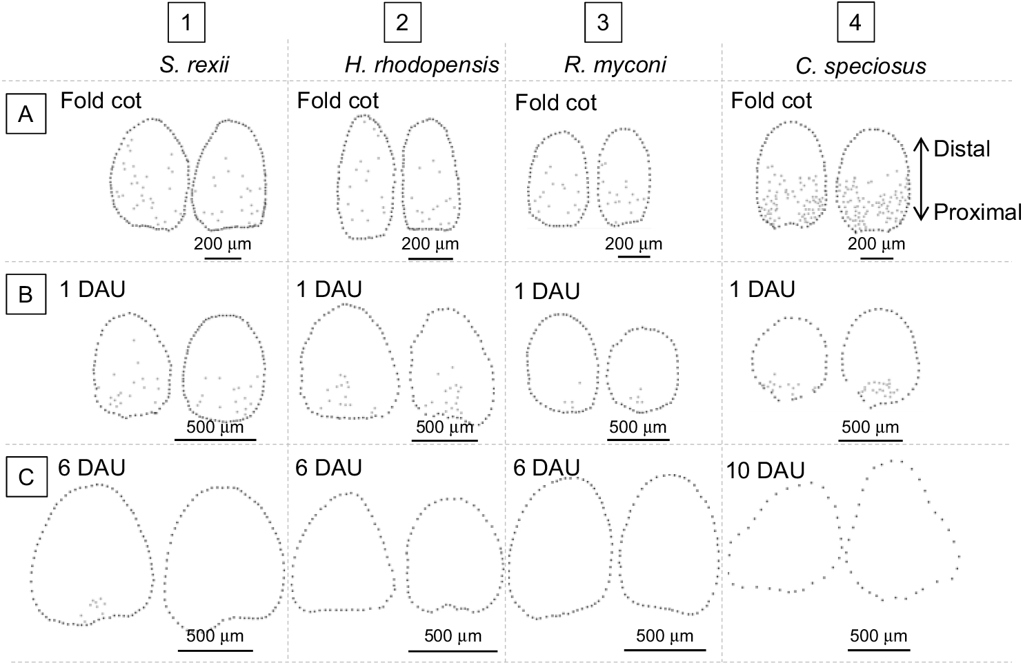
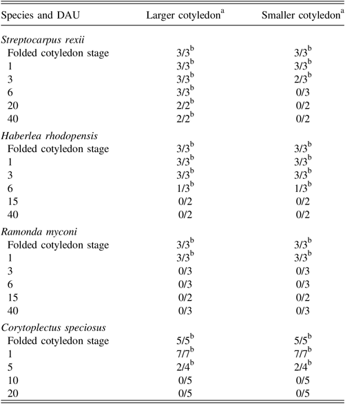
In seedlings with folded cotyledons, dividing cells were observed more or less scattered across both cotyledons (Fig. 4A). Later, at 1 DAU, the arrest front moved towards the base and the dividing cells were focused in the proximal area of the cotyledons (Fig. 4B). At 6 DAU (10 DAU for Corytoplectus speciosus) cell division had ceased in all species except for Streptocarpus rexii, in which cell division was observed continuously in one cotyledon (Table 2, Fig. 4C).
Distribution of planes of newly divided cells stained with aniline blue. A, Folded cotyledon stage; B, cotyledons 1 day after cotyledon unfolding (DAU); C, cotyledons 6 DAU (10 DAU for Corytoplectus speciosus). 1, Streptocarpus rexii; 2, Haberlea rhodopensis; 3, Ramonda myconi; 4, Corytoplectus speciosus.

Detection of cell planes in newly divided cells by aniline blue staining
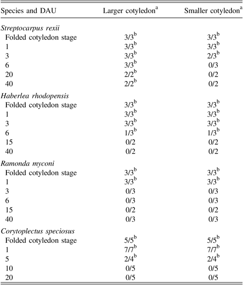
DAU, days after cotyledon unfolding.
a Numbers before the slash are the number of cotyledons showing divisions, and those after the slash are the number of seedlings analysed.
b Stages during which meristems are present in the cotyledons.
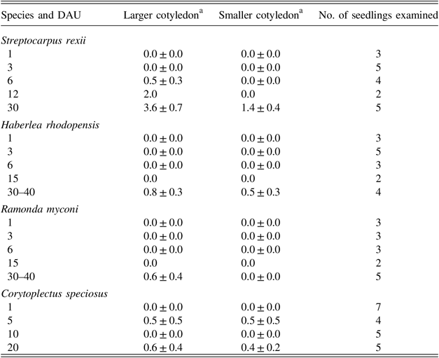
In Streptocarpus rexii, lateral veins were formed in cotyledons at 6 DAU. At 30 DAU, about 3 or 4 veins (mean, 3.6 ± 0.7) were observed in the larger cotyledon and 1 or 2 veins (mean, 1.4 ± 0.4) in the smaller cotyledon (Table 3). In contrast, in Haberlea rhodopensis and Ramonda myconi, most seedlings did not develop lateral veins, only a few showing 1 or 2 veins in cotyledons at 30–40 DAU, similar to Corytoplectus speciosus (see Table 3).
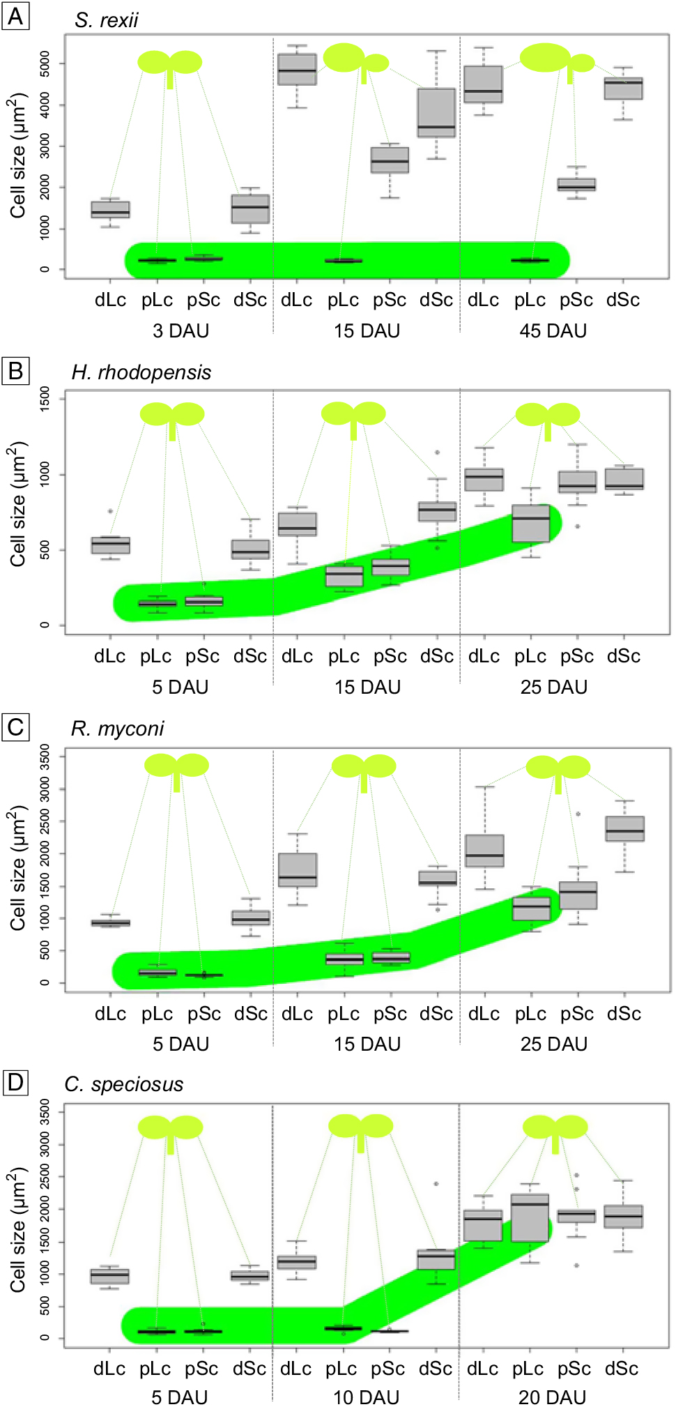
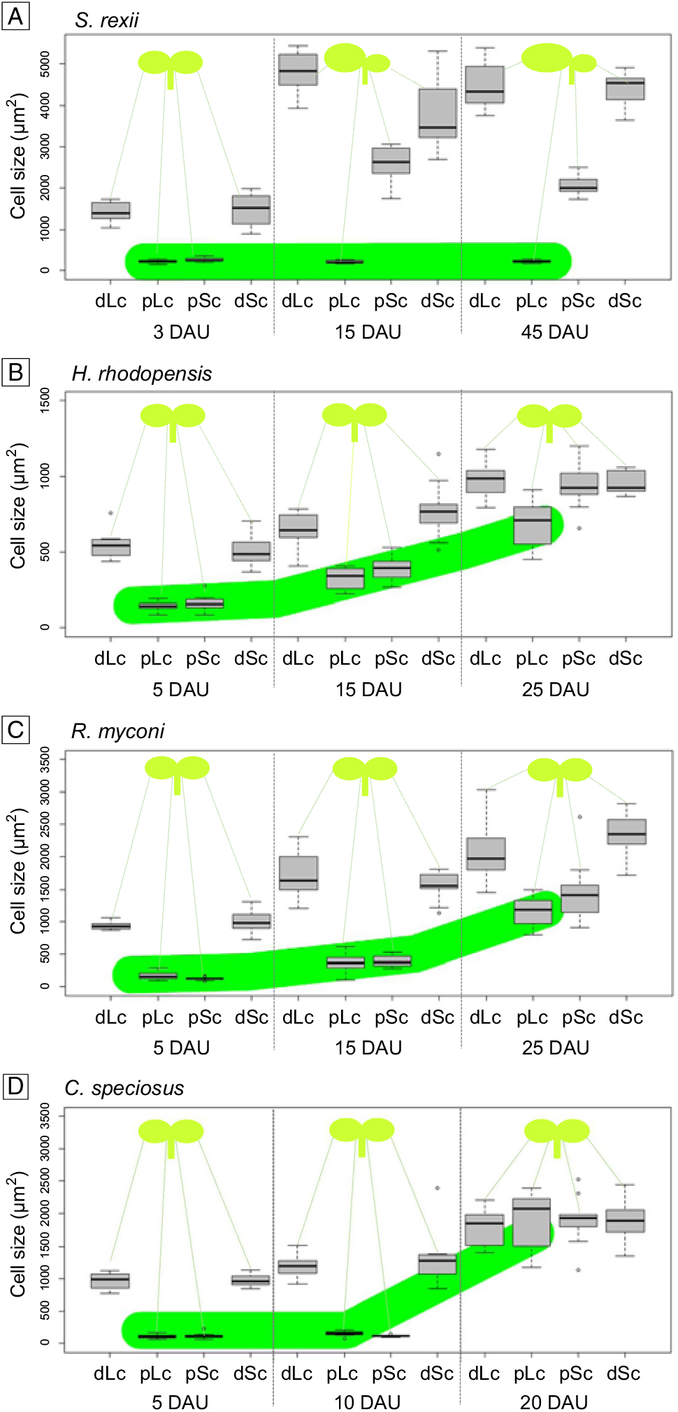
Measurement of epidermal cell size during cotyledon development showed that in both cotyledons of all four species at 3–5 DAU, the proximal region had smaller cells compared with the distal region (Fig. 5). At 15 DAU and 45 DAU in Streptocarpus rexii, only the larger cotyledon still had smaller cells (< 500 μm2) in the proximal region, these not being found in the proximal region of the smaller cotyledon (Fig. 5A). In Haberlea rhodopensis and Ramonda myconi, cell size in the proximal region of both cotyledons had increased at 15 DAU compared with 5 DAU and had further expanded at 45 DAU. Some variation in cell size was observed between the proximal and distal regions (Fig. 5B,C). Cell size in cotyledons of Corytoplectus speciosus at 20 DAU was uniform across and between the cotyledons (Fig. 5D).
Box plots of epidermal cell sizes in cotyledons (n = 10). Cells in proximal and distal positions in the smaller and larger cotyledon were plotted separately. A, Streptocarpus rexii; B, Haberlea rhodopensis; C, Ramonda myconi; D, Corytoplectus speciosus. DAU, days after cotyledon unfolding; dLc, distal part of larger cotyledon; dSc, distal part of smaller cotyledon; pLc, proximal part of larger cotyledon; pSc, proximal part of smaller cotyledon. Broad green lines highlight the cells observed in the proximal region of the larger cotyledons, tracking their cell sizes over time.
Number of lateral veins
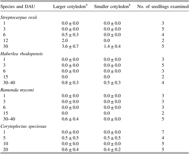
DAU, days after cotyledon unfolding.
a Numbers represent the mean number of lateral veins per cotyledon ± standard error.
Discussion
In this study, we investigated the phenomenon of anisocotyly in two European Gesneriaceae, Haberlea rhodopensis and Ramonda myconi, through the localisation and characterisation of meristematic activities in cotyledons of developing seedlings.
Our results show a cotyledon growth behaviour of the European species that is in-between the typical anisocotyly present in the Old World Streptocarpus rexii and the typical isocotyly of the New World Corytoplectus speciosus. Previous studies of Haberlea and Ramonda have reported their less marked anisocotyly (Hill, Reference Hill1938), but without detailed analysis of the underlying developmental cascades. In our studies, many seedlings of Haberlea rhodopensis and Ramonda myconi showed no anisocotyly and a plumule was observed at an earlier stage than in Streptocarpus rexii. About one-fifth of the seedlings of the European genera showed size differences between the cotyledons in a pair, albeit weak. This unequal size per se would not qualify as anisocotyly. Anisocotyly involves several features, such as continued meristematic activity in a narrowly defined basal region of the cotyledon and its limitation to only one of the two cotyledons in a pair (Burtt, Reference Burtt1970; Jong, Reference Jong1970).
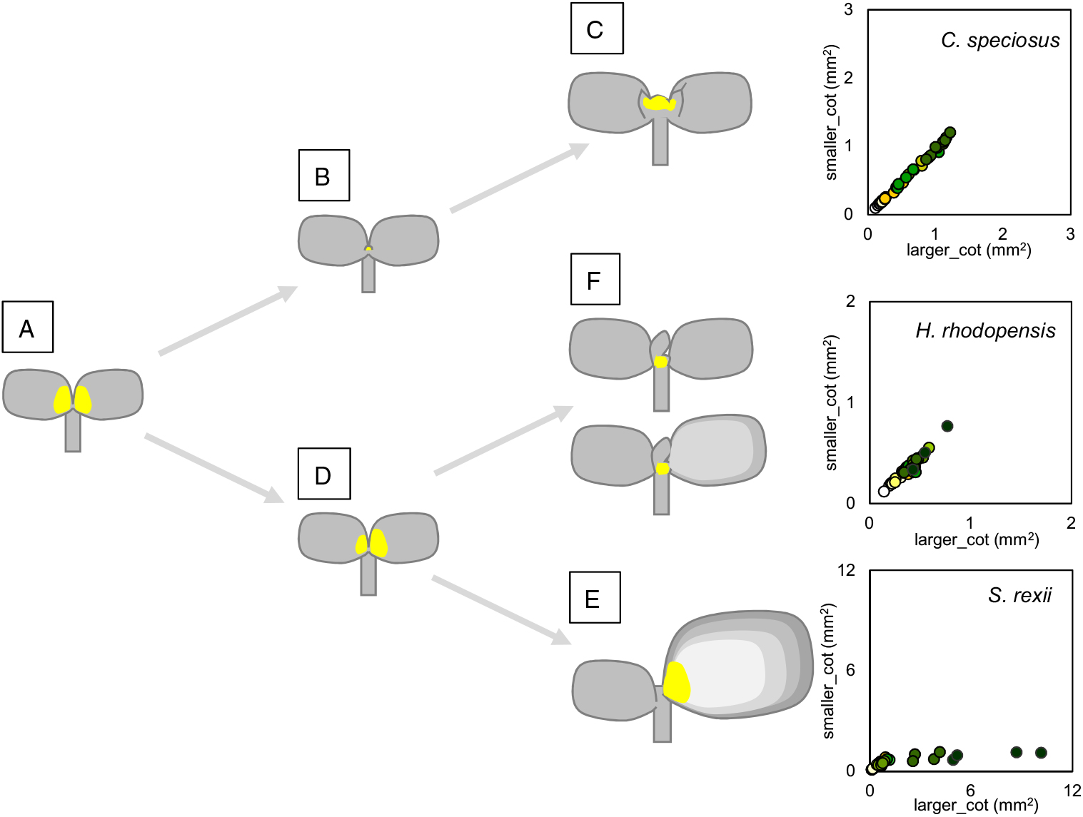
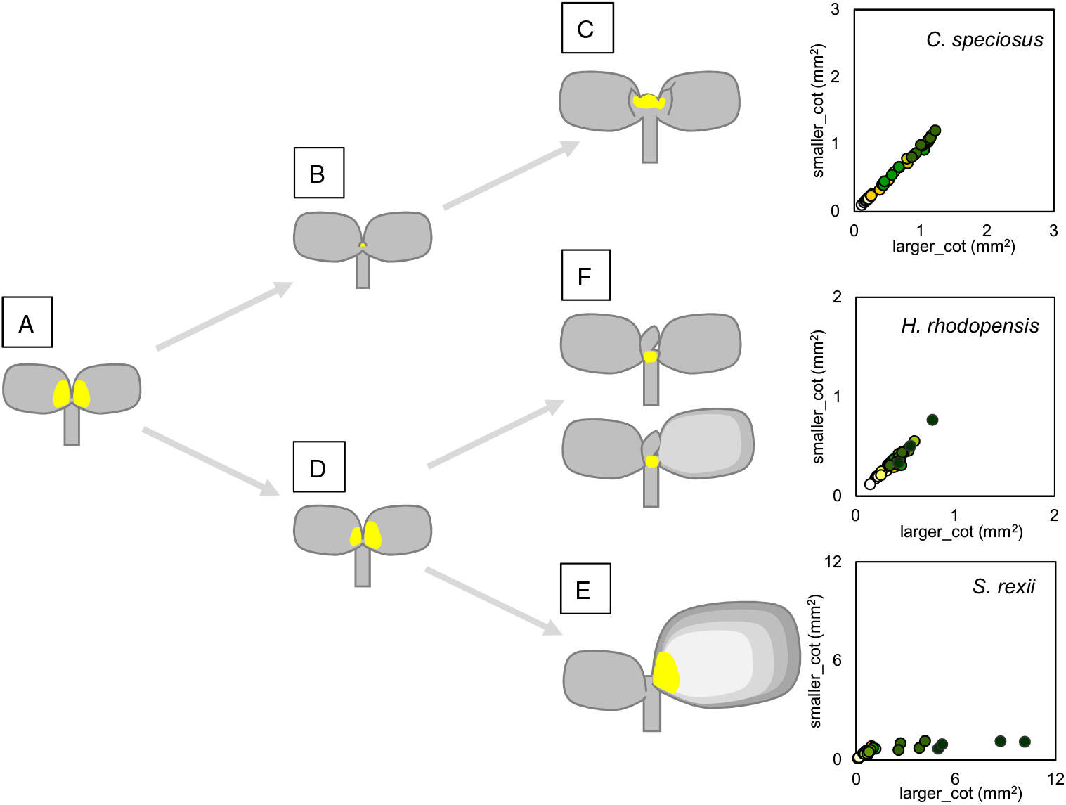
Aniline blue staining showed that all four species initially possessed meristematic activity in the basal region of both their cotyledons, where the cells were, typically for a meristem, very small. Cell sizes in the New World Corytoplectus speciosus, which lacks anisocotyly, became uniform across and between both cotyledons over time, whereas the European species retained smaller cells in the proximal area of cotyledons and, at least in Haberlea, more so in one than in the other cotyledon at 25 DAU (see Figs 4, 5). This may indicate the presence of typical anisocotyly in the European species, although weak and persisting for only a short period. This was reflected in the small size of the cotyledons (<1 mm2) and small size difference between the macrocotyledon and the microcotyledon. In Streptocarpus rexii the ratio can be greater than 5–7 times in seedlings at 40 DAU (see Table 1; Nishii et al., Reference Nishii, Huang, Wang and Möller2017). Considering all the data together, it appears that at least some seedlings of Haberlea rhodopensis and Ramonda myconi possess an anisocotylous phase but of very short duration, and thus anisocotyly is not completely lost in these species, although it is greatly reduced (Fig. 6).
Seedling development of typical isocotylous (Corytoplectus speciosus) and anisocotylous (Streptocarpus rexii) Gesneriaceae species, and European Gesneriaceae (Haberlea rhodopensis). Yellow indicates areas of detected meristem activity. A → B → C, New World Gesneriaceae. A basal meristem exists in both cotyledons just after germination (A) but ceases simultaneously in both cotyledons (B). A plumule develops and a shoot apical meristem produce leaves (C). A → D → E, Old World Gesneriaceae. Both cotyledons maintain cell-division capabilities until the cotyledon fates are determined (A → D). Later, the basal meristem becomes persistent in one cotyledon, which develops into the macrocotyledon (E). A → D → F, European Gesneriaceae. Both cotyledons initially show a basal meristem with variable and limited activities (A → D), which ceases simultaneously soon afterwards, at which point plumule development occurs, creating seedling populations with variable and limited anisocotyly or no anisocotyly (F).
Three Gesneriaceae genera are found in Europe on the Balkan Peninsula and in the Pyrenees: Haberlea, Ramonda and Jancaea Boiss. In recent phylogenetic studies, Jancaea was nested within Ramonda (Petrova et al., Reference Petrova, Moyankova, Nishii, Forrest, Tsiripidis, Drouzas, Djilianov and Möller2015). Jancaea seedlings also appear to show weak anisocotyly for some seedlings within a batch (K. Nishii, personal observation). Thus, all three European Gesneriaceae genera may share a similar cotyledon development with residual anisocotyly.
Anisocotyly has been proposed to be potentially beneficial for the quick expansion of photosynthetic area in the dark forest conditions that many Old World Gesneriaceae inhabit (Burtt, Reference Burtt1970). The European genera, however, occupy alpine habitats and much cooler environments. It is possible that the production of plumules and leaves at an early stage may be beneficial in protecting the shoot apical meristem from damage by low temperatures, reminiscent of winter buds tightly wrapping shoot apical meristems (Moore, Reference Moore1909). However, this hypothesis requires further investigation.
This study was limited to a few Old World genera exhibiting weak anisocotyly and showed that cotyledon size differences, even when small, were based on basal meristem activity in the macrocotyledon. It should be seen as an initial contribution to future broader studies across the subfamily Didymocarpoideae. Such research would allow detailed analyses of the evolutionary history of anisocotyly in the family Gesneriaceae.
Acknowledgements
The authors would like to thank Tsan-Piao Lin and Ling-Long Kuo-Huang at NTU (Taiwan) for helpful comments and discussions on this study, and Yan-Jun Chen (NTU) for technical supervision of the anatomical work. The authors acknowledge the constructive comments of two anonymous reviewers on the manuscript. The authors also thank the Royal Botanic Garden Edinburgh (RBGE) for providing the study material, and the Electron Microscope Laboratory at the Academia Sinica (Taiwan) and Tech-comm at NTU for supporting the SEM work. K.N. was funded by the National Science Council in Taiwan and the Top 100 university program at NTU during the project. The RBGE (Sibbald) Trust, the Sumitomo Foundation (Japan) and the Japan Society for the Promotion of Science (JSPS KAKENHI grant numbers 15K18593 and 18K06375) enabled finalising of the manuscript at RBGE. RBGE is supported by the Scottish Government’s Rural and Environment Science and Analytical Services Division (RESAS).