INTRODUCTION
The blue shark, Prionace glauca, is one of the most abundant shark species worldwide (Compagno, Reference Compagno1984), inhabiting the oceanic epipelagic zone in temperate, subtropical, and tropical waters (Stevens, Reference Stevens1976; Pratt, Reference Pratt1979). It is abundant during the winter and spring between 20° and 40°N in the Pacific Ocean (Strasburg, Reference Strasburg1958). This shark is placental viviparous, with variable fecundity, although the most common number of offspring is between 28 and 54 (Castro et al., Reference Castro, Woodley and Brudek1999), reaching a maximum record of 135 embryos; it is therefore considered the most fecund shark species (Gubanov & Grigor'yev, Reference Gubanov and Grigor'yev1975). The gestation period is 9–12 months (Pratt, Reference Pratt1979) and the size at birth is between 35 and 50 cm total length (TL) (Bigelow & Schroeder, Reference Bigelow, Schroeder, Tee-Van, Breder, Hildebrand, Parr and Schroeder1948; Suda, Reference Suda1953; Strasburg, Reference Strasburg1958; Nakano, Reference Nakano1994; Laughlin & Urogetz, Reference Laughlin and Urogetz1999).
The Mexican Pacific Ocean has a high incidence of pregnant blue sharks (Carrera-Fernández et al., Reference Carrera-Fernández, Galván-Magaña and Ceballos-Vázquez2010), but there have been no records of abnormality in embryos of this species in Mexico. Other morphological abnormalities have been recorded in other elasmobranchs, such as batoids, where the most common deformity is the lack of fusion between the pectoral fins and the head (Bonfil, Reference Bonfil1989; Lamilla et al., Reference Lamilla, Pequeño and Kong1995; Escobar-Sánchez et al., Reference Escobar-Sánchez, Galván-Magaña, Downton-Hoffmann, Carrera-Fernández and Alatorre-Ramírez2009). There has been a record of bicephaly in one juvenile Pacific cownose ray Rhinoptera steindachneri from the western coast of Baja California Sur, Mexico (Castro-Aguirre & Torres-Villegas, Reference Castro-Aguirre and Torres-Villegas1979), and Bornatowski & Abilhoa (Reference Bornatowski and Abilhoa2009) also recorded a bicephalic specimen of guitarfish Rhinobatos percellens off the southern coast of Brazil.
In sharks, the most common abnormalities recorded are albinism and mummification (Rosa-Molinar & Williams, Reference Rosa-Molinar and Williams1983; Sandoval-Castillo et al., Reference Sandoval-Castillo, Mariano-Meléndez and Villavicencio-Garayzar2006; Sandoval-Castillo & Villavicencio-Garayzar, Reference Sandoval-Castillo and Villavicencio-Garayzar2008); some cases of bicephaly in blue sharks were observed by Goto et al. (Reference Goto, Taninuchi, Kuga and Iwata1981) in Japan and by Ferreira et al. (Reference Ferreira, Ferreira and Oliveira2000, Reference Ferreira, Ferreira and Amorim2002) off the Brazilian coast. Recently, Mancini et al. (Reference Mancini, Casas and Amorim2006) reported one abnormal specimen of blue shark from Brazilian waters, an individual with two mouths, and with the body trunk rolled in an anticlockwise direction from the pelvic fin to the caudal fin.
In the Mexican Pacific Ocean the cases of bicephaly in elasmobranchs are scarce; therefore in this paper we describe the first record in Mexico of abnormality in two embryos of P. glauca.
MATERIALS AND METHODS
Two pregnant females of Prionace glauca were captured using long-lines by fishermen off both coasts of Baja California Sur, Mexico: Punta Arenas (24°3′N 109°49′W), located on the Gulf of California, and San Lázaro (24°45′N 112°8′W), located on the western coast of the peninsula of Baja California Sur. For shark identification we used the keys of Compagno (Reference Compagno1984). The first pregnant shark was caught in 1997, 20 miles off San Lázaro, and the second specimen in March 2004 close to Punta Arenas (Gulf of California). The abnormal specimens were donated by the fishermen, but unfortunately they did not record the pregnant shark measurements or other important data; however, for the pregnant shark of Punta Arenas, the fishermen dissected the pregnant shark to observe the uterus and embryos, which were preserved in 10% formaldehyde solution, which was then changed to 70% ethylic alcohol and deposited in the Fish Ecology Laboratory from the Centro Interdisciplinario de Ciencias Marinas-IPN in La Paz, Baja California Sur, Mexico, with the catalogue number: LEPCICIMAR 1997 (San Lázaro embryo) and LEPCICIMAR 03-2004 (Punta Arenas embryo). The malformed embryos were measured and digital photographs were taken. The foetus of Punta Arenas was dissected to observe the internal organs.
RESULTS
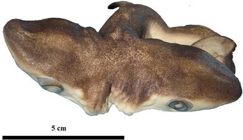
The first abnormal specimen recorded in San Lázaro was a female shark measuring 22.7 cm TL. On the dorsal side there were only one first and second dorsal fin, and one caudal fin; while on the ventral side there was a pair of pectoral and pelvic fins, and one anal fin. It could be considered a normal embryo; however, from the gills slits, two heads were formed. Each head had a mouth and five gills slits on each side of the head, as well as nostrils and eyes (Figure 1).
Anomalous embryo of the blue shark, Prionace glauca from San Lázaro, Baja California Sur, Mexico.

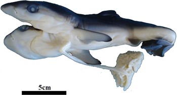
The pregnant female from the Gulf of California (Punta Arenas) had nine embryos in its uterus, one of which presented bicephaly. This embryo was a female measuring 23.8 cm TL. The duplicate heads were formed at the level of the gill slits. Each head had a mouth, a pair of nostrils, eyes and gill slits. However, one head (on the right side), presented only one spiracle. This bicephalic shark showed the common body features of a normal specimen on the dorsal surface: due to the presence of one first and second dorsal fin for each head, it also presented the two lower lobes of the caudal fin. Ventrally there were a pair of pectoral and pelvic fins, one anal fin and one lower lobe of caudal fin; therefore, on the ventral side, the abnormal specimen had the characteristics of one shark.
The specimen also had an umbilical cord (Figure 2). This embryo had two hearts, one of them with hypoplasia, due to the heart on the right side being severely undeveloped. The heart on the left side was normally developed. The stomach and liver on the left side were bigger than the organs on the right side. The undeveloped liver was embedded in the umbilical cord. This umbilical cord was joined to the intestine, which was also joined to the two stomachs. Only one kidney, intestine, rectal gland and epigonal organ (reproductive system) were observed.
Bicephalic specimen of the blue shark, Prionace glauca caught in the Gulf of California (Punta Arenas), Mexico.

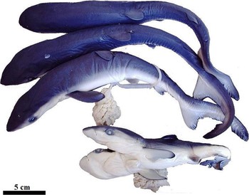
The normal embryos in the uterus of the pregnant female were almost twice the size of the bicephalic specimen. The normal embryos measured an average of 30 cm TL (Figure 3).
Normal pups and anomalous embryo of the blue shark, Prionace glauca from Punta Arenas, Baja California Sur, Mexico.

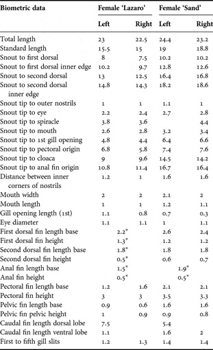
The measurements of each anomalous blue shark are given in Table 1. Some measurements were not possible due to the malformation presented.
Morphometric measurements of abnormal embryos of Prionace glauca, expressed in centimetres (cm).

*, conjoined features.
DISCUSSION
Deformities in sharks during prenatal development usually involve the cephalic region, while postnatal abnormalities involve mainly spinal deformities such as scoliosis, central fusion, and arthritic changes (Bonfil, Reference Bonfil1989). The abnormalities reported do not explain the cause of the deformities, and we can only speculate on the causes, which could be parasitic infection, arthritis, injury, disease, tumours, bad nutrition or genetic abnormality (Heupel et al., Reference Heupel, Simpfendorfer and Bennet1999). We found a genetic abnormality in the blue shark, but we do know its cause.
The uterus area is small for the number of pups and could influence the prenatal development of blue sharks, considering that this shark species is the most fecund of all. Mancini et al. (Reference Mancini, Casas and Amorim2006) explained that the high embryo production in blue sharks, with a maximum reported litter size of 135, could be associated to these abnormalities (Compagno, Reference Compagno1984). In some cases, the small uterine area could prevent placental implantation or cause other effects like mummification (Rosa-Molinar & Williams, Reference Rosa-Molinar and Williams1983). In our study, we deduced that embryonic development continued due to its implantation in the uterus, which provided nutrients for the embryo; however, the transfer of nutrients could have been too low to provide sufficient food for ‘two specimens’ (bicephalic shark), therefore, a different growth was observed in both anomalous embryos, which was seen when compared with the normal embryos: the small size of anomalous blue sharks was evident.
The abnormality recorded is a symmetric bicephaly, with a connection to the xiphoid appendix, which could be caused after or during fertilization by an incomplete zygote division. External factors such as environmental degradation or pollution could influence the rate of occurrence of congenital malformations, but the sharks must be exposed to chemically polluted locations (Grady, Reference Grady1992; Casarini et al., Reference Casarini, Gomes and Tomas1997; Mancini et al., Reference Mancini, Casas and Amorim2006).
We do not consider pollution in the area of Baja California Sur as a possible cause of contaminant exposure in sharks, which could produce an increase in abnormal specimens. Abnormalities during embryonic development could increase the foetal death rate, and although Prionace glauca is a fecund species, it is important to know the factors that can contribute to the high embryonic mortality, considering that shark species have a slow growth.
ACKNOWLEDGMENTS
We thank the fishermen of Punta Arenas and San Lázaro, Baja California Sur, Mexico, who donated the abnormal blue sharks. F.G.-M. thanks the Instituto Politecnico Nacional (COFAA and EDI) for their support in projects on sharks and rays in Baja California Sur. M.C.-F. and O.E.-S. thank PIFI (Plan Institucional de Fomento a la Investigación) and CONACyT (Consejo Nacional de Ciencia y Tecnología) for the scholarships provided.