I. INTRODUCTION
Antibiotics have been widely used for the treatment and prevention of bacterial infections. However, due to their easy access, the misuse and overuse of antibiotics have led to antimicrobial resistance and environment problems around the world. As biorefractory substances, antibiotics are regarded as new pollutants in aquatic environment and have been detected in surface waters in many areas including US, China, some European countries, etc. Massive antibiotics are released through effluents into soil and water, accumulated in drinking water and food, exerting negative influence on people’s health.Reference Wright1–Reference Li, Gao, Shi, Liu and Cai4 Therefore, it is highly desirable to develop an effective way to detect antibiotics in aqueous solution. In the past decades, some analytical methods have been explored for qualitative/quantitative determination of antibiotics such as chromatographic methods, electrophoretic methods, microbiological assays, immunoassays, etc.Reference Lan, Yao, Ping and Ying5 Chromatographic methods are conventional and typical for antibiotic detection, while they are time-consuming and labor intensive.Reference Yang, Yin, Li, Lu, Zhang and Sun6 Electrophoretic methods are more accurate but need complex sample pretreatment.Reference Zhou, Gan, Zhou, Li, Cao and Chen7 Immunoassays are costly with antibodies or enzymes required.Reference Wang, Beier and Shen8
Fluorescent sensing is a powerful tool for the detection of chemical residues.Reference Wang, Zhang and Liang9 But the aggregation-caused quenching effect is a great hurdle for practical applications of many fluorophores in poor solvents or solid states. By contrast, well-designed fluorophores with aggregation-induced emission (AIE) effect show exceptional strong emission upon molecular aggregation due to the restriction of intramolecular motion (RIM) process and are of great significance to technological applications. So far, AIE fluorophores have been used as chemical sensors for the assay of pH, explosive, carbon dioxide, amines, and metal ions with advantages of facile fabrication, ready functionalization, excellent stability, high signal-to-noise ratio, and low detection limit.Reference Wang, Zhao, Lam and Tang10,Reference Hong, Lam and Tang11 Furthermore, various AIE molecules with complex structure were designed and synthesized to obtain efficient fluorescence probes over the past decade. However, the more complex of the molecular structure is, the harder it is to synthesize the molecules. Recently, AIE fluorophores were combined with porous materials to take advantage of their high surface area, tunable pore size, large pore volume, and excellent thermal and chemical stability to obtain new fluorescent sensors.Reference Luo, Xie, Lam, Cheng, Chen, Qiu, Kwok, Zhan, Liu, Zhu and Tang12–Reference Gong, Liu, Wang, Xia, Liu and He14 For instance, metal organic frameworks (MOFs),Reference Zhang, Wang, Liu, Zhang, Tao, Chen and Wei15 porous organic polymers,Reference Wang, Arandiyan, Scott, Bagheri, Dai and Amal16 and mesoporous materialsReference Zhang, Zhang, Wang, Liu, Zhang, Tao and Wei17–Reference Li, Yu and Xu20 have been combined with AIE molecules to develop composite materials. The obtained materials combine the advantages of AIE luminogens and mesoporous materials as emerging fluorescent sensors.Reference Li, Liu, Kwok, Liang, Tang and Yu21,Reference Fan, Li, Yu, Zhang, Cai, Jin and Yu22 For example, Yu’s group combined a representative AIE luminogen, tetraphenylethene (TPE) with mesoporous silica nanoparticles to fabricate AIE functionalized mesoporous material.Reference Miao, Li, Zhang, Yu and Xu23 The obtained materials with AIE characteristics served as a supersensitive and recyclable fluorescent sensor for the detection of picric acid, providing a new candidate for the detection of chemical residues. Tang’s group introduced TPE into MOF via the post-grafting method to form AIE luminogen-functionalized MOF for the detection of volatile organic compounds.Reference Zhang, Feng, Song, Zhou, Chao, Yuan, Tristan, Guo, Hu, Tang, Liu and Zhao24 In this new type of sensor, the movement of the luminogens is restricted by the porous materials because the luminogens are stabilized in the matrix of porous materials, showing amplified sensing performance due to RIM process.
In the above mentioned work, the fluorescent molecules used in these luminogen-functionalized porous materials are mainly focused on the derivatives of TPE, the most representative AIE molecule.Reference Wang, Zhang, Zhang, Zhu and Tang25,Reference Niamnont, Kimpitak, Wongravee, Rashatasakhon, Baldridge, Sieqel and Sukwattanasinitt26 To the best of our knowledge, other common fluorophores are rarely explored to fabricate functionalized porous materials with AIE properties. According to the RIM mechanism of AIE molecules, other easily accessible fluorophores are also expected to show enhanced fluorescence when fixed in the matrix of porous materials for the detection of chemical residuals. Tris(4-bromophenyl)amine (TBPA) is a very common and inexpensive small π conjugated molecule with three benzene rings linked to nitrogen via C–N single bonds. TBPA shows very weak emission itself in solution, but its propeller-type structure makes it expected to show better fluorescence when fixed in the matrix of porous materials. Ordered mesoporous SBA-15 is easy to synthesize with large surface area, high pore volume, two-dimensional hexagonal (p6mm) mesostructure, and ordered parallel channels, which can provide more exposed active sites and enhance mass transport via physical diffusion and strengthen interaction between the sensor and analytes via chemical or electrostatic interaction.Reference Zhang, Zhang, Tao, Chi, Xu and Wei27,Reference Yan, Zhang, Xu and Tian28 In addition, with good chemical stability and reversibility, SBA-15 is a good substrate to modify fluorophores to provide recyclable sensors.
Herein, we report a “fixation-induced emission” (FIE) strategy to develop a fluoresence sensor by post-grafting TBPA on 3-aminopropyltriethoxysilane (APTES)-modified SBA-15. The luminogen-functionalized mesoporous SBA-15 is denoted as SNT. The SNT emits apparent blue light at 523 nm with greatly enhanced emission relative to pure TBPA. In this work, cefalexin was chosen as the model of antibiotics to be detected because it is widely utilized and difficult to degrade in nature.Reference Shukla and Quraishi29 The SNT shows sensitive fluorescence quenching to cefalexin in aqueous solution owing to the photoinduced electron transfer process. In addition, the SNT shows relatively good dispersibility in water as nanoscale particles, which guarantees its better stability and sensitivity for antibiotics detection. Importantly, this probe is recyclable by washing with proper solvents, thus holds potential for practical detection. Compared with previous work, this strategy develops a more economical fluorescent sensor by incorporating inexpensive, easily accessible TBPA into mesoporous SBA-15 and provides a new feasible approach for the detection of antibiotic residues in water.
II. EXPERIMENTAL SECTION
A. Reagents
Poly(ethylene oxide)–poly(propylene oxide)–poly(ethylene oxide) (PEO–PPO–PEO) amphiphilic block copolymer EO20PO70EO20 (Pluronic P123) was purchased from Sigma-Aldrich Corp. (St. Louis, Missouri). APTES and TBPA were purchased from Aladdin Industrial Corporation. Cefalexin was purchased from Shijiazhuang Pharmaceutical Group Company (Shijiazhuang Pharmaceutical Group Company). Tetraethyl orthosilicate, ethanol, acetone, dimethyl sulfoxide (DMSO), and hydrochloric acid (HCl, 36 wt%) were purchased from Tianjin Yongda Chemical Corp (Yongda Chemical Corp). All chemicals were used without further purification. Deionized water was used in all experiments.
B. Preparation of SN
SBA-15 was first synthesized based on the classical method.Reference Chong and Zhao30,Reference Chen, Yu, Wang, Yu, Zhang, Tang and Ma31 To prepare amine-modified SBA-15, the as-made SBA-15 was sealed in an oven at 140 °C for 2 h. Then 1.0 g of SBA-15 was dispersed in 30 mL of toluene, ultrasonically treated for 1 h and vigorously stirred for 1 h. Next, 2 mL APTES was added dropwise to the above solution and stirred at 80 °C for 24 h under reflux. Lastly, amine-modified SBA-15 was obtained after filtering, washing with toluene, and being dried at 60 °C under vacuum, which was denoted as SN.
C. Preparation of SNT
First, 0.25 g of SN and 2 mg of the TBPA were dispersed into 20 mL of DMSO. The slurry was ultrasonically treated for 1 h and vigorously stirred for 24 h at 80 °C. The obtained solution was filtered, washed with ethanol, and dried at 60 °C for 24 h under vacuum. To completely remove the residual uncombined TBPA, the product was washed with acetone several times. The final product was dried in the freezer dryer to obtain SNT.
D. Detection of cefalexin with SNT
2 mg of the as-prepared material SNT was first well dispersed into 2 mL of water and its fluorescence intensity was recorded as I 0 by spectrofluorometer under UV light illumination (305 nm). Immediately, certain amount of cefalexin solution (0.1 mg/mL) was added into the system. Meanwhile, the fluorescence emission spectra for the samples were recorded with the addition of different amounts of cefalexin in water solution until the fluorescence was almost quenched.
E. Characterization
Scanning electron microscopy (SEM) analysis was conducted using a HITACHI S-4800-I scanning electron microscope (Hitachi, Milpitas, California). High-resolution transmission electron micrographs were obtained on a JEOL JEM-2010 electron microscope (JEOL, Tokyo, Japan). Wide-angle X-ray diffraction (XRD) patterns were achieved using a Rigaku D/MAX-2500 XRD system (Cu Kα radiation, λ = 0.15406 nm; Rigaku, Tokyo, Japan) operated at 40 kV and 40 mA. Nitrogen adsorption–desorption isotherm measurements were performed on a Micromeritics TriStar 3020 volumetric adsorption analyzer at 77 K (Micromeritics, Osaka, Japan). The Brunauer–Emmett–Teller (BET) method was utilized to calculate the specific surface area of each sample and the average pore size distribution was derived from the desorption branch of the corresponding isotherm using the Barret–Joyner–Halenda (BJH) method. The total pore volume was estimated from the amount of N2 adsorbed at the relative pressure (P/P 0 = 0.97). The emission spectra were obtained on a F97 pro spectrofluorometer (Shanghai Lengguang Tech, Shanghai, China). The fluorescence microscopy image was obtained using an Inverted Fluorescence Microscope (DMIL LED 364704, Leica, Shanghai, China).
III. RESULTS AND DISCUSSION
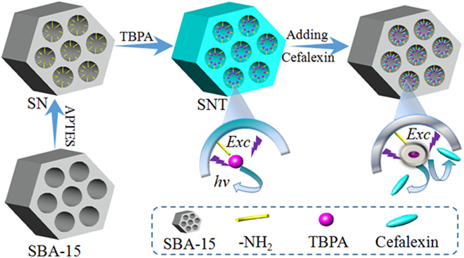
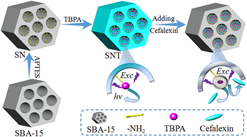
The fabrication process of SNT and sensing for cefalexin is illustrated in Scheme 1. First, the as-made SBA-15 was modified with APTES by the post-grafting method, providing active NH2 groups on SBA-15. The obtained SN was then functionalized with certain amounts of TBPA molecules via covalent bond and TBPA was fixed on rigid SBA-15 to obtain SNT. The SNT shows enhanced fluorescence in water under UV irradiation because intramolecular movement of the fixed TBPA molecules is restricted, which blocks the radiationless relaxation channel and opens the radiative decay pathway. With the increased amount of cefalexin added into SNT solution, the fluorescence intensity of SNT solution gradually became weaker until completely quenched. The fluorescence quenching of SNT can be explained by the photoinduced electron transfer quenching mechanismReference Wang, Zhang, Zhang, Zhu and Tang25,Reference Niamnont, Kimpitak, Wongravee, Rashatasakhon, Baldridge, Sieqel and Sukwattanasinitt26,Reference Deng, Chen, Chien and Hong32 as shown in Scheme 1. Cefalexin is weakly acidic in water as an amphoteric compound with isoelectric point of about 5.0. The SNT is basic due to modified NH2 groups, some of which have been substituted by TBPA and some of which are still remained on SNT. When cefalexin is added to the solution of SNT, it can quickly diffuse into the pores and be effectively adsorbed on the pores of SBA-15 forming a cefalexin-amine complex via acid–base interaction and physical adsorption. The close vicinity between TBPA and cefalexin greatly facilitates the electron transfer process and thus quenches the emission.
Illustration for the fabrication of SNT and detection of cefalexin based on photoinduced electron transfer.

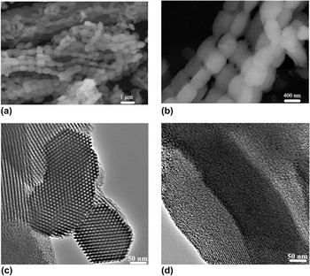
The morphology and structure of SNT are investigated by using SEM and TEM images. As shown in Fig. 1(a), the SNT exhibits adhesive rod-like morphology. In the high-magnified image [Fig. 1(b)], the length and width of SNT are evaluated to be 530 and 320 nm, respectively. Furthermore, the TEM images of SNT are observed. The TEM images [Figs. 1(c) and 1(d)] show more detailed characteristics of SNT with the p6mm mesostructure and ordered parallel channels observed from (100) and (110) orientation,Reference Zhao, Sun, Li and Stucky33 respectively, proving that the mesostructure of SNT is well preserved after functionalization with TBPA.
SEM images (a and b) and TEM images (c and d) of SNT.

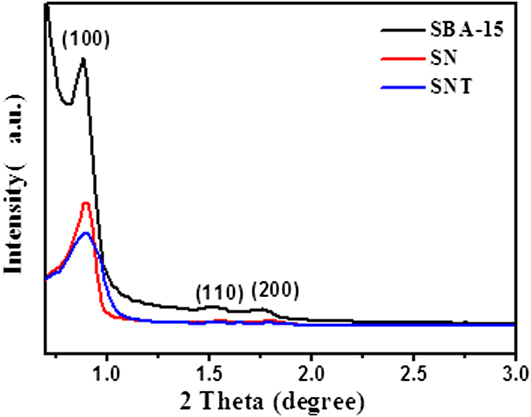
The small-angle XRD patterns of SBA-15, SN, and SNT are depicted in Fig. 2. For SBA-15, the three well-resolved diffraction peaks display at 2θ around 0.9–1.9°, respectively, corresponding to the (100), (110), and (200) planes, which are well consistent with the characteristics of ordered pores and the space group p6mm mesostructure symmetry, indicating a well-defined SBA-15 mesostructure.Reference Marakatti and Peter34 Compared with SBA-15, the peak intensity of SN is significantly weaker, which has a relatively poor-ordered mesophase due to the loading of aminopropyl groups inside the channel. For the SNT sample, which was further modified by TBPA molecules, only one obvious broad peak at 2θ ranging from 0.8 to 0.9 is observed and the peak shifts toward higher angle slightly, implying the mesopore ordering decreases slightly due to the adverse effect of TBPA on the structure of SNT.
Small-angle XRD patterns of SBA-15, SN, and SNT.

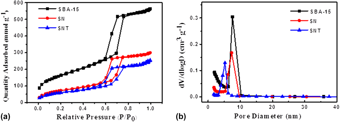
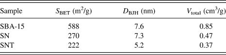
To explore the pore structure of SNT, the N2 adsorption–desorption experiments of SBA-15, SN, and SNT were performed. As shown in Fig. 3(a), the N2 adsorption–desorption isotherms of SBA-15, SN, and SNT all belong to a typical type IV isotherm characteristic with a clear H1 hysteresis loop in a wide P/P 0 range of 0.5–0.8, indicating the presence of highly ordered mesopores and narrow pore size distribution typical of pure SBA-15.Reference Marakatti and Peter34,Reference Thommes, Smarsly, Groenewolt, Ravikovitch and Neimark35 The BET surface area (S BET), pore size (D BJH), and total pore volume (V total) are given in Table I. The corresponding pore size distribution calculated using the BJH method shows that the pore size of SBA-15 centered at 7.8 nm. By contrast, SN and SNT have a smaller pore size distribution centered at 7.3 nm and 5.2 nm, respectively [Fig. 3(b)]. Moreover, both the specific surface area and total pore volume of SN and SNT are reduced in comparison with SBA-15. These results indicate that the order of the mesopore structure of SN and SNT become weaker after modification by –NH2 groups and TBPA, which are well consistent with the results of XRD.
N2 adsorption–desorption isotherms (a) and pore size distribution curves (b) of SBA-15, SN, and SNT.

Textural parameters of SBA-15, SN, and SNT.

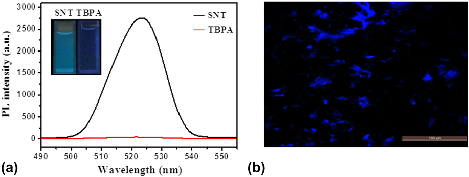
Normally, SBA-15 and SN are not luminescent. But the SNT shows strong fluorescence intensity after modification by TBPA. The emission spectra of SNT in the water solution and the fluorescence microscopy image of SNT powder are shown in Fig. 4. From Fig. 4(a), a strong emission of SNT is observed upon UV irradiation (305 nm), and the maximum photoluminescence intensity is 2754, which is far stronger than pure TBPA. The strong emission was attributed to the FIE effect. As we know, the framework of SBA-15 is rigid. When free TBPA molecules are incorporated on the channel surface of SBA-15 through chemical bond, their intramolecular movement is restricted then the emission is enhanced.Reference Zhang, Zhang, Wang, Liu, Zhang, Tao and Wei17 In addition, under UV irradiation (254 nm) [insert of Fig. 4(a)], the pure TBPA shows no fluorescence emission, while the SNT emits bright blue light, indicating the apparent FIE effect. In Fig. 4(b), the fluorescence microscopy image of SNT powder shows blue fluorescence. This phenomenon indicates that the TBPA molecules have been successfully grafted onto SBA-15 by reacting with the –NH2 groups.
(a) Fluorescence spectra of SNT and TBPA in water solution and (b) fluorescence microscopy image of SNT powder.

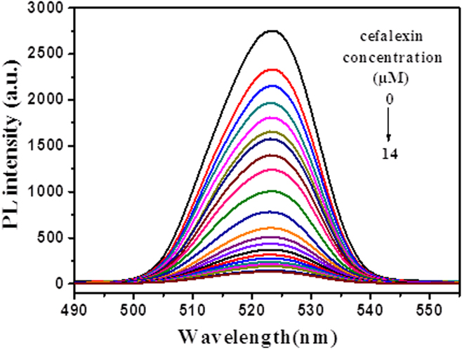
The SNT was used for the detection of cefalexin in water solution. Figure 5 shows the fluorescence emission spectra of SNT after addition of different amounts of cefalexin in water solution. The SNT shows strong emission in water solution, and the fluorescence intensity decreases significantly with increasing of cefalexin amount until the fluorescence almost completely quenched, which can be explained by the photoinduced electron transfer mechanism.Reference Zhang, Zhang, Tao, Chi, Xu and Wei27,Reference Yan, Zhang, Xu and Tian28,Reference Deng, Chen, Chien and Hong32 The emission is almost invisible when the cefalexin concentration is increased to 41 μM.
Fluorescence emission spectra of SNT with the addition of different concentrations of cefalexin in water.

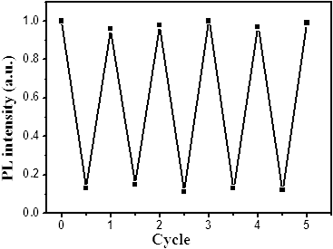
Due to insolubility of SNT in water and common organic solvents, the sensor can be recycled by washing with proper solvents for repeated detection. The repeated detection and recovery experiments are carried out and five-cycle results are recorded. The whole process is as follows: 2 mL of SNT solution (1.0 mg/mL) was used as the sensing sample and its fluorescence emission was recorded under UV irradiation (305 nm). Then cefalexin solution (340 μL, 0.1 mg/mL) was added into the SNT solution immediately and stirred for about 1 min. The fluorescence emission of the mix solution was also recorded. After that, the mixture was centrifuged and SNT was collected and then washed with acetone. After being dried completely in the freezer dryer, the regenerated SNT was subjected to the next detection process. The fluorescence spectra of recycled SNT before and after addition of cefalexin were characterized and the corresponding fluorescence intensity was recorded. The results from five repeated experiments showed that the regenerated SNT still have excellent detection performance (Fig. 6). The PL intensity of the SNT is similar to that of the initial state, proving that the SNT exhibits good recyclability and can be used as recyclable detectors for Cefalexin.
The quenching and recovery test of SNT in water solution.

IV. CONCLUSIONS
In summary, we have prepared the luminogen-functionalized mesoporous silica nanoparticle SNT via post-grafting TBPA on APTES-modified SBA-15. The results show that the SNT has greatly enhanced emission based on FIE strategy and is used as the fluorescent sensor for the detection of antibiotic cefalexin in water. The materials can be regenerated by simply washing and drying, suggesting that they can be used as a recyclable fluorescence probe for antibiotics detection. This strategy can be extended to develop inexpensive, high-performance, recyclable fluorescent sensors by combining fluorophores with mesoporous materials.
ACKNOWLEDGMENTS
This work was financially supported by the National Natural Science Foundation of China (21676070), Hebei Natural Science Foundation (B2015208109), Hebei Training Program for Talent Project (A201500117), Hebei One Hundred-Excellent Innovative Talent Program (III) (SLRC2017034), Hebei Science and Technology Project (17214304D), and the Excellent Going Abroad Experts’ Training Program in Hebei Province.