The Fontan procedure has been performed for almost 50 years and is the standard definitively staged surgery in patients with a functional single ventricle. This surgery enabled the resection and reconstruction of the superior caval vein and inferior caval vein bypassing the heart and allowing the blood to flow passively forward into the pulmonary circulation without right ventricular pumping. As a result of the development of these surgical techniques, the survival of these patients can extend into adulthood. However, because of the unique nature of the Fontan physiology, which consists of persistent central venous hypertension along with chronic liver ischaemia caused by a decreased cardiac output state, there were resulting consequences of long-term complications including plastic bronchitis, protein-losing enteropathy, liver fibrosis, liver nodules, and hepatocellular carcinoma , all of which influence the quality of life. Reference Rychik, Goldberg and Rand1 When systemic venous pressure became elevated, portal deprivation was compensated by a hepatic arterial buffer response. Reference Agnoletti, Ferraro and Bordese2–Reference Eipel, Abshagen and Vollmar4 The liver injury following a Fontan operation is referred to as Fontan-associated liver disease and is recognised by emerging complications in patients worldwide. The prevalence of liver nodules and HCC increases in post-Fontan patients. The incidence of HCC was 1.3% and the survival rate of asymptomatic patients was significantly better than in patients with symptoms at the time of HCC diagnosis. Reference Egbe, Poterucha and Warnes5 Liver biopsy is still the validated gold standard method for the diagnosis of liver nodules. However, this procedure is an invasive tool that can carry a risk of complications. Appropriate non-invasive modalities for the non-inferior diagnosis of FALD have developed, including serologic biomarkers, ultrasound upper abdomen, ultrasound liver elastography, CT scan, and magnetic resonance elastography, which are currently still inconclusive. Several studies reported that a CT scan could detect liver nodules better than upper abdominal ultrasound. Reference Tellez, Rodriguez de Santiago and Minguez6–Reference Horvat, Rocha and Chagas8 Therefore, an abdominal CT scan can early detect the liver nodules before these nodules progress to hepatocellular carcinoma. The aims of this study were to determine the prevalence and associated factors for the presence of liver nodules using a CT scan of the upper abdomen in patients following the Fontan operation.
Materials and methods
A cross-sectional study included 34 patients above the age of 15 following the Fontan operation. All the patients underwent an ultrasound of the upper abdomen, an ultrasound liver elastography, and a CT scan of the upper abdomen after the Fontan procedure. Those patients with underlying liver disease were excluded. Data collection included the age at the Fontan operation, sex, diagnosis of CHD, and the types of the Fontan operation. Laboratory tests consisted of a complete blood count, liver function test, gamma-glutamyl transferase, a fibrosis score that included the aspartate aminotransferase-to-platelet ratio index, and a fibrosis-4 score that was calculated on the basis of age, AST, ALT, and platelet count. Reference Wu, Earing and Aboulhosn9 The cardiovascular evaluation consisted of echocardiography and cardiac catheterisation. The imaging for the evaluation of FALD included an ultrasound of the upper abdomen, ultrasound liver elastography, and a CT scan of the upper abdomen. An ultrasound of the upper abdomen and ultrasound liver elastography would be performed by an experienced radiologist. The ultrasound system used was a GE-E 10 with low-frequency curved array transducers C1-6 (2–5 MHz). Echocardiographic data consisted of the single ventricular morphology, the degree of atrioventricular valve regurgitation, and the ventricular function of the systemic ventricle including an ejection fraction, right ventricular fractional area change, mitral annular planar systolic excursion, tricuspid annular planar systolic excursion, and tissue Doppler imaging. Upper abdominal CT scans were performed in Fontan patients without contraindication to iodine using a standardised protocol on a SIEMENS Healthineers – SOMATOM Force. There were three phases consisting of non-contrast, arterial (25–30 s), portovenous (60 s), and delay phases (3 minutes). Omnipaque 350 was used in the amount of 2 ml/kg in 3 ml/s in each patient. MRI of the upper abdomen with hepatocyte-specific agent would be performed in patients who had been suspected of HCC by an upper abdominal CT scan. The catheterisation data included mean pulmonary artery pressure, Fontan pressure, inferior caval vein pressure, hepatic vein pressure, Nakata index, pulmonary vascular resistance index, and single ventricular end-diastolic pressure. The ultrasound findings of the upper abdomen consisted of liver cirrhosis and liver nodules. FALD was defined as severe hepatic fibrosis (>15 kPa), cirrhosis, or hepatic nodules as documented by an ultrasound of the upper abdomen, ultrasound liver elastography, and a CT scan of the upper abdomen.
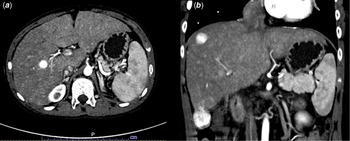
Upper abdominal CT scan of a 16-year-old man with tricuspid atresia/pulmonary atresia, S/P left modified Blalock–Taussig shunt (2006), S/P bidirectional Glenn shunt (2007), S/P extracardiac Fontan with fenestration (2015) demonstrates multiple various size round shaped arterial enhancing lesions with underlying liver cirrhosis and liver congestion. (a) Cross-sectional view. (b) Coronal view.

Statistical analysis
The data were analysed in the form of descriptive statistics with categorical data and quantitative data using SPSS version 23 (SPSS Inc., Chicago, IL, United States of America). Test variables were analysed by the Mann–Whitney U-test and the chi-square test. All of the tests for significance resulted in a p-value <0.05. Logistic regression analysis was used to identify the associated factors of the liver nodules. The intertest agreement was assessed using weighted Cohen’s kappa values that were interpreted as follows: 0.00–0.20, slight agreement; 0.21–0.40, fair agreement; 0.41–0.60, moderate agreement; 0.61–0.80, substantial agreement; and 0.81–1.00, nearly perfect agreement.
Results
Thirty-four patients (17 males) underwent an ultrasound of the upper abdomen, ultrasound liver elastography, and a CT scan of the upper abdomen after the Fontan operation. The median age was 20 years (range 14–36 years). The median age at the time of the Fontan operation was 7 years (range 5–17 years) and the duration following the Fontan operation was 12 years (range 4–22 years). The diagnoses were as follows: tricuspid atresia, 13 cases (37%); double inlet left ventricle, 5 cases (15 %); unbalanced atrioventricular septal defect, 5 cases (15%); heterotaxy syndrome, 2 cases (6%); mitral atresia, 2 cases (6%); pulmonary atresia/intact ventricular septum, 2 cases (6%); and other diagnoses, 5 cases (15%). Twenty-five patients (74%) underwent a bidirectional Glenn shunt prior to the Fontan operation. The majority (91%) of patients had unilateral superior caval vein. The types of Fontan operations consisted of an atriopulmonary connection, 2 cases (6%); lateral tunnel, 1 case (3%); and an extracardiac conduit, 31 cases (91%). Fifteen patients (53%) underwent a fenestration.
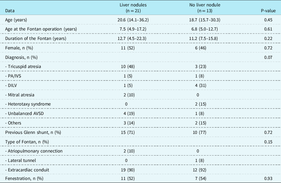
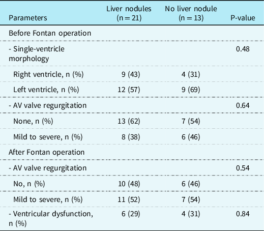
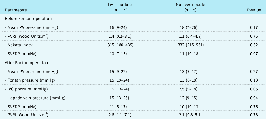
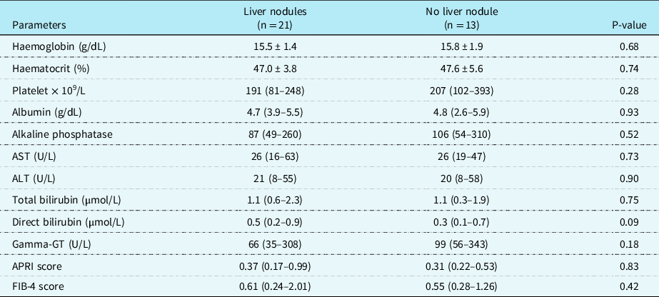
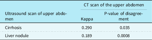
Twenty-one patients (62%) developed liver nodules, as detected by a CT scan of the upper abdomen. Eleven patients had liver nodules larger than 1 centimeter (Fig 1). Only eight patients (24%) revealed the presence of liver nodules in ultrasound of the upper abdomen. Fifteen patients in whom the presence of liver nodules was detected by a CT scan underwent an MRI with hepatocyte-specific contrast. Four patients were diagnosed with HCC based on an MRI with hepatocyte-specific HCC. Microwave ablation was performed in one patient. A comparison of the demographic data, echocardiographic parameters, and cardiac catheterisation data before and after the Fontan operation is shown in Tables 1–4. Also shown in these tables are the laboratory tests contrasting patients both with and without liver nodules. The baseline characteristics of the two groups showed no differences. The post-operative data including cardiopulmonary bypass time, post-operative mean pulmonary artery pressure, arrhythmia, renal failure, and protein-losing enteropathy showed no significant differences between the two groups. The laboratory tests between the two groups were similar. Echocardiography data at pre- and post-Fontan operation, which involved single-ventricle morphology, atrioventricular valve regurgitation, and ventricular dysfunction were not significantly different between the two groups. Results from a haemodynamic study following the Fontan operation showed that only hepatic pressure was the only parameter that was significantly higher in patients who had liver nodules than in those without liver nodules. In a univariate logistic regression analysis, the associated factor for liver nodule was a hepatic vein pressure of more than 13 mmHg [odds ratio 18.0 (95% CI 2.7–120.9)]. Twenty-six patients (76%) were found to have FALD. The agreement between the ultrasound of the upper abdomen and the CT scan in detecting the presence of liver nodules and cirrhosis was low with Kappa = 0.290 and 0.189, respectively, (Table 5).
Comparison of the demographic data in patients following the Fontan operation.

AVSD = atrioventricular septal defect; CPB = cardiopulmonary bypass; DILV = double inlet left ventricle; PA/IVS = pulmonary atresia/intact ventricular septum
Comparison of the echocardiography data in patients following the Fontan operation.

AV = atrioventricular
Comparison of cardiac catheterisation data in patients following the Fontan operation.

IVC = inferior caval vein; PA = pulmonary artery; PVRi = pulmonary vascular resistance index; SVEDP = single ventricular end-diastolic pressure
Comparison of laboratory data in patients following the Fontan operation.

ALT = alanine transaminase; APRI = AST-to-platelet ratio index; AST =Aspartate transaminase; FIB-4 = fibrosis 4; Gamma-GT = gamma-glutamyl transferase
Intertest agreement between an ultrasound and a CT scan of the upper abdomen in detecting cirrhosis and liver nodules.

Discussion
Occurrences of liver disease after the Fontan operation were detected in the early 1980s after the first case report of cardiac cirrhosis following the publication of the Fontan operation. Reference Lemmer, Coran, Behrendt, Heidelberger and Stern10 After conducting additional investigations, more cases of a liver involvement were detected following the Fontan operation. In this study, the presence of liver nodules was detected in 19 patients (62%) by the use of a CT scan of the upper abdomen following the Fontan operation. Liver nodules were detected in only 24% of patients by the use of the ultrasound of the upper abdomen. None of the patients showed any symptoms. Horvat et al conducted a study of 49 patients following their Fontan operations between 2014 and 2016. More than one-third of these patients had liver nodules, as detected by the use of CT or MRI imaging, although the ultrasound did not detect the presence of liver nodules. Reference Horvat, Rocha and Chagas8 In this study, there was very little agreement between the ultrasound of the upper abdomen and the CT scans used in detecting the presence of liver nodules and cirrhosis. For that reason, after 15 or more years following the Fontan operations, patients should undergo a CT scan of their upper abdomens for evaluation of their liver nodules, regardless of whatever symptoms they may have. It is necessary to distinguish between a benign liver nodule and HCC, because the earlier the treatment, the better the survival outcome will be. Possner et al reported that cumulative survival was worse in patients with symptomatic HCC, with a 1-year survival of 31% versus 65% in patients with asymptomatic HCC (p = 0.025). Reference Possner, Gordon-Walker and Egbe11 Yoon JS, et al studied the clinical characteristics between patients diagnosed with HCC and benign arterial phase hyperenhancing nodules. They found that the time elapsed since the Fontan operation, the largest nodule diameter, and serum AFP were significantly greater in patients with HCC than benign nodules. Reference Yoon, Lee and Cho12 In addition, Ohuchi H, et al studied in 339 consecutive Fontan patients demonstrated that elevated APRI and FIB-4 scores together with older age and high plasma levels of AFP could be useful predictors of HCC in patients following Fontan operation. Reference Ohuchi, Hayama, Nakajima, Kurosaki, Shiraishi and Nakai13 Moreover, Egbe AC, et al reported that patients with HCC had a longer duration of post-Fontan operation along with high serum AFP levels. Reference Egbe, Poterucha and Warnes5 The MRI with hepatocyte-specific HCC should be performed when the HCC feature was detected by CT scan, including enhancement during the arterial phase and wash out during portal venous phase together with elevated AFP level. HCC feature on CT scan can also occur in a benign liver nodule in post-Fontan patients due to hepatic arterial buffer response. The MRI with hepatocyte-specific HCC can be differentiated HCC from the benign fibrotic liver nodule using the uptake functioning hepatocyte. Reference Wells, Hough, Fidler, Kamath, Poterucha and Venkatesh14 In this study, three patients were diagnosed with HCC, based on CT scans of their upper abdomens, along with MRI with hepatocyte-specific HCC. Liver biopsy has thus far been the gold standard for the diagnosis of HCC. Nevertheless, it may not be vital if a CT scan of the upper abdomen and an MRI with hepatocyte-specific HCC demonstrates hallmark diagnostic features of HCC. All HCC patients were still asymptomatic. None of the data, including the baseline characteristics, duration after the Fontan operation, type of Fontan operation, laboratory tests, and echocardiography, differed significantly between the presence and absence of liver nodules.
When systemic venous pressure elevated, the patients developed congestive hepatopathy. This had consequently led to indirect hyperbilirubinemia and increased gamma-glutamyl transferase in the early phase. Following liver ischaemia, it will progress to liver fibrosis, cirrhosis, and finally to liver nodules or hepatocellular carcinoma. In this phase, the laboratory shows a transaminitis profile. Portal deprivation was compensated by a hepatic arterial buffer response. In the last phase, the patients have a sign of portal hypertension with synthetic liver dysfunction. Reference Agnoletti, Ferraro and Bordese2–Reference Eipel, Abshagen and Vollmar4 In this study, 26patients (76%) were found to have FALD by the use of ultrasound liver elastography and a CT scan of their upper abdomens. Several cohort studies for early detection of FALD were still considered controversial at the time. Fidai et al demonstrated that laboratory tests including detection of increased haemoglobin, alkaline phosphatase, gamma-glutamyltransferase, bilirubin, APRI, and a low white blood cell count and platelet level can be used for the observation of FALD in adult patients. Reference Fidai, Dallaire and Alvarez15 Chen et al demonstrated that laboratory tests for liver disease in post-Fontan patients had indicated a decrease in platelets and albumin levels, and increased AST, ALT, GGT, and APRI. Reference Chen, Schreiber, Human, Potts and Guttman16 In addition, Smas-Suska et al revealed that a decrease in platelets and increased AST, GGT, APRI, and FIB-4 scores may be used to assess the severity of liver fibrosis. Reference Smas-Suska, Skubera and Wilkosz17 Furthermore, many cohort studies showed that laboratory tests in post-Fontan patients revealed that they had normal symptoms. Reference Wu, Earing and Aboulhosn9,Reference Goldberg, Surrey and Glatz18–Reference Kim, Lee and Jang21 These inconsistent findings could be explained by different multiple factors which contribute to liver injury, including the severity and duration of hypoxia prior to the Fontan operation, time elapsed from the Fontan surgery, or degree of hepatic buffer response mechanism. In this study, no factor was associated with FALD. Therefore, there is no biochemical marker that is generally accepted to assess liver disease after a Fontan operation.
There was no correlation of single-ventricle morphology, atrioventricular valve regurgitation, or single ventricular dysfunction with the liver nodules in this study. Goldberg et al revealed that ventricular function and atrioventricular valve regurgitation were not associated with the collagen deposition and the presence of liver nodules. Reference Goldberg, Surrey and Glatz18 During cardiac catheterisation following the Fontan operation, only the hepatic vein pressure in patients with liver nodules was significantly higher than in those with no liver nodules. In a univariate logistic regression analysis, the associated factor for liver nodules was found to be a hepatic vein pressure of more than 13 mmHg (odds ratio = 18). It could be explained by the persistent high central venous pressure that had been directly transmitted to the hepatic vein, leading to liver congestion, inflammation, and fibrosis, all of which ultimately developed into liver nodules. Poterucha et al reported a high hepatic vein pressure of 17 ± 3 mmHg in 30 Fontan patients. Reference Poterucha, Johnson and Qureshi22 Kiesewetter et al demonstrated that the extent of cirrhosis was correlated with the hepatic vein pressure. Reference Kiesewetter, Sheron and Vettukattill23 Wu et al demonstrated that the liver histopathology of 68 patients following the Fontan operation led to a high prevalence of portal and centrilobular hepatic fibrosis. Reference Wu, Jonas and Opotowsky24 The limitation of this study was the small number of patients, who following their Fontan operations, underwent CT scans of their upper abdomens.
Conclusions
Liver nodules are commonly found in adult Fontan patients. High hepatic pressure was the only factor associated with liver nodules in post-Fontan patients. A CT scan should be used as the observation modality for the assessment of liver nodules in all elderly patients following their Fontan operations.
Acknowledgements
None.
Financial support
This research received no specific grant from any funding agency, commercial, or not-for-profit sectors.
Conflict of interest
The authors have no conflicts of interest to declare.
Ethical standards
The authors assert that all procedures contributing to and reported within this work comply with the ethical standards of the relevant national guides.