Introduction
Otosclerosis is a progressive disorder of otic capsule bone metabolism. In its advanced form, it can lead to footplate obliteration, retrofenestral disease and cochlear ossification. In 1.6 per cent of otosclerosis patients, there is a development of a progressive profound sensorineural hearing loss from ossification of the round window membrane or cochlear scalae.Reference Castillo, Polo, Gutierrez, Reyes, Royuela and Alonso1,Reference Kabbara, Gauche, Calmels, Lepage, Escude and Deguine2 Ossification of the round window membrane is seen in 60 per cent of patients with advanced otosclerosis.Reference Marchioni, Soloperto, Bianconi, Guarnaccia, Genovese and Presutti3 With indications for cochlear implantation expanding in the present era, advanced otosclerosis has become an increasingly common aetiology for hearing loss that is not amenable to conventional hearing amplification and when speech recognition scores meet cochlear implantation candidacy. There are a heterogeneous group of studies that have reported reasonable outcomes for cochlear implantation in patients with advanced otosclerosis.Reference Assiri, Khurayzi, Alshalan and Alsanosi4 Advanced otosclerosis poses surgical challenges with regards to cochlear implantation in the presence of round window or cochlear ossification. Abnormal bone formation may necessitate surgical drilling in the vicinity of the round window for access or the use of an alternative approach, such as subtotal petrosectomy.Reference Vashishth, Fulcheri, Rossi, Prasad, Caruso and Sanna5 Electrode placement may also be difficult because of cochlear ossification, leading to a non-optimal position.
High-resolution computed tomography (CT) and T2-weighted magnetic resonance imaging (MRI) have been used to identify obstruction in such cases. Although high-resolution CT has been used in detecting otosclerosis with a sensitivity approaching 90 per cent, it is less sensitive in detecting luminal obstruction. High-resolution CT for detecting cochlear luminal obstruction has been reported in the literature with a sensitivity between 33–76.5 per cent and specificity of 88–100 per cent.Reference Frau, Luxford, Lo, Berliner and Telischi6,Reference Bettman, Beek, Van Olphen, Zonneveld and Huizing7 Some comparative studies have not found a significant difference between MRI or high-resolution CT in detecting obstruction. Bettman et al. suggested CT to be equivalent to MRI in predicting cochlear patency.Reference Bettman, Beek, Van Olphen, Zonneveld and Huizing7 Obliteration of the round window niche is a frequent finding in otosclerosis patients and can normally be predicted on high-resolution CT.Reference Castillo, Polo, Gutierrez, Reyes, Royuela and Alonso1,Reference Marshall, Fanning, Symons, Shipp, Chen and Nedzelski8–Reference Rotteveel, Proops, Ramsden, Saeed, van Olphen and Mylanus10 The majority of studies evaluating the use of imaging to predict outcomes for cochlear implantation in otosclerosis have only assessed round window ossification. Although both CT and MRI have been shown to be moderately sensitive and very specific for luminal obstruction, there is a lack of evidence correlating pre-operative imaging with post-implant hearing outcomes. Vashishth et al.Reference Vashishth, Fulcheri, Rossi, Prasad, Caruso and Sanna5 observed good auditory outcomes despite radiological evidence of cochlear ossification and did not identify any difference between those with versus those without ossification.
The current study was conducted to review patients with otosclerosis who underwent cochlear implantation, analyse post-implantation auditory outcomes, and determine whether these can be predicted based on pre-operative imaging, intra-operative features and final electrode position. The secondary aims of this study were to identify pre-operative features on imaging that would help predict possible surgical difficulties, assist in the decision-making when selecting the appropriate electrode array and optimise electrode placement intra-operatively. An additional aim was to determine the incidence of post-operative facial nerve stimulation.
Materials and methods
Institutional ethics review board approval was obtained. Patients were identified through the cochlear implant audiological database, sorted by aetiology of hearing loss. Retrospective chart review of those patients in the database was conducted to confirm that all patients had a history of otosclerosis and received a cochlear implant between 2014 and 2020 to allow for sufficient 12-month follow up. Patients were identified as having a diagnosis of otosclerosis confirmed with previous stapes surgery and/or radiological evidence of otosclerosis.
All advanced otosclerosis patients included in the study also required pre-operative imaging (CT and/or MRI) and a post-operative cone-beam CT scan to assess electrode position. All patients had pre-operative high-resolution CT of the petrous temporal bone. Some patients did not have pre-operative MRI because of a history of previous stapedectomy and unknown MRI compatibility of the stapes prosthesis. Demographic data as well as intra-operative implantation details were collected and retrospectively reviewed to determine if there were difficulties in performing the cochleostomy or electrode insertion. Any evidence of facial nerve stimulation was recorded at the time of switch-on and 3 months and 12 months post-implantation.
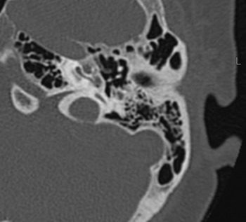
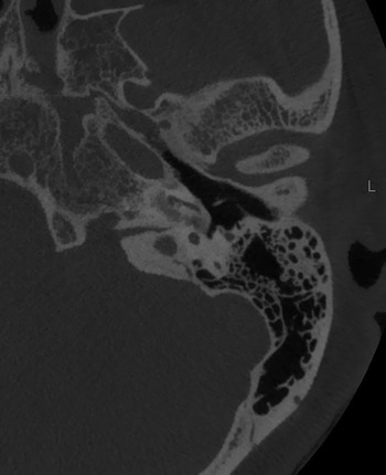
Pre-operative and post-operative radiographic scoring was performed independently by two senior otologists. Where there was discordant data, cases were reviewed together until a consensus was achieved. Pre-operative radiographic scoring was scored as ‘0’ for normal imaging, ‘1’ for round window obstruction (Fig. 1) and ‘2’ for any evidence of narrowing of the cochlear lumen (Fig. 2) or reduced scala signal (Fig. 3). Most patients had pre-operative MRI, but where this was not possible (i.e. presence of non-compatible stapes prosthesis), the pre-operative CT scan was utilised to assess any evidence of scala narrowing. Post-operative electrode position was graded as ‘0’ for scala tympani insertion and a ‘2’ for scala vestibuli insertion. In circumstances where the scala position was ambiguous, without the use of image segmentation to further characterise it, a grading of ‘1’ was given in order to give more accurate subgroup analysis.
High-resolution cone-beam computed tomography of left temporal bone demonstrating round window ossification with fenestral and retrofenestral otosclerosis. The basal turn (incompletely visualised in this image) also narrowed.

High-resolution cone-beam computed tomography of left petrous temporal bone demonstrating abnormally narrowed basal turn of cochlea.

High-resolution T2-weighted magnetic resonance imaging demonstrating abnormally narrowed right basal turn with loss of signal in the region of the scala tympani. Normal left side for comparison.

Intra-operative scoring was graded a ‘0’ for no surgical difficulty, ‘1’ for difficult identification of the round window requiring subjective determination for optimal cochleostomy position, and ‘2’ for difficult electrode insertions (such as extending drilling of scala, opening of scala vestibuli, usage of depth gauge or partial insertion). All surgical procedures were performed under general anaesthesia with facial nerve monitoring. A mastoidectomy with facial recess approach was performed. The receiver-stimulator package was placed in a subperiosteal pocket. Electrode insertions were performed atraumatically via the round window into the scala tympani when possible. In the presence of significant intra-operative ossification of the scala tympani, a drill-out was performed until luminal patency was encountered or scala vestibuli insertion was performed. The standard technique was to identify the round window and either perform a round window approach or an extended round window approach. In the absence of an identifiable round window, such as an obliterated round window, the stapes was utilised as a landmark to perform a cochleostomy 2 mm inferiorly and slightly posteriorly. Neural response telemetry was performed at the conclusion of the operation. Post-operative CT was performed post-procedure to confirm electrode position.
Patients underwent speech perception assessment before and after the procedure. Testing included open-set monosyllabic word testing in quiet (consonant-vowel-consonant words) scored based on correct phonemes and words. Separate ear and binaural testing were completed when there was useful residual hearing in both ears. Post-operative assessments were performed 3 and 12 months after implantation. The pure-tone average was calculated as a five-frequency average hearing loss (250, 500, 1000, 2000 and 4000 Hz). Where there was no response at the limit of the audiometer, 125 dB was recorded. Audiological variables of interest included: duration of deafness in the implanted ear, pure tone average and pre-implant phoneme scores for the implanted ear. Duration of deafness was defined as duration of severe (more than or equal to 70 dB or worse) hearing loss. This was calculated based on evidence of hearing thresholds in the severe range. In the absence of hearing threshold data, duration of deafness was calculated from the time point that the recipient reported last being able to use the phone. The post-operative phoneme scores at 3 months and 12 months were used as a measure of hearing outcome.
Speech perception scores were compared with a larger cohort (n = 1414) of post-lingually deaf adults with bilateral hearing loss who received implants in Melbourne from January 2000 to June 2020 with otherwise normal cochlear anatomy. This larger cohort excluded those with documented otosclerosis as the aetiology, non-English speakers, and some patients with psychiatric or medical conditions precluding formal testing.
All statistical analysis was conducted using Stata 13 (Stata-Corp, College Station, USA) and Minitab (Minitab, State College, USA) statistical software. Values of p < 0.05 were considered statistically significant, and t-tests were performed for speech perception score comparisons. Where the data did not follow normal distribution, non-parametric tests, such as the Mann–Whitney U-test and Wilcoxon signed-rank test were performed. Chi-square tests and Pearson pairwise correlations were performed to look for associations between pre-operative imaging scores, intra-operative findings, and final post-operative electrode position. In the analysis of post-operative electrode position, data were analysed as both independent and as binary terms, either scala tympani or not (the indeterminate cases were considered not scala tympani insertions). For pairwise comparisons, an r-value of more than 0.5 was considered strong, 0.3–0.5 moderate and 0.1–0.3 weak correlation.Reference Cohen11
Results
Demographic data
A total of 48 patients were included in this study, and 52 ears were implanted from 2014 to 2020. Forty-four patients had unilateral implantations during the study period, and 4 patients had sequential bilateral implantations. The advanced otosclerosis group was not significantly different for age at implantation (mean, 65.1 years; standard deviation (SD), 13.8 years) than the larger group (mean, 65.6 years, SD, 15.6 years; p = 0.8). The majority of patients received a CI512 (Contour Advance® electrode) implant (71 per cent; see Table 1). None of the patients required revision surgery or experienced implant-related complications.
Demographic data of otosclerosis cohort

*Four patients received sequential bilateral implantations during the study period. CT = computed tomography; MRI = magnetic resonance imaging
Cohort comparison
Pre-operative speech perception and duration of deafness
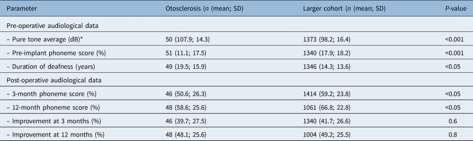
The mean duration of deafness in the implanted ear was longer for the advanced otosclerosis cohort (mean, 19.5, SD, 15.6 years) compared with the larger cohort (mean, 14.3 years, SD, 13.6 years; p < 0.05). The mean pre-implantation phoneme scores were also worse in the advanced otosclerosis group (11.1 per cent vs 17.9 per cent; p < 0.01) (Table 2).
Pre- and post-operative audiological data (otosclerosis, larger cohort)

*All frequencies (250, 500, 1000, 2000, 4000 Hz); no response = 125 dB. SD = standard deviation
Post-operative speech perception
Three-month post-operative phoneme scores on the cochlear implantation side were used to determine audiological outcome. There was a statistically significant (p < 0.001) improvement in mean speech perception scores (50.6 per cent, SD 26.3) compared with pre-operative scores (11.1 per cent, SD, 17.5). This cohort showed significantly poorer results when compared with the larger group of adults from the Melbourne clinic (n = 1414; mean, 59.2 per cent; SD, 23.8; p < 0.05). Similarly, 12-month mean post-operative phoneme scores were also significantly improved from the pre-operative period in the otosclerosis group (58.6 per cent, SD 25.3; p <0.001), but scores were worse than for the larger cohort (mean, 66.8 per cent, SD, 22.8; p < 0.05). Improvements in speech perception from pre-implant to 3 months (39.5 vs 41.4 per cent) and 12 months (47.6 per cent vs 48.9 per cent) post-implant were not significantly different between the two cohorts. There was a moderate correlation between duration of deafness and 12-month post-phoneme scores (r (44) = −0.45; p = 0.002).
Radiographic scoring
Pre-operative imaging scoring and correlation with intra-operative findings
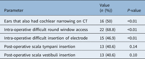
Sixty-two per cent of ears demonstrated evidence of otosclerotic changes at the round window on pre-operative imaging (32 of 52), and 33 per cent of ears were assessed as having cochlear changes on pre-operative imaging (17 of 52). Sixteen ears (31 per cent) had evidence of both. Intra-operatively, it was difficult to identify the round window in 44 per cent of ears, and this required subjective determination for the optimal cochleostomy position (23 of 52). Twenty-nine per cent had difficult electrode array insertions (15 of 52).
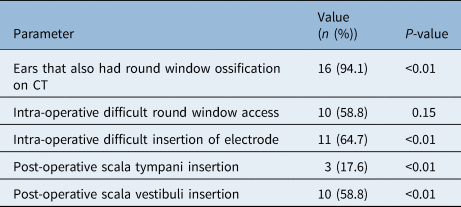
There was a strong correlation between the pre-operative assessment of both the round window and cochlea and the intra-operative assessment of these structures (Table 3 and 4). A normal round window on pre-operative imaging was associated with a normal round window intra-operatively in 95 per cent of the cases, and an abnormal round window on pre-operative imaging was associated with difficulty in identification of the round window intra-operatively in 68.8 per cent of cases (χ2 = 20.28; p < 0.01). There was a significant correlation between pre-operative findings of cochlear narrowing or abnormal signal and intra-operative difficulty in electrode insertion, with 64.7 per cent of the cases that demonstrated narrowing or signal abnormality on pre-operative imaging also having difficulty with electrode insertion intra-operatively (χ2 = 13.7; p < 0.001). There was also a moderate correlation between pre-operative imaging assessment of the round window and intra-operative difficulty in electrode insertion (r (50) = 0.44; p = 0.001). When an abnormal round window was demonstrated on pre-operative imaging, almost half (46.9 per cent) of the cases had difficulty on electrode insertion intra-operatively (χ2 = 10.1; p < 0.001).
Ears identified with round window ossification on pre-operative CT*

* n = 32. CT = computed tomography
Ears identified with evidence of cochlear narrowing on pre-operative imaging*

* n = 17. CT = computed tomography
Pre-operative imaging and post-operative electrode position
There was no significant correlation between pre-operative assessment of the round window and post-operative electrode position (Table 5). A normal round window on pre-operative imaging correlated with a scala tympani position on post-operative imaging in 68 per cent and an abnormal round window on pre-operative imaging correlated with a scala tympani position in 41 per cent of cases (χ2 = 3.36; p = 0.07).
Pre-operative radiological findings correlated with final electrode position

*n = 17; *n = 27; *n = 8
There was a moderate correlation between pre-operative assessment of the cochlea and post-operative electrode position (r (50) = 0.46; p = 0.001) with only 13 per cent of cases with a pre-operative abnormal cochlea demonstrating scala tympani placement (χ2 = 13.13; p < 0.01).
Intra-operative findings and post-operative electrode position
Twenty-seven ears were scala tympani insertions (52 per cent), and 17 ears were scala vestibuli insertions (33 per cent). In 8 ears, the position was indeterminate (15 per cent). There was a 92 per cent agreement in scoring between the two otologists (48 of 52).
There was no significant correlation between the intra-operative assessment of the round window and the post-operative electrode position (r (50) = 0.17; p = 0.22). An uncomplicated round window access correlated to a scala tympani position in 60.7 per cent of cases, and a scala tympani position was seen in 40 per cent of cases with intra-operative difficulty identifying the round window (χ2 = 2.0; p = 0.16).
There was a strong correlation between the intra-operative difficulty in electrode insertion and post-operative electrode position (r (50) = 0.60; p < 0.001). Intra-operative difficulty of insertion correlated to a scala tympani placement in only 7 per cent of cases, with the remaining being either indeterminate or scala vestibuli placement (χ2 = 16.0; p < 0.01).
Correlation between hearing outcomes with imaging and operative findings
There was no significant correlation between pre-operative or intra-operative assessment of the round window or cochlea and pre-implant or 12-month post-implant hearing outcomes (Table 6).
Correlation between pre-operative or intra-operative assessment and hearing outcomes

Correlation between hearing outcomes and final electrode placement
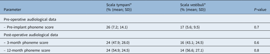
There was no significant correlation between final electrode placement and pre-implant or 12-month post-implant hearing outcomes (Table 7).
Speech perception scores comparing scala tympani and scala vestibuli insertions in the otosclerosis cohort

*n = 17. SD = standard deviation
Facial nerve stimulation
No patients were identified as having any facial nerve stimulation post-operatively at any point from switch-on during the follow-up period.
Discussion
Speech perception
Patients with advanced otosclerosis demonstrated slightly poorer speech perception scores than the larger cohort, but the degree of improvement pre- to post-operatively was not significant between groups (Fig. 4). The advanced otosclerosis cohort has a lower start point with significantly poorer pre-operative speech perception in the implant ear. This group performed slightly worse pre-operatively and at 3 and 12 months post-operatively, but the degree of improvement was not significantly different in comparison to the larger cohort (Table 2). This study confirms patients with advanced otosclerosis still derive significant benefit post-implantation. Interestingly, Vashishth et al. did not demonstrate audiological outcomes in patients with ossification to be any worse than patients without ossification in their cohort of 36 patients with otosclerosis.Reference Vashishth, Fulcheri, Rossi, Prasad, Caruso and Sanna5
Boxplot of speech perception scores for otosclerosis cohort (O) and large comparison cohort (L). post-3m = 3 months post-implantation; post-12m = 12 months post-implantation

The duration of deafness was significantly correlated with hearing outcomes in the otosclerosis cohort at 12 months. Because the advanced otosclerosis group had worse hearing at time of implantation compared with the larger cohort, this could suggest that these patients wait longer prior to implantation in comparison to non-otosclerosis patients. This may be explained by the fact that advanced otosclerosis patients are all offered stapedectomy and amplification prior to implantation. The majority of patients in the advanced otosclerosis cohort had previously undergone stapedectomy. Advanced otosclerosis patients may have worse speech perception scores because of a longer period of auditory deprivation and more extensive sensorineural loss resulting from the disease process itself.
Predicting intra-operative difficulty
Summary of results
Pre-operative imaging had a strong correlation with intra-operative imaging findings for both cochlea and round window assessment. Additionally, there was a strong correlation between post-operative electrode position and intra-operative difficulty of electrode insertion and a moderate correlation between post-operative electrode position and pre-operative imaging assessment of the cochlea. There was no significant correlation between post-operative electrode position and pre-operative or intra-operative round window assessment.
From this information, we can infer that narrowing or signal change has a more significant effect on the final electrode position, whereas round window ossification or obstruction is not as important. Multiple previous studies have demonstrated the safety and efficacy of cochlear implants in round window ossification or obstruction,Reference Castillo, Polo, Gutierrez, Reyes, Royuela and Alonso1,Reference Marshall, Fanning, Symons, Shipp, Chen and Nedzelski8–Reference Rotteveel, Proops, Ramsden, Saeed, van Olphen and Mylanus10 yet to date there has been minimal investigation of the effect of cochlear narrowing on the outcomes for cochlear implants.Reference Vashishth, Fulcheri, Rossi, Prasad, Caruso and Sanna5
Ultimately, pre-operative imaging of the cochlea provides the best prediction of final electrode position and should be carefully assessed by surgeons and radiologists to identify challenging cases. Although this study did not demonstrate a significant difference in speech scores with regards to final electrode position, a review of the literature supports scala tympani insertion for optimal outcomes. Different grading systems for otosclerosis on high-resolution CT have been describedReference Marshall, Fanning, Symons, Shipp, Chen and Nedzelski8,Reference Rotteveel, Proops, Ramsden, Saeed, van Olphen and Mylanus10 based on location of otosclerotic lesions. Difficult electrode insertions tend to have cochlear involvement on CT, although this has not previously been demonstrated to be statistically significant in the literature.Reference Merkus, van Loon, Smit, Smits, de Cock and Hensen9,Reference Ramsden, Rotteveel, Proops, Saeed, van Olphen and Mylanus12 Scala vestibuli insertions are sometimes necessitated in severe cases where there is significant osteoneogenesis of the scala tympani lumen.Reference Merkus, van Loon, Smit, Smits, de Cock and Hensen9 Vashishth et al.Reference Vashishth, Fulcheri, Rossi, Prasad, Caruso and Sanna5 had a low threshold to perform a subtotal petrosectomy approach (37 per cent of ears) after pre-operative radiological evidence of cochlear luminal obstruction or ossification on high-resolution CT or MRI. None of the patients in our otosclerosis cohort required a subtotal petrosectomy approach for electrode insertion.
Predicting audiological outcome
There was no significant correlation between hearing outcomes and pre-operative radiology, intra-operative assessment or post-operative electrode position. The results of the data analysis suggest that pre-operative imaging findings do not predict post-operative speech perception outcomes. There is conflicting limited evidence regarding this topic. Rotteveel et al. found that patients with more extensive otosclerotic changes on CT had rapidly progressive hearing loss and problematic insertion of the electrode array, although this was not statistically significant.Reference Rotteveel, Proops, Ramsden, Saeed, van Olphen and Mylanus10 Other studies have shown no difference in speech scores between patients with and without ossification.Reference Matterson, O'Leary, Pinder, Freidman, Dowell and Briggs13,Reference Semaan, Gehani, Tummala, Coughlan, Fares and Hsu14 Vashishth et al. found patients without ossification did better in all auditory parameters with a trend towards significance, although only sentence scores at 24 months were found to be significantly better in patients without ossification.Reference Vashishth, Fulcheri, Rossi, Prasad, Caruso and Sanna5 This study did not demonstrate any significant difference in post-operative audiological outcomes between scala vestibuli and scala tympani electrode insertions, although previous studies have shown that full electrode insertions into the scala tympani are associated with superior speech perception.Reference O'Connell, Hunter and Wanna15 We have previously demonstrated scala vestibuli insertion and translocations were associated with poorer speech perception outcomes.Reference Shaul, Dragovic, Stringer, O'Leary and Briggs16 It may be that a size limitation in our advanced otosclerosis cohort could not identify a potential difference in speech perception scores between scala vestibuli and tympani insertions. However, we recommend that all patients should have full scala tympani insertions based on available evidence in the literature. If pre-operative imaging demonstrates pathology of the scala, and there is significant difficulty in electrode insertion, a scala vestibuli insertion can still provide reasonable post-operative outcomes.Reference Lin, Marrinan, Waltzman and Roland17,Reference Steenerson, Gary and Wynens18 There is limited data in the literature regarding audiological outcomes following scala vestibuli insertions.
Facial nerve stimulation
There was no evidence of facial nerve stimulation in our otosclerosis cohort. Some authors have reported an increased rate of facial nerve stimulation post-implantation in this population.Reference Niparko, Oviatt, Coker, Sutton, Waltzman and Cohen19–Reference Rayner, King, Djalilian, Smith and Levine23 Cochlear implantation surgery in patients with otosclerosis has been shown to be more challenging, with higher rates of partial electrode insertions, misplacements and facial nerve stimulation,Reference Rotteveel, Proops, Ramsden, Saeed, van Olphen and Mylanus10 which occur in up to 38 per cent of patients with advanced otosclerosis.Reference Ramsden, Rotteveel, Proops, Saeed, van Olphen and Mylanus12 The fundamental pathogenesis of facial nerve stimulation is that the electrical current intended for the spiral ganglia can cause unwanted stimulation of the nearby facial nerve, resulting in symptoms ranging from simple awareness to severe facial spasm. The incidence of facial nerve stimulation in the general cochlear implantation population varies from 0.9 to 14 per cent. The otospongiosis in otosclerosis may result in reduced impedance in the bone, facilitating a shunt of current from the electrode to the facial nerve.Reference Seyyedi, Herrmann, Eddington and Nadol24 Marshall et al. found facial nerve stimulation occurred in 17 per cent of patients in the otosclerosis group, necessitating deactivation of at least one electrode.Reference Marshall, Fanning, Symons, Shipp, Chen and Nedzelski8 A recent systematic review investigating the factors influencing aberrant facial nerve stimulation following cochlear implantation found that recipients with otosclerosis were also more likely to experience facial nerve stimulation (odds ratio, 13.73, 95 per cent confidence interval (CI), 3.57–52.78; p < 0.01).Reference Van Horn, Hayden, Mahairas, Leader and Bush25
• Cochlear implantation in patients with far advanced otosclerosis is well-tolerated
• There have been reported increased rates of facial nerve stimulation post-implantation in this population
• This study demonstrated that contemporary implantation in patients with far-advanced otosclerosis was not associated with post-operative facial nerve stimulation
• There is little evidence regarding the predictive value of pre-operative imaging on degree of intra-operative difficulty, final electrode position and post-operative audiological outcomes
• This study demonstrated that certain features on pre-operative computed tomography (CT) in advanced otosclerosis can help predict degree of intra-operative difficulty and final electrode position
• Although this study found no correlation between post-operative speech outcomes and final electrode position, this may be because of the limited number of patients in the advanced otosclerosis group
• Advanced otosclerosis patients were found to be implanted later with worse pre-operative speech discrimination scores and longer duration of deafness prior to implantation
In addition to the disease process itself, choice of electrode can impact on the rates of facial nerve stimulation. Van Horn et al. demonstrated that lateral wall electrodes have a higher odds ratio than peri-modiolar electrodes with respect to facial nerve stimulation (odds ratio, 3.92; 95 per cent CI, 1.46–10.47; p = 0.01). Lateral wall electrodes also tend to require higher electrical intensity to stimulate the spiral ganglion within the cochlear modiolus and are positionally closer to the facial nerve, which abuts the lateral wall of the otic capsule. Peri-modiolar electrodes theoretically shield against lateral spread of the current and reduce the likelihood of facial nerve stimulation.Reference Frijns, Kalkman and Briaire26 We had previously demonstrated, in an older cohort of patients with otosclerosis (1986 to 2004), that 14 of 35 patients implanted with a straight electrode experienced facial nerve stimulation during mapping sessions, whereas none of the 24 implanted with a Contour electrode had facial nerve stimulation.Reference Matterson, O'Leary, Pinder, Freidman, Dowell and Briggs13 In this study, there was a deliberate decision to implant peri-modiolar implants in our advanced otosclerosis cohort where possible (98 per cent, 51 of 52), which could explain why no patients experienced facial nerve stimulation.
Conclusion
Patients with severe-to-profound sensorineural hearing loss and otosclerosis benefit significantly from cochlear implantation. Our advanced otosclerosis cohort had, on average, worse hearing thresholds, poorer speech perception and longer duration of deafness in the implanted ear in comparison with the larger cohort of post-lingually deaf patients. However, the degree of improvement between cohorts was similar. Based on pre-operative speech perception scores and duration of loss, most patients in this cohort could have benefited from cochlear implantation many years earlier, when their duration of deafness was shorter and anatomical changes associated with otosclerosis were less advanced. Implanting these patients earlier could reduce the complexity of surgery and maximise the chance of achieving a satisfactory final electrode position. The majority of our advanced otosclerosis cohort received peri-modiolar implants, with no facial nerve stimulation observed post-implantation.
Anatomical changes of the otic capsule can result in increased difficulty of surgery, reduced depth of insertion and non-optimal final electrode position. Often these difficulties can be anticipated on review of pre-operative imaging. Careful review of imaging can be done reliably and assist in preparing for surgery and electrode choice. Evidence of cochlear luminal narrowing on pre-operative imaging is associated with increased difficulty of insertion at the time of surgery as well as final electrode placement in a non-scala tympani position. The results of this study did not demonstrate any difference in hearing outcomes at 12 months for patients with advanced otosclerosis who had scala vestibuli versus scala tympani insertions. However, the sample size in this cohort may have been too small to demonstrate a statistically significant difference.
Competing interests
None declared.