INTRODUCTION
Emerging diseases are of increasing concern for both humans and wildlife, and determining the identity of their causative agents and potential impacts on their hosts will be important for developing control measures (Daszak et al. Reference Daszak, Cunningham and Hyatt2001). This is especially true for amphibians, which are declining precipitously worldwide due, in large part, to emerging diseases. The pathogens and parasites most commonly implicated in amphibian declines and mortality events include the chytrid fungus (Batrachochytrium dendrobatidis), ranaviruses, Saprolegnia spp. fungi, and Ribeiroia spp. trematodes, which cause limb deformities (Green et al. Reference Green, Converse and Schrader2002; Jancovich et al. Reference Jancovich, Davidson, Parameswaran, Mao, Chinchar, Collins, Jacobs and Storfer2005; Johnson and Lunde, Reference Johnson, Lunde and Lannoo2005; Lips et al. Reference Lips, Brem, Brenes, Reeve, Alford, Voyles, Carey, Livo, Pessier and Collins2006). However, mortality and morbidity events in North America have also been attributed to Ichthyophonus sp., Amphibiothecum (Dermosporidium) spp., and a Perkinsus-like organism, all of which are fungus-like mesomycetozoan organisms (Jay and Pohley, Reference Jay and Pohley1981; Green and Sherman, Reference Green and Sherman2001; Green et al. Reference Green, Converse and Schrader2002, Reference Green, Feldman and Wimsatt2003; Feldman et al. Reference Feldman, Wimsatt and Green2005; Raffel et al. Reference Raffel, Dillard and Hudson2007).
Outbreaks of another mesomycetozoan, Amphibiocystidium ranae, have been associated with declining populations of Rana lessonae in Italy, although the role of the parasite in these declines is still undetermined (Pascolini et al. Reference Pascolini, Daszak, Cunningham, Tei, Vagnetti, Bucci, Fagotti and Di Rosa2003). Amphibiocystidium spp. have been reported for over a century in frogs and salamanders within Europe, where the parasite has been found in adults of 3 types of anurans (Rana temporaria, Rana esculenta, and Alytes obstetricians) and 2 newts (Triturus marmoratus and Triturus cristatus) (Pascolini et al. Reference Pascolini, Daszak, Cunningham, Tei, Vagnetti, Bucci, Fagotti and Di Rosa2003). Amphibiocystidium spp. infections have not previously been reported in New World amphibians (Pascolini et al. Reference Pascolini, Daszak, Cunningham, Tei, Vagnetti, Bucci, Fagotti and Di Rosa2003), although Carini (Reference Carini1940) described a similar infection, Dermosporidium hyalarum, in Brazilian frogs. Green et al. (Reference Green, Converse and Schrader2002) reported a Dermocystidium-like organism in North American ranid tadpole livers, but this was later shown to be more closely related to Perkinsus spp. parasites of marine shellfish (Green et al. Reference Green, Feldman and Wimsatt2003). Another similar parasite, Amphibiothecum penneri, was described in American toads but is now considered to be outside the genus Amphibiocystidium based on morphological and phylogenetic evidence (Feldman et al. Reference Feldman, Wimsatt and Green2005). The lack of any reports of Amphibiocystidium spp. in North America is striking given that these parasites produce clearly visible macroscopic subcutaneous cysts (Pascolini et al. Reference Pascolini, Daszak, Cunningham, Tei, Vagnetti, Bucci, Fagotti and Di Rosa2003).
Little is known about the extent or pathogenicity of Amphibiocystidium spp. in natural populations, aside from sparse prevalence data from a few sites and sampling dates (Poisson, Reference Poisson1937; Broz and Privora, Reference Broz and Privora1951; Pascolini et al. Reference Pascolini, Daszak, Cunningham, Tei, Vagnetti, Bucci, Fagotti and Di Rosa2003). Several authors have reported general malaise and mortality of infected individuals – which they attributed to the infection (Moral, Reference Moral1913; Gambier, Reference Gambier1924) – but other authors have not observed any noticeable effect and, to date, no study has compared mortality rates of infected amphibians to uninfected controls (Pérez, Reference Pérez1907; Guyenot and Naville, Reference Guyenot and Naville1922; Pascolini et al. Reference Pascolini, Daszak, Cunningham, Tei, Vagnetti, Bucci, Fagotti and Di Rosa2003). In contrast, the closely related and better characterized Dermocystidium spp., which infect salmonid fishes, are known to cause high mortality and have been associated with fish kills in the U.S. Pacific Northwest (Olson et al. Reference Olson, Dungan and Holt1991).
Here we present evidence of widespread Amphibiocystidium sp. infection in adults of a North American amphibian species, the eastern red-spotted newt (Notophthalmus viridescens), first reported as a Candida-like cyst-producing organism by Raffel (Reference Raffel2006). We describe the unique pathology of this organism in newts, provide morphological and phylogenetic evidence for its relationship to other mesomycetozoans, and describe the seasonal distribution of this infection in multiple newt populations across the northeastern United States. We also present preliminary evidence of mortality and morbidity due to infection and the potential importance of secondary infections as a source of mortality.
MATERIALS AND METHODS
Observations and collections of newts
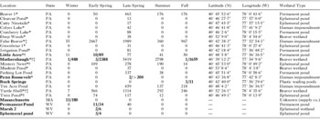
Newts were sampled or observed for visible signs of infection from 19 wetland locations in central Pennsylvania between 2002 and 2006, from 3 locations in the MeadWestvaco Wildlife and Ecosystem Research Forest (Cassity, West Virginia; N38°48′45″, W80°3′45″) in April and May 2004, and from a population in Hampshire County, Massachusetts in December 2006 (Table 1). The Pennsylvania and West Virginia newts were sampled with a combination of dip-nets and minnow traps. The Massachusetts newts were obtained from a biological supply company. For the Pennsylvania populations, a subsample of up to 10 newts was collected for dissection on each sampling date for each of the ponds included in a 2004 spatial survey or a 2003–2005 seasonal survey (Table 1). Newts examined for a 2005–2006 mark-recapture study were not collected; any additional newts were collected or observed for unpublished experiments and surveys (Table 1). The numbers and locations of subcutaneous cysts were recorded for all Pennsylvanian newts.
Observations of red-spotted newts (Notophthalamus viridescens) infected with Amphibiocystidium ranae, compared to the total numbers of newts observed in different seasons and locations
(Observations are divided into winter (Dec, Jan, Feb), early spring (Mar, Apr), late spring (May, Jun), summer (Jul, Aug) and fall (Sep, Oct, Nov). Numbers indicate the total number of newts observed. Ponds in which the infection was found are highlighted in bold type, and bolded entries each have an additional number to indicate how many of infected newts were observed in each season and pond (# infected/total # observed).)

* 2004 late spring survey; † 2003–2005 seasonal survey; ‡ 2005–2006 mark-recapture study.
Newts collected for surveys in Pennsylvania were euthanized by decapitation within 3 h of collection and dissected for internal parasite examination. Blood smears were produced and examined as described by Raffel et al. (Reference Raffel, Rohr, Kiesecker and Hudson2006, Reference Raffel, Dillard and Hudson2007), and 25 mg of the liver was ground up and cultured for bacteria on TSA (trypticase soy) blood agar, as described by Raffel (Reference Raffel2006). Bacterial isolates were identified using the Biolog® system (Biolog Inc., Hayward, California). Culturing of cyst contents was attempted on 6 occasions, variously using TSA (trypticase soy) blood agar, corn meal agar or YPD (yeast peptide dextrose) agar. See Raffel (Reference Raffel2006) for further details concerning collection and dissection methods for the Pennsylvanian populations. Following dissection, newts were fixed and stored in 70% ethanol. Eleven of the Massachusetts newts, including 3 containing visible subcutaneous cysts, were preserved in 70% ethanol within 24 h of death and dissected. Subcutaneous and liver cysts were counted for all dissected newts.
Mortality data collection
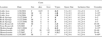
Upon arrival at Southern Illinois University on 18 December 2006, the Massachusetts newts were divided into groups of approximately 35 individuals and transferred to a set of 20 gallon aquariums each containing 10 gallons of deionized water treated using ASTM (1988) methods (0·03 g/l calcium sulfate, 0·03 g/l magnesium sulfate, 0·048 g/l sodium bicarbonate, and 0·002 g/l potassium chloride) and aged under aeration for 24 h prior to addition to aquaria. Aquaria were maintained at 23·8°C with a photoperiod of 16L:8D and water was changed every other day. Newts were fed approximately 0·25 ml of captive-raised blood worms (Chironomus tentans) per newt following each water change, and tanks were checked for food consumption at 3 and 24 h following each feeding. Newts with visible signs of infection were observed on the day of arrival and immediately placed in a separate aquarium. A subset of 120 uninfected newts was removed for use in a separate experiment on 28 December 2006. Mortality data were recorded for all other newts until 18 January 2007. Eleven newts were preserved in 70% ethanol within 24 h of death and necropsied for confirmation of the causative agent, including 3 newts with visible subcutaneous cysts (Table 2).
Results for all dissected newts, including the location, date of euthanasia/mortality, newt sex, numbers of subcutaneous and liver cysts, types of cysts observed. Bolded type indicates the cyst type from which spore measurements were taken), mean spore diameter±s.d. (μm), mean size of the large spore inclusion±s.d. (μm), and approximate numbers of granules observed within spores (min-max)

* Granules difficult to count due to poor sample preservation; † sampled for genetic analysis.
Histology
Of the 15 infected newts which were collected for dissection during surveys or experiments, cysts from 12 were further examined histologically. Tissue containing 1 or more cysts was fixed in 10% buffered formalin for 24 h prior to paraffin embedding and sectioning. Cysts were sectioned at 10 μm thickness with a Shandon Finesse® Paraffin microtome (Thermo Electron Corporation, Waltham, Massachusetts) and stained with haematoxylin and eosin.
Diameters of spores and their inclusions from all cysts examined histologically were measured using light micrographs (1000× magnification). Spores were selected for measurements by zooming into a 25 μm2 region of the cyst and measuring all spores that were in focus (6–22 spores depending on the sample) using Image-J image analysis software (Wayne Rasband, National Institutes of Health, USA). Length and diameter of 54 cysts from 3 newts, including all 3 morphologies, were measured using a dissecting microscope. Length was measured as the maximum linear dimension of the cyst and diameter was measured halfway between the cyst ends and perpendicular to the longest dimension (not necessarily the maximum diameter of the cyst due to the dumbbell shape of type C cysts, described below). Photomicrographs were taken using a Nikon Coolpix® 4500 camera (Melville, New York).
Genetic analysis
Cysts from 1 Little Acre newt and 2 Massachusetts newts were microdissected for DNA analysis. Samples were frozen and ground in liquid nitrogen. DNA was extracted with the Qiagen DNeasy® Blood and Tissue kit, using the spin-column protocol for purification of total DNA from animal tissues. DNA was eluted from spin columns with 100 μl of elution buffer. Undiluted DNA was used as template for polymerase chain reactions (PCR). PCR was performed in 20 μl reactions using GoTaq (Promega) per manufacture's instructions with 1·5 mm MgCl2 and 2 μm of each primer. Touchdown PCR was performed using the following parameters: an initial 2 min denaturation at 94°C, followed by cycles of denaturation at 94°C for 2 min, annealing at 60°C for 30 s (decreasing 1°C each cycle for 10 cycles to reach 50°C), and extension at 72°C for 1·5 min. These cycles were followed by 30 cycles with an annealing temperature of 50°C and a final 5 min extension at 72°C. The PCR products were electrophoresed on 2% agarose gels to ensure that a single product was produced. For DNA sequencing, a 5 μl aliquot of each PCR reaction was incubated for 37°C for 30 min with 5 U ExoI and 1 U shrimp alkaline phosphatase (USB) in an 8 μl reaction vol. The reaction was stopped by heating the reactions to 80°C for 15 min. DNA sequencing was performed with the same primers used for PCR in 10 μl reactions using the ABI Prism (ABI/Perkin Elmer) Dye Terminator Cycle Sequencing Ready Reaction Kit according to the manufacturer's instructions.
Primer design
We used a modification of the universal non-metazoan primers (Bower et al. Reference Bower, Carnegie, Goh, Jones, Lowe and Mak2004), 18s-EUK581-F (5′-GTGCCAGCAGCCGCG-3′) and 18s-EUK1134-R (5′-TTTAARKTTCAGCCTTGSG-3′), designed to amplify a 544-base pair fragment of 18s rDNA from protists without amplifying animal DNA (modifications in bold type). Specificity is provided by the final base of the reverse primer (Bower et al. Reference Bower, Carnegie, Goh, Jones, Lowe and Mak2004). To amplify a longer region of the 18s rDNA gene, we designed an additional forward primer, 18s-EUK581L-F (5′-ATCAACTTTCGRTGGTAAGGTA-3′), based on an alignment of 16 mesomycetozoan 18s rDNA sequences (GenBank Accession numbers AY550245, AY692319, AY772001, AY772000, AF533941, Y19155, AF070445, AJ130859, Y16260, AY267346, AF436886, AF192386, U25637, AF232303, U21337, U21336). We verified the specificity of the reverse primer by alignment with an amphibian (toad) sequence (M59386).
Phylogenetic analysis
Mesomycetozoean 18s rRNA sequences were assembled and aligned with the program MUSCLE (Edgar, Reference Edgar2004). After trimming hanging ends, the resulting alignment was comprised of sequences ranging from 778 to 1780 nt in length. Accession numbers: Dermocystidium sp. CM-2002, AF533950; Rhinosporidium sp. ex. Canis familiaris, AY372365; Rhinosporidium seeberi 1, AF158369; Rhinosporidium seeberi 2, AF118851; Dermocystidium salmonis, U21337; Rhinosporidium cygnus from Florida swans, AF399715; Amphibiocystidium ranae strain 2-04, AY692319; Dermocystidium sp., U21336; Amphibiocystidium ranae, AY550245; Sphaerothecum destruens isolate BML, AY267345; Choanoflagellate-like sp., L29455; Sphaerothecum destruens isolate WA, AY267344; Sphaerothecum destruens isolate SK, AY267346; Dermocystidium percae, AF533941; Amphibiothecum penneri 1, AY772001; Amphibiothecum penneri 2, AY772000; Amoebidium parasiticum 1, Y19155; Paramoebidium sp. CMJ-2003 isolate KS61W6, AY336708; Sphaeroforma arctica, Y16260; Amoebidium parasiticum 2, AF274051; Pseudoperkinsus tapetis, AF192386; Ichthyophonus irregularis, AF232303; Ichthyophonus hoferi, U25637. The program MODELTEST (Posada and Crandall, Reference Posada and Crandall1998) was employed to select the optimal model of nucleotide substitution for the alignment. Both a hierarchical likelihood ratio test and Akaike information criterion found the TrN+I+ Γ model of substitution to best fit the data. A maximum likelihood (ML) tree, based on this model, was then inferred using the PAUP* (version 4.0) package (Swofford, Reference Swofford2003). The support for each node was determined with bootstrap resampling analysis based on 1000 pseudo-replicates of neighbour-joining trees estimated under the ML substitution model. The phylogeny was midpoint rooted, consistent with specifying the members of the order Ichthyophonida as an outgroup.
Alternative phylogenetic topologies, based off the ML tree, were constructed with the program TreeView version 1.5.3 (Page, Reference Page1996). The likelihoods of each of these topologies were compared with the Shimodaira-Hasegawa (Reference Shimodaira and Hasegawa1999) topology test (RELL distribution with 1000 bootstrap replicates), also implemented in PAUP*.
Statistical analyses
Differences in prevalence among seasons were analysed for the Little Acre, Mothersbaugh and West Virginia Permanent Pond populations by binomial regression analysis using the data presented in Table 1. To determine the degree of aggregation of infection intensity in Little Acre, the distribution of cyst counts from newts caught in early spring was compared to expected values for the Poisson and negative binomial distributions using chi-square goodness of fit tests (Parasite load categories 1–9 and 10–166 were binned to ensure expected values of ⩾5), and the variance to mean ratio was calculated for comparison with published values for other parasites. The locations on the body (head, back, throat, stomach, forelimbs, hindlimbs or tail) of 262 cysts were recorded for 20 of the infected newts observed or collected in Pennsylvania, and the total number of cysts observed on each body part was compared to expected numbers (the proportion surface area of the body part times the total number of cysts observed) using a chi-square goodness of fit test as described by Raffel et al. (Reference Raffel, Dillard and Hudson2007). We calculated the proportion of the total skin surface area accounted for by different body parts by analysis of newt photos using Image-J®. The effect of cyst type on cyst length and diameter was analysed with one-way analysis of variance using available cyst measurements, and pairwise differences between cyst types were assessed by Tukey's test for honestly significant differences (family-wise error rate of 0·05).
Mortality due to infection in the Massachusetts population was estimated using a survival analysis regression on the time to death of infected versus uninfected newts, with censoring for the 120 newts which were removed from the experiment and for those still alive at the end of the experiment. A model with logistic errors fitted significantly better than one with exponential errors (AIClogistic=205·0, AICexponential=305·6). Statistical analyses were conducted using the ‘R’ statistical software system (www.cran.r-project.org).
RESULTS
Visible subcutaneous cysts were observed in newts from 4 Pennsylvania populations, 3 populations in West Virginia and in a shipment of newts collected in Massachusetts (Fig. 1A). Infections were most commonly observed in early spring but were also observed in fall and winter, with infections being found in 4 of the 9 populations sampled in early spring (Table 1). There was a significant effect of season on cyst prevalence in Little Acre (χ2=25·2, d.f.=4, P<0·001), Mothersbaugh (χ2=11·2, d.f.=4, P<0·001) and the Permanent Pond in West Virginia (χ2=20·6, d.f.=4, P<0·001), with early spring having the highest prevalence in all 3 locations (Table 1). Infections were seldom observed in late spring and summer for any population despite these being the most extensively sampled seasons (Table 1).
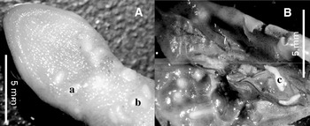
Macroscopic appearance of the three cyst types, shown (A) under the skin (ventral view of throat) and (B) in the liver. Representative cysts of each type are indicated by lowercase letters (a=Type A; b=Type B; c=Type C).

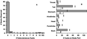
Cyst counts were highly aggregated in the Little Acre population, with a variance to mean ratio of 133·0 (mean=2·58, variance=300·3, Fig. 2A). The observed distribution differed significantly from a Poisson distribution (χ2=689, d.f.=5, P<0·001), but not from a negative binomial distribution (χ2=0·2, d.f.=2, P>0·8, Fig. 2A). Subcutaneous cysts were observed on every body part, but with significantly more cysts than expected on the stomach (χ2=174·7, d.f.=6, P<0·001, Fig. 2B), even when the single newt with 163 cysts was removed from the analysis (χ2=17·0, d.f.=6, P=0·009, Fig. 2B). Of 95 newts randomly selected for dissection from Little Acre pond during the seasonal survey, 2 had visible subcutaneous cysts (Table 2) and 3 apparently uninfected newts each had a single cyst in the liver (1 in 10 dissected newts on 5 October 2004, 1 in 8 on 28 January 2005 and 1 in 10 on 8 June 2005), providing an estimated false negative rate for presumptive diagnosis of 3·2% due to internal infections. Liver cysts were also found in 2 of the 11 dissected Massachusetts newts and 3 newts from Penn Roosevelt, all of which also had multiple subcutaneous cysts (Table 2, Fig. 1B). Cyst samples from all 12 preserved newts were confirmed by histology to be Amphibiocystidium sp. (the Little Acre newt liver samples were not available for histology). Syntype specimens were submitted to the U. S. National Parasite Collection (Accession numbers: 99608-99619).
Distributions of cyst counts. (A) Aggregated distribution of visible cyst counts for newts caught in early spring from Little Acre pond. Open diamonds indicate the expected values for the negative binomial distribution in each cyst count category. (B) Distribution of subcutaneous cysts on the newt body. Grey bars indicate the total number of cysts observed on each body part (sums of counts from 20 newts). White bars indicate cyst numbers when the single most heavily infected newt was excluded from the analysis. Black lines indicate the expected cyst counts based on the relative proportion of surface area for each body part.

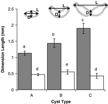
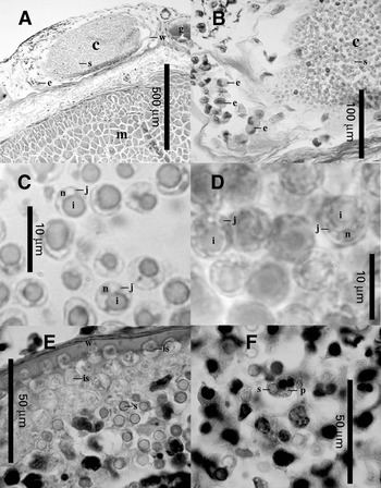
Subcutaneous cysts appeared as raised bumps under the skin, which appeared white under the translucent skin of the throat and abdomen. Cyst shape was variable, including unbent elongated egg- to rice-shaped cysts (type A, Fig. 1A), curved C-shaped cysts (type B, Fig. 1A), and bent dumbbell-shaped cysts with incompletely restricted globules at the ends (type C, Fig. 1C). These resembled descriptions of cyst stages by Broz and Privora (Reference Broz and Privora1951), except that types A and B were larger than they described, and type C cysts were not as acutely bent. Cysts of types A and B were observed on newts from all infected populations except for the Rock Springs population (Table 2, Fig. 1A). Type C cysts were only observed in the Massachusetts newts (Table 2, Fig. 1B). Cyst length and diameter both varied significantly among the cyst types (respectively: F=44·9, d.f.=[2, 47], P<0·001; F=5·3, d.f.=[2, 47], P=0·008), with type A and type C cysts having the smallest (~1 mm) and largest (~2 mm) lengths, respectively, and type B cysts having significantly larger diameters (~0·5 mm) than type A or type C cysts (Fig. 3). When dissected, cysts were found to contain white pus composed of many spherical hyaline spores, each containing a nucleus and a large inclusion body comprising most of the cell (Fig. 4). Each cyst was bounded by a cyst wall attached to the host connective tissue (Fig. 4A, E). Spores frequently contained additional smaller (~1 μm diameter) inclusions in the cytoplasm, hereafter referred to as ‘granules’, ranging from 0–5 granules in many of the type A cysts to >20 in the type C cysts (Table 2, Fig. 3C, D). Spore diameter was generally larger in type C cysts than in types A or B, but was consistent within individual cysts (Table 2, Fig. 3C, D). The diameter of the single large inclusion tended to be greater in cysts containing larger spores (Table 2). Although most cysts lacked membrane-delimited chambers surrounding spores (Fig. 3A), we observed that the margin of one cyst consisted of individual spores similar to the immature spores described by Poisson (Reference Poisson1937) in that they were contained within small chambers, which appeared to disintegrate toward the centre of the cyst (Fig. 3E).
Differences in size between the three cyst types. Grey bars indicate cyst length (straight-line=length of the longest dimension) and white bars indicate cyst diameter (measured halfway between the cyst ends and perpendicular to the longest dimension). Different lowercase letters indicate significant differences among cyst types. Error bars represent 95% confidence intervals.

Light micrographs of cysts and spores stained with haematoxylin and eosin, including (A) a typical cross-section of a subcutaneous cyst in the dermis and (B) higher magnification of the same cyst showing an aggregation of eosinophils. Typical spores from (C) a type A cyst and (D) a type C cyst, illustrating the larger size and greater number of granules for spores of the type C cyst. The lower panels depict (E) developing spores in the cyst periphery and (F) phagocytes attacking spores of a ruptured cyst. Specific features are indicated with lowercase letters (c=cyst; e=eosinophil; g=skin gland; i=large inclusion; is=immature spore; j=small granular inclusion; m=muscle; n=nucleus; p=phagocyte; s=spore; w=cyst wall).

Visibly infected newts from the Massachusetts shipment had a significantly lower survival rate than uninfected newts (Coef.=−9·4, χ2=21·1, d.f.=1, P<0·001), with a life expectancy of only 19·9 days after arrival compared to 29·3 days for the (visibly) uninfected newts.
The most heavily infected newt observed in the Pennsylvania populations (163 cysts) also had a severe Pseudomonas aeruginosa infection (>2500 colonies from 12·5 mg liver), whereas all 9 other newts (including the other infected newt with 10 subcutaneous cysts) caught at Little Acre at the same time-point had little to no evidence of bacterial infection (3 or fewer colonies). The most heavily infected newt also had elevated numbers of lymphocytes and neutrophils (361 lymphocytes and 1105 neutrophils per 5000 erythrocytes) relative to the other 9 newts (51–161 lymphocytes and 4–27 neutrophils per 5000 erythrocytes). The 2 infected newts also had significantly less food in their stomachs than the other 8 newts, as shown using a one-tailed t-test assuming unequal variances (2·0±2·0 mg vs 21·3±8·2 mg respectively [±s.e.]; t=2·29, d.f.=8, P=0·026).
Amphbiocystidium viridescens n. sp
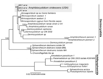
Phylogenetic analysis of Mesomycetozoean 18S rRNA regions showed that the subcutaneous and liver cysts observed in widely separated populations of red-spotted newts were all caused by phylogenetically similar parasites (Fig. 5). Although many clades within the Mesomycetozoeans cannot be sufficiently separated or their relative positions clearly resolved, due to limited sequence availability and a mix of long and short branches, it is apparent that the 3 isolates sequenced in this report (Genbank Accession numbers: EF493028-EF493030) belong to the order Dermocystida. The most likely solution is that these 3 isolates form a clade distinct from other members of the order, though additional sequence data would be needed to verify this conclusion due to low bootstrap values (Fig. 5). Indeed, the low bootstrap values make it clear that current sequence data are insufficient to strongly support or refute any of the genus designations within the order Dermocystida.
ML phylogenetic tree of partial 18s rRNA sequences from the Mesomycetozoea. The sequences characterized in this study are highlighted. The tree is midpoint rooted, consistent with an Ichthyophonida outgroup and branch lengths are drawn to scale. Only highly-supported or relevant nodes within the Dermocystida are depicted with bootstrap values.

The red-spotted newt isolates resembled A. ranae morphologically, with subcutaneous cysts varying in shape from elongated ellipsoids to curved ‘U’-shaped cysts with globular ends and containing spores with large inclusions and granules (Guyenot and Naville, Reference Guyenot and Naville1922; Broz and Privora, Reference Broz and Privora1951). Although there was no available phylogenetic evidence to determine its relationship with A. pusula, which infects European newts (Pérez, Reference Pérez1913), the presence of curved cysts distinguishes this parasite from all other closely-related subgroups except A. ranae. The elongate cyst shape and absence of chambers containing multiple spores distinguishes this parasite from Amphibiothecum penneri, the only other member of this clade to infect North American amphibians (Jay and Pohley, Reference Jay and Pohley1981; Feldman et al. Reference Feldman, Wimsatt and Green2005). The presence of cysts in the liver represents an apparently unique pathology for members of this group that infect fish and amphibians (Pascolini et al. Reference Pascolini, Daszak, Cunningham, Tei, Vagnetti, Bucci, Fagotti and Di Rosa2003). This, together with the fact that this parasite represents both a new host record and a new geographical range for Amphibiocystidium, seems to warrant the designation of a new species.
Pascolini et al. (Reference Pascolini, Daszak, Cunningham, Tei, Vagnetti, Bucci, Fagotti and Di Rosa2003) postulated that Dermocystidium, Dermomycoides and Dermosporidium spp. infecting amphibians are monophyletic and placed them into the new genus Amphibiocystidium, a grouping supported by Pereira et al. (Reference Pereira, Di Rosa, Fagotti, Simoncelli, Pascolin and Mendoza2005). Feldman et al. (Reference Feldman, Wimsatt and Green2005) cast doubt upon this hypothesis by providing phylogenetic evidence for placement of Dermosporidium penneri (now Amphibiothecum penneri) basal to the Amphibiocystidium, Dermocystidium, and Rhinosporidium clade. We tested this hypothesis by constructing 2 alternative trees, based on our ML tree, which repositioned only the Amphibiothecum subgroup either immediately basal to the Amphibiocystidium, Dermocystidium, and Rhinosporidium subgroups or immediately basal to Amphibiocystidium, Dermocystidium, Rhinosporidium, and D. percae. The likelihoods of these two alternative topologies were not significantly better than that of the original ML tree (P=0·109, 0·087), indicating that, with the current data, the branching order of subgroups cannot be stated with certainty. Alternative trees with the red-spotted newt isolates forming a sister clade to A. ranae were not significantly different from our ML tree (P=0·159, 0·206). Thus, we follow the recommendation of Pascolini et al. (Reference Pascolini, Daszak, Cunningham, Tei, Vagnetti, Bucci, Fagotti and Di Rosa2003) and tentatively name the new species Amphibiocystidium viridescens based on its morphological resemblance to A. ranae and its use of an amphibian host.
Presumptive identification based on the presence of visible cysts appears to be accurate provided that cysts are examined by a trained observer. All 12 randomly selected infected newts were confirmed to be infected with A. viridescens upon histological examination, but this only provides sufficient power to conclude that the false positive rate for presumptive diagnosis is less than 22% (based on a binomial test with α=0·05). Nevertheless, the disease signs are distinguishable from other parasites which infect red-spotted newts. The most likely parasites to confuse with A. viridescens are Clinostomum sp. trematodes, which encyst under the skin and grow to a similar size, and an unidentified metacercarial trematode that commonly encysts in the newt liver (Raffel, Reference Raffel2006). Both trematodes form spherical cysts distinct from A. viridescens, but identifications by untrained observers should be confirmed by histology.
DISCUSSION
To our knowledge this is the first report of Amphibiocystidium sp. infection in a North American amphibian species, though a separate genus designation for Amphibiothecum penneri, a parasite of the American toad (Bufo americanus), might be unwarranted unless additional sequence data are found to support the separation. The lack of previously published accounts of this parasite in red-spotted newts is striking given the visible signs of A. viridescens infection, its widespread distribution in red-spotted newt populations, the early description of Amphibiocystidium spp. in European amphibians (Pérez, Reference Pérez1907), and the large number of studies which have been devoted to red-spotted newt ecology and physiology (e.g. Holl, Reference Holl1932; Russell, Reference Russell1951; Gill, Reference Gill1979; Rohr et al. Reference Rohr, Madison and Sullivan2002; Sever, Reference Sever2002; Muzzall et al. Reference Muzzall, Peterson and Gillilland2003). Other common parasites of newts with visible signs, such as Clinostomum sp. and Ichthyophonus sp., have been known in North American amphibians for many decades (Hopkins, Reference Hopkins1933; Goodchild, Reference Goodchild1953). The seasonal distribution of A. viridescens and its low prevalence in most populations might limit the ability of researchers to detect this parasite. However, our detection of this parasite in 3 different states over a 5-year period suggests that it should have been noted by previous researchers if its historical incidence was comparable to current levels.
Although A. viridescens might be a newly acquired parasite of red-spotted newts, it is unclear where the parasite would have originated, given the geographical separation of A. viridescens from other members of the genus Amphibiocystidium. Spillover of Dermocystidium sp. infection from fish seems unlikely since most of the infected newts were observed in fishless ponds. In addition, we are aware of an unpublished observation of heavily infected newts collected by Thomas Pauley on 28 February 1990 in Grandview State Park (Raleigh Co., W.V.), which we confirmed based on photos of infected newts and histological preparations (James Joy, personal communication). We suggest that a simpler explanation for the emergence of A. viridescens in red-spotted newts is a recent increase in incidence of a previously rare endemic parasite. Examination of museum specimens would be necessary to address whether the parasite was present in red-spotted newts prior to 1990.
The winter and early spring peaks in prevalence that we observed for A. viridescens are consistent with published accounts of Amphibiocystidium spp. in Europe, where infections have generally been reported in winter and early spring. Over 3 years of sampling Triton spp. in France, Pérez (Reference Pérez1907, Reference Pérez1913) consistently found A. pusula infections in February and March but not at any other time of year. Over 2 years of sampling, Poisson (Reference Poisson1937) found a consistently higher (25–30%) prevalence of A. armoriacus infecting T. palmatus in March and April than in the rest of the field season. Guyenot and Naville (Reference Guyenot and Naville1922) reported A. ranae infection in 12 of 200 R. temporaria caught in November 1921 and A. pusula infection of several T. cristatus over the winter of 1918–1919; and Remy (Reference Remy1931) reported an infected R. esculenta individual in February 1924. Broz and Privora (Reference Broz and Privora1951) reported early-stage infections of A. ranae infecting R. temporaria in all seasons, but only found late-stage cysts in April. This consistent seasonal pattern differs from the seasonality found for the majority of red-spotted newt parasites, which mostly have peak infection rates in the late spring and summer (Raffel, Reference Raffel2006). However, winter peaks in prevalence are not uncommon amongst fungal pathogens of fish and amphibians such as Saprolegnia spp. and Batrachochytrium dendrobatidis, for which high winter infection rates have been attributed to cold-induced immune suppression coupled with the low optimal growth temperatures of the pathogens (Bly et al. Reference Bly, Lawson, Szalai and Clem1993; Berger et al. Reference Berger, Speare, Hines, Marantelli, Hyatt, McDonald, Skerratt, Olsen, Clarke, Gillespie, Mahony, Sheppard, Williams and Tyler2004).
The considerable variability in cyst size and shape, spore size and numbers of granules in spores from different isolates of A. viridescens supports the argument of Pascolini et al. (Reference Pascolini, Daszak, Cunningham, Tei, Vagnetti, Bucci, Fagotti and Di Rosa2003) that these are poor diagnostic characteristics for distinguishing Amphibiocystidium species. The type C cysts of A. viridescens resemble the mature cysts of A. ranae (Guyenot and Naville, Reference Guyenot and Naville1922; Pascolini et al. Reference Pascolini, Daszak, Cunningham, Tei, Vagnetti, Bucci, Fagotti and Di Rosa2003), and type A cysts might be difficult to distinguish from the ‘spherical’ and ‘egg-shaped’ cysts of A. pusula and A. hyalarum, respectively (Pérez, Reference Pérez1913; Carini, Reference Carini1940). Broz and Privora (Reference Broz and Privora1951) described a range of cyst sizes and types similar to our 3 cyst types in A. ranae, attributing the differences to different developmental stages. The steady increase in cyst size from type A through to type C cysts observed in this study support their conclusion, as do the larger and more granular spores we observed in type C cysts.
This developmental progression is not unlike the development of closely related Dermocystidium salmonis, which transmits by tiny flagellated zoospores that develop within spores from granules similar to those in Amphibiocystidium spp. spores (Olson et al. Reference Olson, Dungan and Holt1991). D. salmonis spores each possess a large inclusion similar to those seen in A. viridescens, which become progressively smaller as the zoospores mature (Olson et al. Reference Olson, Dungan and Holt1991). Zoospores have not been observed in Amphibiocystidium spp. infections (Pascolini et al. Reference Pascolini, Daszak, Cunningham, Tei, Vagnetti, Bucci, Fagotti and Di Rosa2003), but this is not surprising given the 2-week incubation period before release of zoospores in D. salmonis (Olson et al. Reference Olson, Dungan and Holt1991). Several authors have described progressive weakening of the skin covering cysts of Amphibiocystidium spp., leading to cyst rupture and the release of spores onto the skin surface (Pérez, Reference Pérez1913; Broz and Privora, Reference Broz and Privora1951). Perhaps zoospore development follows cyst rupture in Amphibiocystidium spp., so that zoospores are released from spores resting in the benthos. This mechanism would help explain the high numbers of cysts on the ventral surface of newts observed in this study, since the source of infection would be underneath the newt.
This is, to our knowledge, the first report of Amphibiocystidium sp. infection in the liver. Since many authors have examined this parasite solely through external examination and biopsies (e.g. Pascolini et al. Reference Pascolini, Daszak, Cunningham, Tei, Vagnetti, Bucci, Fagotti and Di Rosa2003) and the actual number of specimens dissected is unclear in other studies (Moral, Reference Moral1913; Pérez, Reference Pérez1913; Remy, Reference Remy1931; Broz, Reference Broz1944), it seems possible that liver infections may have simply been missed by other researchers. The related parasite Rhinosporidium seeberi, associated with human disease, has been reported from the liver and other soft tissues in very rare disseminated infections (Branscomb, Reference Branscomb2002). However, spores or other identifiable stages have not been observed outside cysts for any Amphibiocystidium species (Pascolini et al. Reference Pascolini, Daszak, Cunningham, Tei, Vagnetti, Bucci, Fagotti and Di Rosa2003), and an analysis of the parasite distribution in the Little Acre population provided no support for the hypothesis that A. viridescens replicates within the host to produce additional cysts. This hypothesis predicts that the distribution of cyst counts in the host population should be more highly aggregated than in macroparasitic infections. The distribution of cysts in the Little Acre population was aggregated but fit a negative binomial distribution, as do most macroparasitic helminth infections (Shaw et al. Reference Shaw, Grenfell and Dobson1998). The observed log-variance of 2·5 in this study was higher than the expected value of 1·9 for macroparasites with the same log-mean intensity, but within the range observed in a large sample of helminth and arthropod parasites (Shaw and Dobson, Reference Shaw and Dobson1995). Although we cannot rule out disseminated infection based on this analysis, the result is consistent with the hypothesis that each cyst represents a discrete infection event, despite the high numbers of cysts observed in some individuals. Perhaps infectious stages of A. viridescens access the liver through the bile duct following ingestion of mature spores by foraging newts, as an alternative to the hypothesis of disseminated infection.
Previous studies have asserted that Amphibiocystidium spp. are relatively benign parasites of amphibians (Pérez, Reference Pérez1913; Guyenot and Naville, Reference Guyenot and Naville1922; Broz and Privora, Reference Broz and Privora1951), and although A. ranae was associated with population declines of Rana lessonae, the lack of systemic post-mortem examinations has made it difficult to determine whether the parasite contributed to declines (Pascolini et al. Reference Pascolini, Daszak, Cunningham, Tei, Vagnetti, Bucci, Fagotti and Di Rosa2003). Our finding of significantly higher mortality in the visibly infected newts from Massachusetts suggests that A. viridescens can reduce red-spotted newt survival, though we cannot rule out the possibility of an additional mortality factor in the infected newts' tank or that the stress of shipping newts and placing them in a new environment exacerbated the disease. Newts in this shipment also had substantially suppressed appetites compared to newts in previous shipments (Bommarito, T., personal observation), consistent with the low appetite in an infected Triton cristatus observed by Moral (Reference Moral1913). Preliminary evidence suggests that infected newts also experience decreased appetite in natural ponds, since the most heavily infected newts in Little Acre pond had less food in their stomachs than uninfected newts. In addition, the single most heavily infected newt in the seasonal survey was co-infected with Pseudomonas auruginosa, suggesting that the proximate cause of mortality due to A. viridescens infection might be secondary infections.
The incidence of A. viridescens in red-spotted newt populations might be higher than suggested by the low prevalence recorded in this study, since parasites with short infectious periods are less likely than persistent parasitic infections to be detected in horizontal surveys. While the length of the infectious period in natural ponds remains unknown, red-spotted newts which survive the infection appear to lose their cysts in a relatively short amount of time. The one infected newt, still alive at the end of the mortality study apparently cleared its infection within 25 days of arrival (Bommarito, T., personal observation). In addition, an infected newt collected in 2004 from a Pennsylvania site also lost its single subcutaneous cyst after being held in the lab for approximately 2 weeks, although the site of infection still appeared inflamed (Raffel, T., personal observation). The one internally ruptured cyst observed in this study was infiltrated by a large number of eosinophils and other phagocytic cells. These observations are consistent with published accounts of A. pusula and A. ranae cysts, which are infiltrated by neutrophils shortly following cyst rupture, and healed in a matter of days (Pérez, Reference Pérez1913; Broz, Reference Broz1944). The timing of cyst rupture appears to be controlled more by the parasite than by the host, since the host response to intact cysts is usually minimal (Pascolini et al. Reference Pascolini, Daszak, Cunningham, Tei, Vagnetti, Bucci, Fagotti and Di Rosa2003), although we observed an eosinophilic response to one intact cyst – consistent with that described by Broz (Reference Broz1944) for A. ranae.
Although the significance of A. viridescens to red-spotted newt populations remains an open question, its apparently recent emergence in North America is troubling. Other pathogens and parasites also seem to be increasing in incidence and geographical range amongst North American amphibians, including deformity-inducing trematodes (Johnson et al. Reference Johnson, Lunde, Zelmer and Werner2003), ranaviruses (Chinchar, Reference Chinchar2002), and the chytrid fungus associated with amphibian declines (Green et al. Reference Green, Converse and Schrader2002; Weldon et al. Reference Weldon, du Preez, Hyatt, Muller and Speare2004), which has recently been found for the first time in red-spotted newts (Padgett-Flohr et al. Reference Padgett-Flohr, Bommarito and Sparling2007). Reasons for the emergence of these parasites remain unclear, but human-induced environmental changes and transportation of infected carriers of the diseases appear to be important (Johnson and Chase, Reference Johnson and Chase2004; Jancovich et al. Reference Jancovich, Davidson, Parameswaran, Mao, Chinchar, Collins, Jacobs and Storfer2005). Red-spotted newts and other North American amphibians play crucial roles in North American ecosystems (Kurzava and Morin, Reference Kurzava and Morin1998), and determining the potential causes and severity of emerging infections will be important for ensuring the future conservation of these species.
We would like to thank P. Hudson and J. Rohr of Penn State University, T. Pauley of the WV Herpetological Museum, J. Joy and R. Gain of Marshall University, and the Cooperative Wildlife Research Lab of Southern Illinois University-Carbondale for their time, consultation and supplies. Histological sectioning was performed by R. Haldeman of the Penn State EM Facility. C. Barry, Z. Loughman, J. Sias, J. Falkenbach, J. Dillard, R. LeGros, and R. Huang provided significant assistance with field observations and dissections. M. Lopez performed the bacterial identification with the help of F. Zambito from the PSU Animal Diagnostic Lab. W. Marshall and S. Feldman provided advice about primer selection, and K. Vandegrift and S. Lass provided comments on early drafts of the manuscript. This work was supported in part by a National Science Foundation (NSF) Fellowship to T. Raffel, NSF Dissertation Improvement Grant #0508847, and NSF Grant #0516227. Funding for the Massachusetts newts was provided by the Barton Springs Salamander Conservation Fund and the City of Austin, Texas. Funding for West Virginia sampling was provided by the MeadWestvaco Corporation.