Introduction
Infective endocarditis is an inflammatory condition affecting the endocardium, heart valves, or adjacent intracardiac structures, typically caused by microbial infection. Infective endocarditis is rare in children, but infections often lead to serious consequences. Globally, the annual incidence of infective endocarditis is estimated to be 3–10 cases per 100,000 population. The annual incidence of infective endocarditis in children in the United States of America is 0.3–3.3 per 100,000 people. A retrospective analysis of childhood infective endocarditis in Queensland (2009 –2014) reported an estimated annual incidence of 0.84 per 100, 000. Reference Mahony, Lean and Pham3 While medical advancements have led to significant improvements in the prognosis of infective endocarditis, reported mortality rates exhibit considerable variation globally. For instance, mortality rates as low as 10% have been documented in some developed nations. Reference Mahony, Lean and Pham3–Reference Becher, Gossling and Fluschnik7 However, there is currently a lack of accurate, large-scale studies specifically assessing the prognosis of infective endocarditis within the global paediatric population.
A significant proportion of paediatric patients diagnosed with infective endocarditis had known predisposing conditions or risk factors. Potential factors include artificial valves or artificial materials, structural CHD (cyanosis, small or residual defect), history of infective endocarditis infection, hypertrophic cardiomyopathy, cardiac devices (permanent cardiac pacemakers, implantable cardioverter-defibrillators (ICDs), catheter placement), abuse of intravenous medication, acquired valvular heart disease with stenosis or insufficiency. CHD remains the greatest risk factor for paediatric infective endocarditis. Mahony et al. showed that underlying heart disease in 35–60% of children with infective endocarditis. Reference Mahony, Lean and Pham3 There is an increased risk of developing infective endocarditis in children with CHD, especially those with cyanotic heart disease. Children with complex cyanotic heart disease have the highest risk of infective endocarditis, especially those who have received surgical interventions.
The clinical manifestations of infective endocarditis patients are various, often involving multiple organs, and there are some difficulties in clinical diagnosis. The clinical symptoms and characteristics of young children are often atypical. The most common pathogenic microorganisms are Staphylococcus aureus and the viridans group of streptococci, followed by coagulase-negative Staphylococcus, Streptococcus pneumoniae, HACEK (Haemophilus influenzae/haemophilus parainfluenzae, Aggregatibacter actinomycetemcomitans, Aggregatibacter aphrophilus, Cardiobacterium hominis, Eikenella corrodens, Kingella kingae), Enterococcus, and so on. Reference Sharland, Butler and Cant1 Reference David and Henry8 The literature reports that viridans group of streptococci represents about 32 to 43% of pathogenic bacteria, about 27% of Staphylococcus aureus to 33%, and the HACEK group about 6%. Reference Ferrieri, Gewitz and Gerber9,Reference Yagupsky10 Clinical manifestations caused by Staphylococcus are often acute, but those caused by Streptococcus are often subacute (up to weeks or even months). Infective endocarditis may present atypically when caused by intracellular pathogens, such as Bartonella spp. and Coxiella burnetii.
There is a paucity of data pertaining to infective endocarditis in children. Adequate attention to the aetiology and therapeutic regimen of the disease and adequate pharmacological supervision are very necessary. From the perspective of clinical pharmacists, this study retrospectively analysed the case data of children diagnosed with infective endocarditis in our hospital, and summarised the treatment plan, medication monitoring and medication education of such children, so as to provide a reference for more efficient clinical treatment of such diseases.
Materials and methods
Data Source
Patients diagnosed with infective endocarditis were identified through the hospital’s electronic medical record system using “infective endocarditis” as the keyword in discharge diagnoses, covering the period from May 2016 to May 2023. Diagnostic criteria were established in accordance with the Recommendations for Diagnostic Criteria of Paediatric Infective Endocarditis promulgated by the Chinese Medical Association. 11 Antimicrobial selection followed pathogen susceptibility and European Society of Cardiology/American Heart Association infective endocarditis guidelines. Reference Habib, Lancellotti and Antunes12,Reference Lamas13
Inclusion criteria: Infective endocarditis explicitly documented in discharge diagnosis; Availability of complete medical records including echocardiography reports and microbiological tests; Patients aged <18 years.
Exclusion criteria: Suspected infective endocarditis cases without confirmatory evidence; Cases with missing critical clinical data (e.g. incomplete laboratory results or imaging reports); Patients aged ≥ 18 years.
A total of 12 paediatric patients were included in this study. Data collected included patient demographics, presenting signs and symptoms, laboratory and microbiological findings, complications, primary medications administered, and treatment outcomes. Treatment outcomes were classified as microbiological cure, clinical improvement, or treatment failure.
Statistical methods
Statistical analysis was conducted using SPSS software (version 25.0). Descriptive epidemiological methods were employed to analyse the data. Data for continuous variables exhibiting a normal distribution are presented as mean ± standard deviation. Categorical data are summarised as frequencies and percentages (%).
This study was approved by the Ethics Committee of Hebei Children’s Hospital (No. 202222-89). As a retrospective analysis using fully anonymised clinical data, the requirement for written informed consent was waived. All procedures complied with the privacy protection principles of the Declaration of Helsinki.
Results
A total of 15 cases were initially identified. After excluding 3 cases (unconfirmed diagnosis or incomplete core data), 12 patients were included. Baseline characteristics of the cohort are presented below:
Basic information: There were 12 cases in total, 4 males (33.33%); 8 females (66.67%), male: female ratio = 1:2. Mean age at onset (6.93 ± 4.95) years; 10 cases (83.33%) were treated surgically; 2 cases (16.67%) were treated conservatively. Nine cases achieved microbiological cure, 2 exhibited clinical improvement and were discharged, and 1 experienced treatment failure resulting in death. Mean hospitalisation days (41.85 ± 18.19) days; median hospitalisation days: 43.00 days, 7 days, and 81 days.
CHD: Out of the total cases, 10 (83.33%) presented with CHD or cardiovascular malformations, while 2 (16.67%) had no underlying cardiac disease. Among those with malformations, specific diagnoses included: ventricular septal defects (5 cases), patent ductus arteriosus (3 cases), and atrial septal defect (1 case). In addition, 5 cases involved congenital mitral valve prolapse or insufficiency, 2 cases had congenital tricuspid insufficiency, and 1 case presented with a coronary malformation. Regarding the multiplicity of malformations, 5 cases had single-site malformations, 2 cases had two malformations, and 3 cases had three or more malformations.
Infection in other sites: 10 cases of pneumonia; 5 cases of bloodstream infection; 2 cases of bacterial meningitis; and 1 case of skin and soft tissue infection.
Pathogen testing: Multiple blood cultures were performed for all cases to identify the causative pathogen. About 8 cases were detected with bacteria, and the positive detection rate was 61.54%, 8 / 12. All 8 culture-positive cases demonstrated multiple positive blood cultures.
1 Microbiology
The distribution of 28 bacteria detected in the blood culture and vegetation culture is as follows: 27 (96.43%, 27 / 28) Gram-positive strains, 1 (3.57%, 1 / 28) Gram-negative strain. Methicillin-resistant Staphylococcus aureus (MRSA) has the highest percentage with 13 strains; 6 Abiotrophia defectiva; 5 Streptococcus mitis; 2 Streptococcus sanguinis; 1 Bacillus cereus; 1 Klebsiella oxytoca.
2 Antibiotic resistance
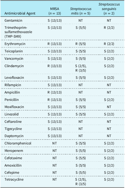
Among the isolated pathogens, 27 were Gram-positive and 1 was Gram-negative. Gram-positive bacteria were shown in Table 1. Staphylococcus aureus was MRSA. Overall, all three Gram-positive species (MRSA, Streptococcus mitis, and Streptococcus sanguinis) demonstrated universal susceptibility (100%) to critical antimicrobial agents such as vancomycin, teicoplanin, and linezolid. This finding highlights the continued efficacy of these drugs against the investigated Gram-positive isolates from blood and surgical specimens. Streptococcus isolates demonstrated consistent susceptibility (100%) to penicillin, amoxicillin, and other beta-lactams such as meropenem, cefotaxime, and cefepime. A Klebsiella oxytoca isolate, recovered from a vegetation culture, demonstrated ampicillin resistance but was susceptible to all other tested antimicrobial agents.
Antimicrobial susceptibility profiles of gram-positive isolates from blood and surgical specimens

Note: S: Susceptible; R: Resistant. NT: Not Tested. (n/N): Indicates the number of susceptible (S) or resistant (R) isolates out of the total number of isolates tested for that specific antimicrobial agent. For example, “S (13/13)” means all 13 isolates were susceptible. “S (2/5), R (3/5)” means 2 isolates were susceptible and 3 isolates were resistant out of 5 tested.
3 Types of antimicrobial drugs
Antimicrobial drug utilisation in the 12 patients included in this study was 100% (12/12): semisynthetic penicillin ± enzyme inhibitor 58.33% (7/12); first/second-generation cephalosporins 41.67% (5/12); and third-generation cephalosporins ± enzyme inhibitor 58.33% (7/12); fourth-generation cephalosporins were utilised at 8.33% (1/12); glycopeptides (vancomycin/norvancomycin/ticlopidine) were utilised at 75.00% (9/12); linezolid was utilised at 41.67% (5/12); and carbapenems (meropenem/imipenem cilastatin) were utilised at 50% (6/12).
4 Drugs that increase myocardial energy metabolism
The use of these drugs was 100% (12/12), sodium phosphocreatine (100%, 12/12), carnosine (33.33%, 4/12), nicotinamide (16.67%, 2/12), and fructose diphosphonine (8.33%, 1/12).
5 Cardiotonic Agents
Among the 12 patients, drug administration was observed in 83.33% (10/12). This included cardiac glycosides (e.g. digoxin) in 75.00% (9/12) of patients; catecholamines (dopamine and dobutamine) in 25.00% (3/12); and phosphodiesterase inhibitors (Milrinone) in 58.33% (7/12).
6 Duration of antimicrobial use
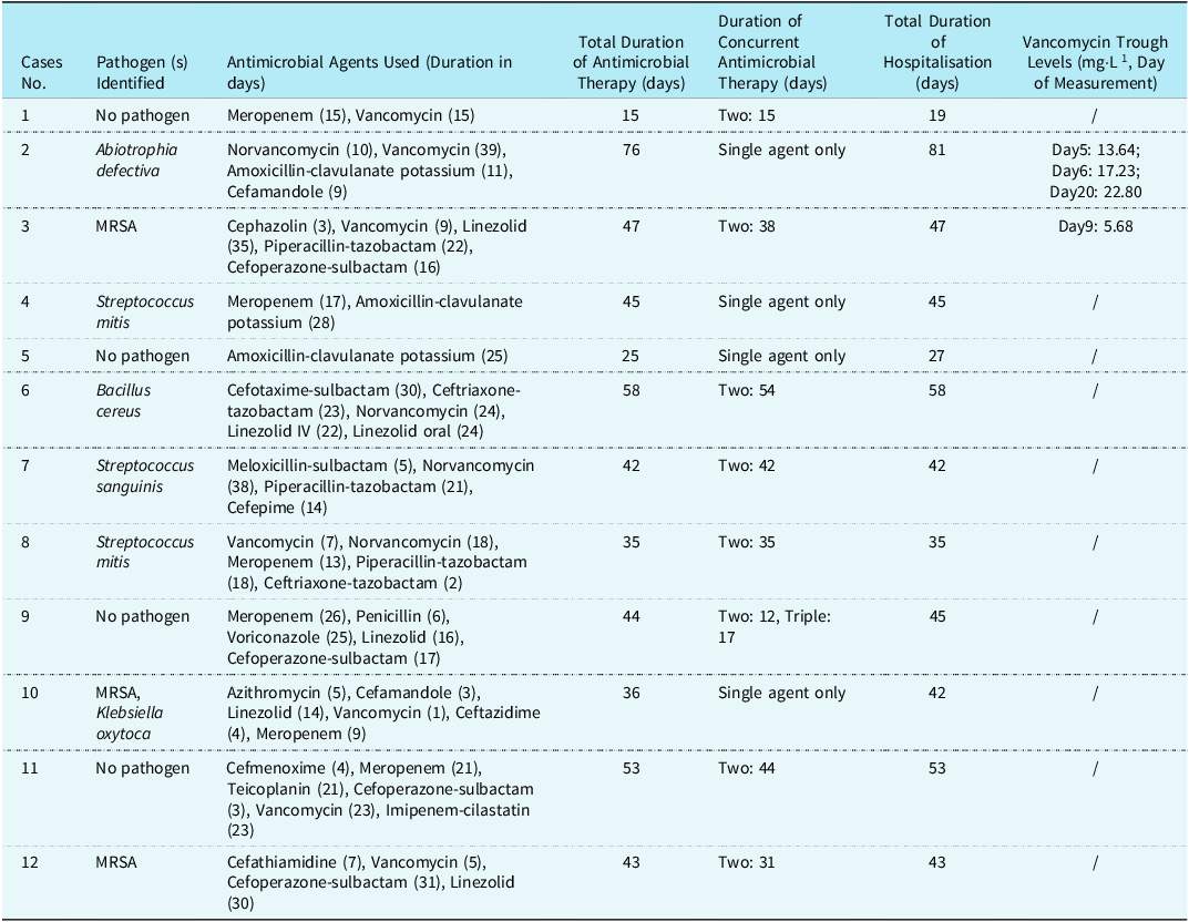
In all cases, the average length of hospital stay was 45 days (standard deviation = 16 days), and the average duration of antimicrobial use was 43 days (standard deviation = 16 days). Eight patients received combination therapy with two or more antimicrobial agents, while four patients received monotherapy. During hospitalisation, patients received an average of 4 different antimicrobial agents per person, often involving regimen changes. Details of medication days are shown in Table 2. Most patients (11/12, 91.7%) received multiple antibiotics. Among these patients, the longest duration of dual antibiotic therapy was 38 days (Case 3), and that of triple antibiotic therapy was 17 days (Case 9). Only one patient (Case 5) received monotherapy. The most commonly used antibiotics included vancomycin (7 patients, 58.3%), linezolid (5 patients, 41.7%), and meropenem (5 patients, 41.7%). Other frequently administered antibiotics were cefoperazone-sulbactam (4 patients), piperacillin-tazobactam (3 patients).
Antimicrobial use and clinical characteristics in study patients

Note: “Two: X” means using two drugs simultaneously for X days. “Triple: Y” means using three drugs simultaneously for Y days. “/” means no monitoring was performed.
Discussion
Our findings on the microbiological profile of infective endocarditis generally align with established literature and the 2015 AHA guidelines. Gram-positive bacteria were identified in all eight patients with positive aetiological findings (66.67%, 8/12), with Streptococcus spp. and Staphylococcus spp. each accounting for 37.5% (3/8) of these isolates. This predominance of Gram-positive cocci indicates no significant temporal shift in the pathogen spectrum of infective endocarditis. Reference Baltimore, Gewitz and Baddour15 While previous reports suggest no statistically significant difference in mortality rates between infective endocarditis caused by Streptococcus oxysporus or Staphylococcus aureus, Reference Thoresen, Jordal and Lie16 our small sample size precluded a statistically meaningful analysis of prognostic differences between the streptococcal and staphylococcal infection groups. Notably, the single fatal case in our cohort involved Streptococcus mitis.
Effective antimicrobial selection for infective endocarditis is critical, particularly given the propensity of pathogens to form platelet-fibrin biofilms, which render them metabolically slow and less susceptible to cell wall-active agents. Reference Pernow, Shannon and Öberg17 Consequently, guidelines advocate for prolonged, high-dose β-lactams and glycopeptides, often combined with aminoglycosides for bactericidal effect. 14 However, reflecting a cautious approach in Chinese paediatric populations due to potential adverse effects, aminoglycosides were not first-line agents at our centre and were not administered to the enrolled children. Instead, β-lactams (e.g. meropenem) and glycopeptides (vancomycin, norvancomycin) were administered at doses within the mid-range of recommended therapeutic levels. For instance, meropenem was given at 60–120 mg·kg−1·d⁻1 intravenous q8h, vancomycin at 36–40 mg·kg⁻1·d−1 intravenous bid/tid, and norvancomycin at 20 mg·kg−1·d−1 (or 800 mg·d−1) intravenous bid. Therapeutic drug monitoring for vancomycin, performed in two children, maintained trough concentrations within 5–25 mg·L−1. Overall, eight children achieved microbiological cure or clinical improvement, with one fatality attributed to cerebral herniation.
Infective endocarditis caused by Bacillus cereus is rare, particularly in paediatric patients, with systematic data limited primarily to case reports. Reference Ikeda, Yagihara and Tatsuno18 This ubiquitous Gram-positive bacillus naturally produces β-lactamase, conferring resistance to penicillins and cephalosporins. While vancomycin and clindamycin are typically recommended as first-line empirical therapies, often supplemented by agents such as imipenem or fluoroquinolones, Reference David and Henry8 our Case 6 presented a unique challenge as drug sensitivity results were unavailable. Guided by literature recommendations, we opted for a combination of surgery and prolonged norvancomycin/linezolid therapy. This approach ultimately led to microbiological cure and patient discharge, highlighting a successful management strategy in the absence of specific susceptibility data.
Abiotrophia defectiva, an inactive, catalase-negative, Gram-positive streptococcus, is a recognised cause of culture-negative infective endocarditis, despite its rarity. Reference Lancaster, Patel and Tamboli21,Reference Rudrappa and Kokatnur22 Its association with severe outcomes, including valvular destruction, heart failure, and high embolic rates, underscores the need for effective management. The organism typically shows resistance to penicillin and macrolides, with variable susceptibility to ampicillin/amoxicillin, necessitating susceptibility testing. Current guidelines suggest vancomycin or ceftriaxone/ampicillin plus gentamicin as empirical regimens, Reference Kiernan, O'FLAHERTY and Gilmore23–Reference Liao, Teng and Hsueh26 with carbapenems (e.g. meropenem, imipenem) also showing promise in limited reports. Reference Alberti, Hindler and Humphries27 Consensus on optimal treatment duration for A. defectiva infective endocarditis is lacking, though some reports suggest 6 weeks. Reference Cochicho Ramalho, Marques Santos and Abecasis28 In our Case 2, multiple positive blood cultures confirmed A. defectiva infective endocarditis, and the patient achieved microbiological cure following surgery and extended (desmethyl) vancomycin therapy for 76 days.
Vancomycin was used in 9 cases, and limited by the late introduction of the assay in our hospital, there were only 2 patients, for whom trough concentration monitoring was performed 4 times. Monitoring results: 5–10mg·L-1 1 time; 10-20 mg·L-1 2 times; >20mg·L-1 1 time. There were no adverse drug reactions such as drug-related renal damage, red man syndrome, or other adverse drug reactions during the medication period.
Linezolid was administered to 5 patients for a duration of 16 to 35 days. The drug was administered intravenously and orally. During the observation period in this study, no suspected adverse drug reactions were noted.
Cardiac energy metabolism is often compromised in infective endocarditis and associated heart failure. Reference Del Franco, Ambrosio and Baroncelli29 Sodium creatine phosphate supplementation is recognised for its cardioprotective role, particularly in myocardial ischaemia and heart failure, and is widely used in China for these indications. Reference Chen30–34 In our cohort, nearly all patients received sodium creatine phosphate. While this study noted good tolerability with no adverse drug reactions attributable to odium creatine phosphate, its retrospective and uncontrolled design, coupled with the absence of a control group, precluded any definitive assessment of its therapeutic efficacy in infective endocarditis patients. Future prospective, controlled trials are needed to evaluate the potential benefits of sodium creatine phosphate in this specific population.
The use of digoxin in paediatric heart failure, particularly in infective endocarditis contexts, is challenging due to its narrow therapeutic index and significant pharmacokinetic variability Reference Zhuang, He and Jia35,Reference Yang, Wang and Chai36. Our experience underscores the importance of meticulous dose individualisation and therapeutic drug monitoring to ensure efficacy while minimising toxicity. Despite these challenges, careful monitoring of serum digoxin levels, particularly after steady-state achievement, facilitated safe administration in our cohort, with no observed drug toxicity.
Stringent nutritional requirements cause fastidious oral/upper respiratory microbiota (e.g. Granulicatella, HACEK group) to frequently evade conventional culture, Reference Albano, Bagarello and Giordano37 potentially underestimating HACEK-associated infective endocarditis. mNGS provides reliable pathogen detection, particularly in culture-negative infective endocarditis. Reference Fortini and Mcneil38 Here, three standard-cultured aseptic site specimens were negative and not genomically analysed. For suspected infective endocarditis, prolonged culture and mNGS can enhance pathogen identification for targeted therapy, preventing empirical antimicrobial changes. 39
Finally, while traditional infective endocarditis management has long favoured exclusive intravenous antimicrobial therapy, growing evidence supports early transition to oral regimens. Reference Broom, Broom and Adams40,Reference Linde, Fassler and Meissner41 A meta-analysis by Phillips et al. Reference Phillips, Wald-Dickler and Davar42 reported non-inferior outcomes with oral transition compared to intravenous-only therapy, with the latter incurring longer hospitalisations and higher rates of catheter-related adverse events. Consequently, early conversion to oral antimicrobials, supported by therapeutic drug monitoring, is advisable once steady-state concentrations are reached. Reference Bock, Theut and Van Hasselt43 However, paediatric oral dosing presents unique challenges due to wide weight variation, limited suitable dosage forms, and the common need for dose splitting, which increases safety risks and necessitates detailed medication education and supervision.
This study shared the paediatric patients with infective endocarditis treated in our hospital in recent years. Comparison of the pathogenic bacteria detected in the blood or vegetations was basically consistent with literature reports, with slight differences in terms of resistance.
Conclusions
The detected organisms were mainly MRSA, and no vancomycin-resistant staphylococci or streptococci were found. Except for one case of in-hospital death due to brain herniation and multi-organ failure, all of them were microbiological cured. There are still many deficiencies in the therapeutic process, such as too frequent replacement of antimicrobial drug use and insufficient basis for replacement; late aetiological search; insufficient pharmacological monitoring and inadequate medication education. Medical personnel should focus on the development of individualised treatment plans, continuous monitoring of efficacy and monitoring of adverse drug reactions, and cooperate with each other to gradually improve the level of care for children with infective endocarditis.
Limitations
This study has several limitations that warrant consideration. As a retrospective, single-centre study, our findings are based on a limited sample size and the specific clinical practices of our institution. This inherent design limits the generalizability of our results and may introduce selection bias or unmeasured confounding factors. Therefore, while this article describes our centre’s experience in endocarditis treatment, caution should be exercised when applying these findings, particularly given the potential for regional and individual differences in disease presentation and management.
Acknowledgements
The authors thank the patients, families, and staff of Hebei Provincial Children’s Hospital. Special gratitude is extended to Pharmacist Xueqin Zhang for her pivotal contributions to data collection coordination and ethical compliance oversight, along with the clinical documentation team at Hebei Provincial Children’s Hospital.