INTRODUCTION
Leishmaniases are a complex of insect-borne tropical diseases presented in 2 broad clinical forms (i) cutaneous leishmaniasis (CL) that can further develop into mucosal, diffuse and disseminated subforms and (ii) visceral leishmaniasis (VL). Together, CL and VL impose serious health problems in 88 countries, where 12–15 million people are estimated to be affected (World Health Organization, 2009). Whereas most of the 20 parasite species known to cause the disease in man are agents of the more benign CL, Leishmania donovani and L. chagasi (syn. L. infantum) are the causative agents of VL, a fatal disease characterized by hepatosplenomegaly, immunosuppression, anaemia and haemorrhages.
There are no prospects for anti-leishmanial vaccines becoming available soon and, although drugs remain the mainstay of disease control, current treatment options are limited. The first line drugs for CL and VL in most affected countries remain the pentavalent antimonials sodium stibogluconate (Pentostam) and meglumine antimoniate (Glucantime). Other recommended drugs include pentamidine, paromomycin and amphotericin B and its lipid formulations. All those drugs have limitations that include parenteral administration by injection, high cost, toxicity, variable efficacy, and/or restricted supplies (Croft et al. Reference Croft, Seifert and Yardley2006). Besides, in countries like India the emergence of antimony-resistant parasites has rendered those drugs obsolete for VL treatment (Sundar, Reference Sundar2001). The former anti-cancer drug miltefosine has been approved in India for the oral treatment of antimony-resistant VL cases, but teratogenesis and induction of resistance are current concerns (Perez-Victoria et al. Reference Perez-Victoria, Sanchez-Canete, Seifert, Croft, Sundar, Castanys and Gamarro2006).
Susceptibility of mice to CL and VL has been associated with the expansion of parasite-specific T-helper 2 and regulatory T cells that may impair the initiation of effector responses (Launois et al. Reference Launois, Louis and Milon1997; Belkaid, Reference Belkaid, Blank and Suffia2006), and immunomodulatory drugs such as tucaresol can reduce L. donovani infection in C57BL/6 mice (Smith et al. Reference Smith, Yardley, Rhodes and Croft2000). Previously, we described the immunosuppressive effect and the efficacy of the leaf extract of Kalanchoe pinnata (Kp) in controlling CL in mice and man (Rossi-Bergmann et al. Reference Rossi-Bergmann, Costa, Borges, Da Silva, Noleto, Souza and Moraes1994; Da Silva et al. Reference Da Silva, Costa, Mendonça, Silva, Moraes and Rossi-Bergmann1995; Torres-Santos et al. Reference Torres-Santos, Da Silva, Costa, Santos, Almeida and Rossi-Bergmann2003). The protective effect of Kp in L. amazonensis-infected mice is not due to a direct anti-parasitic effect. Rather, it has been attributed to its Th2-suppressive effect in vivo that allowed an effective nitric oxide-dependent parasite killing by macrophages during infection (Da Silva et al. Reference Da Silva, Costa and Rossi-Bergmann1999).
Attempts to identify the active constituent(s) of Kp in CL led to the flavonoid quercitrin (quercetin 3-O-α-L-rhamnopyranoside) that is an important, although not the sole, active Kp component with potent anti-allergic/Th2-suppressive and anti-leishmanial properties (Muzitano et al. Reference Muzitano, Cruz, Almeida, Silva, Kaiser, Guette, Rossi-Bergmann and Costa2006a; Cruz et al. Reference Cruz, Da-Silva, Muzitano, Silva, Costa and Rossi-Bergmann2008; Muzitano et al. Reference Muzitano, Falcão, Cruz, Bergonzi, Bilia, Vincieri, Rossi-Bergmann and Costa2009). Although active components have been identified, crude preparations of this medicinal plant are used as such in different parts of the world to treat a variety of conditions, such as arthritis and skin and gastric ulcers (VanWyk and Wink, Reference VanWyk and Wink2004). Of particular interest, since the liver of VL patients is badly affected by the intense parasitism, was the finding that the leaf juice of K. pinnata is hepatoprotective to CCl4-treated rats (Yadav and Dixit, Reference Yadav and Dixit2003).
Thus, using the murine model of infection with L. chagasi, we proposed here to evaluate whether the anti-leishmanial effect of the crude Kp extract is extensive to VL, the most serious form of the disease.
MATERIALS AND METHODS
Kalanchoe pinnata extract (Kp)
Non-blooming Kalanchoe pinnata (syn. Bryophylum pinnatum; Crassulaceae) specimens were collected form the outskirts of Rio de Janeiro city, and the species certified according to the voucher no. 292.697 deposited at Rio de Janeiro Botanical Garden (Brazil). An aqueous extract was prepared as previously described (Da Silva et al. Reference Da Silva, Costa, Mendonça, Silva, Moraes and Rossi-Bergmann1995). Briefly, fresh leaves were minced, heated in distilled water at 20% (w/v) for 30 min at 50°C, and the aqueous extract therein named Kp was lyophilized and reconstituted with phosphate buffered saline (PBS) prior to use.
Animals
BALB/c mice originally purchased from Jackson Laboratory (Bar Harbor, Maine, USA) were bred and maintained at our own facilities in light/dark cycle and controlled temperature, using sterilized bedding, filtered water and pelleted food. Female animals aged 8 weeks and weighing 20 g were used in the present study after approval (no. 1004) by the Animal Ethics Committee of the Medical Sciences Center of the Federal University of Rio de Janeiro.
Infection and treatment
Mice (6/group) were i.v.-injected with 107 promastigotes of Leishmania chagasi (MHOM/BR/1975/M2682) at the stationary phase of growth. From day 1 to day 29 of infection, they received daily doses of Kp (400 mg/kg of body weight) or 200 μl of PBS alone by intragastric gavage. Positive controls were given intraperitoneal injections with 72 mg/kg body weight of Pentostam (sodium stibogluconate Welcome Laboratories, UK, equivalent to 20·2 mg of SbV/mg/kg body weight) on alternate days (Baillie et al. Reference Baillie, Dolan, Alexander and Carter1989).
Parasite burden
On day 30 of infection mouse spleens and livers were isolated for individual determination of the parasite burden by limiting-dilution analysis. The whole organs were individually weighed and a weighed fragment was homogenized in a tissue grinder in DMEM medium containing 20% heat-inactivated fetal calf serum (HIFCS). Serial dilutions were done in quadruplicates in 96-well flat-bottomed microtitre plates. After 12 days at 25°C, the load of amastigotes in each organ was determined from the reciprocal of the highest dilution at which promastigotes were detected.
Parasite-specific cytokines and antibodies
On day 30 of infection, spleen cell suspensions were prepared from individual mice and plated at 5×10⁶/ml in DMEM medium containing 10% HIFCS, 20 mm HEPES, 50 μm 2-mercaptoethanol, 2 mm L-glutamine, and 20 μg/ml of gentamycin in 24-well tissue culture plates. Cells were cultured for 48 h in the presence of freeze-thawed promastigotes of L. chagasi (LcAg, 50 μg protein/ml), and the levels of IFN-γ and IL-4 in the supernatants were measured by ELISA following the manufacturer's instructions (R & D Systems, Minneapolis, USA). The parasite-specific IgG levels were measured in the sera of individual mice by direct ELISA using LcAg-coated microplates (Gomes et al. Reference Gomes, Pinto, Guedes, Melo, Loma, Larraga, Lopes and Rossi-Bergmann2007).
Nitric oxide production
On day 30 of infection, the production of nitric oxide (NO) was measured in the supernatants of LcAg-stimulated cell cultures obtained as above for cytokines, using the Griess method (Green et al. Reference Green, Wagner, Glogowski, Skipper, Wishnok and Tannenbaum1982). Briefly, 50 μl of cell culture supernatants were mixed with 50 μl of Griess reagent (1% sulphanilamide and 0·1% N-1-naphthylethylenediamine dihydrochloride in 2·5% o-phosphoric acid) and incubated at room temperature for 10 min. The nitrite concentrations were determined at 540 nm wavelength using a standard sodium nitrite curve.
Statistical analyses
The results shown are representative of 2 independent experiments. Statistical differences between groups were determined by the Student's t-test, and were considered significant when P<0·05.
RESULTS
Oral Kp prevents parasite growth in infected mice
Fig. 1 shows that the oral treatment of L. chagasi-infected mice with 400 mg/kg of Kp prevented parasite growth in both the spleen and the liver by 4-fold and 6-fold, respectively, as compared with animals receiving PBS alone. Treatment efficacy was comparable with that obtained with the Pentostam control given by the i.p. route.
Effect of drug treatment on parasite burden. BALB/c mice were intravenously infected with 107Leishmania chagasi promastigotes and from day 1 onwards they were daily treated by the oral route with Kp (400 mg/kg) or on alternate days by the i.p. route with Pentostam (72 mg/kg). Controls received PBS alone. On day 30 of infection, the numbers of parasites were quantified in individual livers (top) and spleens (bottom) by limiting dilution analysis. Arithmetic means±s.d. (n=6). * P<0·05 as compared with PBS.

Oral Kp suppresses IgG antibody and IL-4 production but does not affect IFN-γ
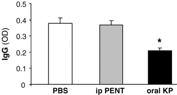
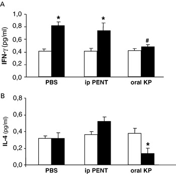
Changes in the production of specific antibodies and the classical Th1 (IFN-γ) and Th2 (IL-4) cytokines were assessed on day 30 of infection. The results in Fig. 2 show that Kp but not Pentostam treatment reduced the parasite-specific IgG serum levels. Contrary to Pentostam that did not affect the capacity of spleen cells to respond to parasite antigens with increased IFN-γ production, Kp treatment impaired the IFN-γ response (Fig. 3A). Whereas the production of IL-4 was increased in Pentostam-treated animals, it was significantly decreased (P<0·05) with Kp treatment (Fig. 3B). Together, these results show that orally administered Kp selectively depresses IgG and IL-4 but not IFN-γ production.
Effect of drug treatment on antibody production. Mice were infected and treated as for Fig. 1. On day 30 of infection, the level of parasite-specific IgG was determined by ELISA in individual sera, and expressed as the optical density at 495 nm. Means±s.d. (n=5). * P<0·01 as compared with PBS.

Effect of drug treatment on cytokine production. Mice were infected and treated with Kp or control drug Pentostam as for Fig. 1. Controls received PBS. On day 30 of infection, the production of IFN-γ (A) and IL-4 (B) by spleen cells cultured for 48 h in medium alone (white bars), or in the presence of 50 μg/ml of LcAg (black bars) was determined in the supernatants by ELISA. Arithmetic means±s.d. (n=6). * P<0·05 as compared with the respective unstimulated samples.

Both Kp and Pentostam treatments resulted in effective NO production
To evaluate the suppressive effect of Kp in IgG and IL-4 production, which allowed a more effective anti-leishmanial response despite an unchanged IFN-γ response, NO production was concomitantly measured in the supernatants of parasite antigen-stimulated spleen cells. As seen in Fig. 4, cells from both i.p. Pentostam- and oral Kp-treated groups spontaneously produced more NO than untreated (PBS) controls (P<0·05), and this was intensified (P<0·01) after stimulation with parasite antigens independent of the type of treatment.
Effect of drug treatment on nitric oxide production. Mice were infected and treated with Kp or control drug Pentostam as for Fig. 1. Controls received PBS. On day 30 of infection, the levels of nitrite were measured in the supernatants of their spleen cells cultured for 48 h in medium alone (black bars), or in the presence of 50 μg/ml of LcAg (white bars). Arithmetic means±s.d. (n=6). * P<0·05 and ** P<0·01, as compared with the respective bars of PBS.

DISCUSSION
In this study, we used the mouse model of infection with L. chagasi to explore the extension of the anti-leishmanial efficacy of Kp to the more serious and more difficult to treat visceral form of leishmaniasis. Oral treatment with Kp significantly (P<0·05) reduced the splenic and hepatic parasite burdens of L. chagasi-infected mice to levels similar to that achieved with the antimonial drug Pentostam given by the i.p. route for the same period of time. Treatment was initiated at an early stage of VL infection so as not to miss any drug effect. A dose of Kp higher than Pentostam (400 mg/kg vs 72 mg/kg) was used to compensate for the gastrointestinal digestion and excretion of the extract, and its non-defined nature. Pentostam was administered parenterally because antimonials are not orally active unless complexed with special carriers like β-cyclodextrin (Demicheli et al. Reference Demicheli, Ochoa, Silva, Falcão, Rossi-Bergmann, Melo, Sinisterra and Frezard2004). Considering the differential clinical picture, the same oral Kp regimen was effective in BALB/c mice cutaneously infected with L. amazonensis in the footpad (Da Silva et al. Reference Da Silva, Costa, Mendonça, Silva, Moraes and Rossi-Bergmann1995), indicative of the broad spectrum of Kp.
Although fractionation of the Kp extract leads to loss of anti-leishmanial activity, quercitrin was identified as an abundant and important component of an active flavonoid fraction of Kp both in intracellular amastigotes and in mice infected with L. amazonensis (Muzitano et al. Reference Muzitano, Cruz, Almeida, Silva, Kaiser, Guette, Rossi-Bergmann and Costa2006a, Reference Muzitano, Tinoco, Guette, Kaiser, Rossi-Bergmann and Costab). The activity of quercetin (the quercitrin aglycone) was previously described in hamsters infected with L. donovani (Sen et al. Reference Sen, Mandal, Saha, Mukhopadhyay and Biswas2005, Reference Sen, Mukhopadhyay, Ray and Biswas2008). In those studies, the authors did not investigate the immunomodulatory role of quercetin, rather, they attributed the therapeutic effect in VL to inhibition of exogenous iron uptake by the intracellular parasite, and also to its anti-anaemic property as an antioxidant. Thus, it seems that quercetin has more than one favourable effect in VL. Contrary to quercitrin, negligible amounts of quercetin are present in the Kp extract. However, quercetin is one of the main flavonoid metabolites that appear in the blood circulation after oral administration of Kp (Muzitano et al. Reference Muzitano, Falcão, Cruz, Bergonzi, Bilia, Vincieri, Rossi-Bergmann and Costa2009). Therefore, despite the higher reported in vitro anti-parasitic activity of quercitrin (Muzitano et al. Reference Muzitano, Cruz, Almeida, Silva, Kaiser, Guette, Rossi-Bergmann and Costa2006a), the in vivo relevance of quercitrin-derived quercetin should be considered.
The mode of action of Kp in VL seems to be related to the activation of nitric oxide intermediates, as in L. amazonensis infection (Da Silva et al. Reference Da Silva, Costa and Rossi-Bergmann1999). Although Kp suppresses Th2 responses, such as specific IgE, IL-5 and IL-10 production, leading to protection against fatal allergic reaction to ovoalbumin in mice (Cruz et al. Reference Cruz, Da-Silva, Muzitano, Silva, Costa and Rossi-Bergmann2008), a direct correlation between anti-leishmanial and immunomodulatory Kp activities had not been addressed in vivo. In the present study, we found a decrease in total parasite-specific IgG not found in Pentostam-treated animals, implying that the immunomodulatory effect of Kp was not due to the lower parasite load alone. It is worth emphasizing that, although a correlate of the state of immunity, antibody responses are not a marker of protection in VL (Kar, Reference Kar1995). Interestingly, not only the production of Th2-type IL-4 cytokine was decreased in Ag-stimulated spleen cells but also the Th1-type IFN-γ production was decreased. This was not due to cytotoxicity since in vitro antigen stimulation resulted in increased NO production in Kp-treated animals, compatible with the NO enhancing effect of Kp (Da Silva et al. Reference Da Silva, Costa and Rossi-Bergmann1999) that may have ultimately led to the decreased parasite load. It is feasible that down regulation of macrophage-deactivating cytokines including IL-4, and possibly IL-10 and TGF-β (Vouldoukis et al. Reference Vouldoukis, Bécherel, Riveros-Moreno, Arock, da Silva, Debré, Mazier and Mossalayi1997), may have accounted for the enhanced NO production, whose increase could in turn have counter-regulated IFN-γ production (Taylor-Robinson, Reference Taylor-Robinson1997). Another possibility is the enhancement of IFN-γ receptors with consequent increased sensitivity to IFN-γ, as demonstrated for Miltefosine in L. donovani-infected macrophages (Wadhone et al. Reference Wadhone, Maiti, Agarwal, Kamat, Martin and Saha2009).
Visceral leishmaniasis is normally associated with severe immunosuppression that not only imparts host fighting against the disease but also responsiveness to antimonial treatment (Goto and Lindoso, Reference Goto and Lindoso2004). Thus, discovery of new drugs that upmodulate and render the host more prone to kill the intracellular parasites are urgently needed to treat this disease.
This study demonstrates that the previously reported oral activity of Kp in CL is applicable to VL using the murine model of infection with L. chagasi, indicating its broad effect in leishmaniasis. Although a crude extract of partially defined composition, the growing knowledge of its Th2-suppressive effect (Cruz et al. Reference Cruz, Da-Silva, Muzitano, Silva, Costa and Rossi-Bergmann2008), anti-leishmanial flavonoidic components (Muzitano et al. Reference Muzitano, Cruz, Almeida, Silva, Kaiser, Guette, Rossi-Bergmann and Costa2006a, Reference Muzitano, Tinoco, Guette, Kaiser, Rossi-Bergmann and Costab, Reference Muzitano, Falcão, Cruz, Bergonzi, Bilia, Vincieri, Rossi-Bergmann and Costa2009), and low toxicity (Da Silva et al. Reference Da Silva, Costa and Rossi-Bergmann1999; Torres-Santos et al. Reference Torres-Santos, Da Silva, Costa, Santos, Almeida and Rossi-Bergmann2003; Yadav et al. Reference Yadav and Dixit2003) supports further development of Kp as a new oral and low-cost treatment for leishmaniasis.