Introduction
The genus Elaphostrongylus (Nematoda; Metastrongyloidea; Protostrongylidae; Elaphostrongylinae) currently includes three species of parasites that usually infest ruminants from the family Cervidae: E. cervi (Cameron, Reference Cameron1931), a parasite of red deer (Cervus elaphus) and other Cervus spp., E. rangiferi (Mitskevich, Reference Mitskevich1958) in reindeer (Rangifer tarandus), and E. alces (Stéen et al., Reference Stéen, Chabaud and Rehbinder1989) in moose (Alces alces).
Elaphostrongylus cervi has been recorded in many countries of Europe, in Asia and in New Zealand. Adult worms live in the intramuscular connective tissue and beneath the epimysium of the skeletal muscles; mature parasites and larvae at their third and fourth stage can also be present in the subdural and subarachnoid spaces of the central nervous system (CNS) (Kutzer & Prosl, Reference Kutzer and Prosl1975). The eggs are deposited in small veins and are carried by blood to the lungs where dorsal-spined first-stage larvae (L1) reach the bronchial tree. These are swallowed and excreted with faeces. The L1 develop to the infective third stage (L3) in different snail intermediate hosts (Řezáč et al., Reference Řezáč, Palkovič, Holasová and Bušta1994; Kuligowska & Demiaszkiewicz, Reference Kuligowska and Demiaszkiewicz2010) that can be accidentally ingested by definitive hosts, mainly during late summer. Both E. cervi and E. rangiferi migrate via the general circulation from the intestinal wall to the CNS, where they develop to the adult stage in the subarachnoid spaces and subsequently migrate into the skeletal muscles. The prepatent period can last from 3 to 6 months or more (Gajadhar et al., Reference Gajadhar, Tessaro and Yates1994; Handeland et al., Reference Handeland, Gibbons and Skorping2000a). Even though in most cases elaphostrongylosis in cervids has no symptoms, three clinical forms have been described: (1) nervous disorders such as ataxia, paralysis and blindness caused by the presence of worms in the central nervous system; (2) verminous pneumonia resulting from migration of large numbers of larvae through the lungs; and (3) chronic ill-health (Gajadhar et al., Reference Gajadhar, Tessaro and Yates1994). Dorsal-spined larvae of E. cervi cannot be reliably differentiated from those of other species in the subfamily Elaphostrongylinae. Therefore, recovery and identification of the adult nematodes are necessary for a definitive diagnosis in regions where more than one of these species exist. In geographic locations where no other elaphostrongylinae nematodes of deer occur, the presence of dorsal-spined larvae in the faeces of cervids can be considered diagnostic of E. cervi infection. Recently it has become possible to differentiate larvae of Elaphostrongylinae by polymerase chain reaction (PCR) (Gajadhar et al., Reference Gajadhar, Steeves-Gurnsey, Kendall, Lankester and Stéen2000; Huby-Chilton et al., Reference Huby-Chilton, Chilton, Lankester and Gajadhar2006) and serological techniques (Duffy & Burt, Reference Duffy and Burt2002).
Elaphostrongylus cervi can infect and cause neurological disease in sheep and goats grazing in the same areas as red deer. Their migratory route is the same. Clinical signs, normally associated with the migration of nematodes in the CNS parenchyma, can be mild or severe. They include spinal ataxia, asymmetrical paraparesis and delayed pelvic limb posture reflexes, reduced sensitivity, reduced vision, circling, mental confusion, tetraparesis, head tilt and lowered palpebra and ear. Experimental studies have shown that there is a gradual die-off of the parasite during its development in the CNS of small ruminants and that, as it does not produce eggs, first-stage larvae are not present in the bronchial mucus and faeces of such unnatural hosts (Pusterla et al., 1997, Reference Pusterla, Caplazi and Braun2001; Handeland et al., Reference Handeland, Gibbons and Skorping2000b; Scandrett & Gajadhar, Reference Scandrett and Gajadhar2002).
In Italy E. cervi has been recorded in a population of hunted red deer from Tuscan-Emilian Appennines (Morandi et al., Reference Morandi, Galuppi, Nicoloso, Benazzi, Tampieri and Simoni2006), in a single red deer and in a goat showing neurological disease respectively from the provinces of Trento (Bregoli et al., Reference Bregoli, Natale, Cova, Vascellari and Pasolli2006) and Varese (Sironi et al., Reference Sironi, Zanzani, Villa and Manfredi2006). This data prompted our investigation into the actual presence and spread of E. cervi in the deer population from the province of Varese and the consequent risk of cerebrospinal nematodiasis in goats and sheep. In fact, suspected cases of elaphostrongylosis in dairy goats had been reported by veterinarians and goat farmers in the northern area of Varese. Furthermore, the presence of E. cervi in the nearby Swiss Canton Ticino was already known (Pusterla et al., Reference Pusterla, Caplazi, Hertzberg, Ehrensperger and Braun2001). Thus the present study was aimed at confirming E. cervi infection in the deer and in small ruminants showing signs of neurological disease in the area of Varese.
Materials and methods
Study area and sampling
The province of Varese is located in the north-western part of the Lombardy region. The area is flat in the southern part of the province and becomes gradually hilly and then mountainous going north. Several species of wild ruminants, in particular red deer, have established and given rise to significant populations here. In the specific study area (Val Veddasca and Val Dumentina; 46°3′N in latitude and 8°47′E in longitude) there are also many small, family-run farms where goats and sheep are bred and where they graze freely from April to November.
Faecal samples were obtained from 31 adult red deer (18 males and 13 females) from the area, which had been hunted or killed in road accidents. The samples were collected from August to October 2008. Faecal samples and tissue samples of the central nervous system (CNS) were taken from five goats (four Nera di Verzasca, one Camosciata delle Alpi) and one sheep (Bergamasca) with a history of neurologically related lameness. They were all females over 18 months of age that had grazed in the area the previous summer. Clinical examination was carried out before slaughter when possible. Furthermore, a questionnaire, about the presence of neurological syndromes in their goats, was submitted to several goat breeders (n = 8) who kept their animals at pasture in the studied area. At the same time, a total of 97 faecal samples were collected, before anthelmintic treatment, from two goat flocks that had sporadic cases of neurological disorders (spinal ataxia and asymmetrical paraparesis), in order to check the parasitological status of the animals.
Parasitological analyses, gross pathology and histology
All faecal samples were analysed by a modified Baermann technique to recover the L1 of Protostrongylidae (Garippa et al., Reference Garippa, Manfredi and Otranto2010). The obtained sediment was observed with a light microscope to find larvae, which were then identified according to their morphological and morphometric features, which were compared with descriptions from the following authors: Baruš & Blažek (Reference Baruš and Blažek1973), Kutzer & Prosl (Reference Kutzer and Prosl1975), English et al. (Reference English, Watt and Corrigal1985), Demiaszkiewicz (Reference Demiaszkiewicz1986), Řezáč (Reference Řezáč1990), Vicente & Gortázar (Reference Vicente and Gortázar2001), Morandi et al. (Reference Morandi, Galuppi, Nicoloso, Benazzi, Tampieri and Simoni2006) and Panayotova-Pencheva & Alexandrov (Reference Panayotova-Pencheva and Alexandrov2008). At least ten larvae per animal were measured by Axiovision® software (http://www.zeiss.it). Furthermore, part of the sediment was preserved in 70% ethanol to extract the parasitic DNA for PCR assay.
Samples of CNS from the six small ruminants that had been slaughtered were fixed in 10% buffered formalin for at least 6–7 days. At trimming, CNS samples were examined grossly and by a stereomicroscope. Then, specimens of cervical, thoracic and lumbo-sacral spinal cord, and of the brain, if present, were sectioned at a 0.3 cm thickness and embedded in paraffin. Six to eight tissue blocks for each animal were obtained in two different embedding processes. Sections of thickness 4 μm were stained with haematoxylin and eosin (H&E).
Molecular techniques
Genomic DNA was extracted from larvae of six red deer faecal samples. These larvae were obtained by the Baermann technique and showed protostrongylid L1 positivity at microscopical examination. Three hundred microlitres of each Baermann sediment sample were added to 900 μl of TNNT lysis buffer (0.5% Tween 20, 0.5% Nonidet P-40, 10 mm NaOH, 10 mm Tris (pH 7.2)) and 50 μl of 20 mg/ml proteinase K (Lachaud et al., Reference Lachaud, Chabbert, Dubessay, Dereure, Lamothe, Dedet and Bastien2002). Samples were then incubated at 56°C for 18 h. Proteinase K was inactivated by incubation at 95°C for 10 min. DNA was isolated by standard basic phenol–chloroform extraction followed by ethanol precipitation (Sambrook et al., Reference Sambrook, Fritsch and Maniatis1989). Then the DNA was subjected to PCR using conserved oligonucleotide primers:
NC1-forward: (5′-ACGTCTGGTTCAGGGTTGTT-3′) and
NC2-reverse: (5′-TTAGTTTCTTTTCCTCCGCT-3′)
that amplified a 597 base pair (bp) fragment of the second internal transcribed spacer region (ITS-2) of the ribosomal RNA gene of the genus Elaphostrongylus (Gajadhar et al., Reference Gajadhar, Steeves-Gurnsey, Kendall, Lankester and Stéen2000). PCR reactions were carried out in a total volume of 20 μl containing 1 μl of genomic DNA for each sample amplification, 1 × Taq buffer containing 1.5 mm MgCl2, 0.2 mm dNTPs, 0.5 U XtraTaq polymerase (GeneSpin S.r.l, Milan, Italy) and 1 μm of each primer. PCR reactions were performed on an Mastercycler Gradient thermal cycler (Eppendorf AG, Hamburg, Germany) under the following conditions: 95°C for 4 min; 35 cycles of 95°C for 1 min, 60°C for 1 min, 72°C for 1 min and final elongation step at 72°C for 10 min.
Amplification products were run on 1.5% ethidium bromide agarose gel followed by UV-visualization. To verify the specificity of the reactions, the amplicons were purified from the gel and sequenced by the standard dideoxynucleotide method using an ABI PRISM 310 Genetic Analyzer (Applied Biosystems Inc., Foster City, California, USA). The obtained sequences were aligned to expected target sequences using ClustalW (http://www.ebi.ac.uk/clustalw).
Results
Parasitological analyses
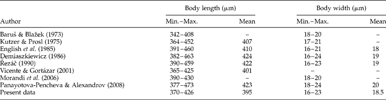
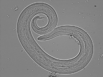
Protostrongylid L1 were detected in 14 red deer faecal samples out of 31 (45.2%, 95% CI = 27.8%, 63.7%). Morphometric and morphological features were consistent with the Elaphostrongylinae subfamily. Larvae were 370–426 μm (mean 395 μm) long, 16–23 μm (mean 18.5 μm) wide, with a typical dorsal-spined tail (table 1, fig. 1). In addition, three red deer faecal samples out of 31 (9.7%, 95% CI = 2.5%, 26.9%) showed similar larvae that were 241–334 μm long, probably belonging to Varestrongylus sagittatus. First-stage larvae of Muellerius spp. (74.7%, 95% CI = 64.3%, 83%) were detected in goat faecal samples from the two examined flocks and in two out of six slaughtered animals. No L1 of Elaphostrongylinae was present in goat faecal samples.
Comparative morphometric data of first-stage larvae of E. cervi.
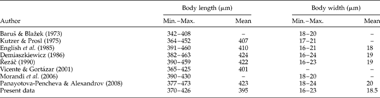
–, Data not given.
First stage larva of Elaphostrongylinae with the characteristic dorsal-spined tail.

Pathological examination
Clinical signs in sheep and goats consisted of spinal ataxia, asymmetrical paraparesis and delayed pelvic limb posture reflexes. Some of the animals were no longer able to rise, or tried to walk on their carpal joints, and showed reduced sensitivity.
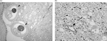
No gross lesions were observed in the spinal cords or brains from the one sheep and five goats examined at slaughter. Histological examination revealed diffuse white matter demyelination of the spinal cord, consistent with copper deficiency, in one out of six animals (a goat), whereas severe degenerative and regressive neurological lesions were seen in five out of six animals (the sheep and four goats). Large areas with axonal degeneration, demyelination, focal necrosis and lymphocytic perivascular cuffs were found in many spinal cord sections (fig. 2). Gitter cells and, to a lesser degree, eosinophils and lymphocytes, were frequently present in necrotic areas. In one case, neuritis and ganglioneuritis with focal infiltration of lymphocytes and eosinophils were also seen. Nematode sections were detected in the sub-pial spaces in two goats (one Nera di Verzasca and one Camosciata delle Alpi) (fig. 2).
Transverse sections of the spinal cord of an infected goat to show (a) two sections of E. cervi (arrowed) and (b) wallerian degeneration, with swelling of several axons (arrowhead), demyelination (thin arrows) and slight perivascular cuffing with few lymphocytes (thick arrow).

Molecular analysis
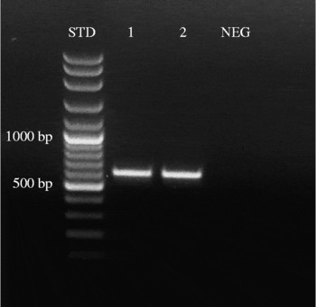
The presence of Elaphostrongylus DNA was confirmed by PCR in two out of the six red deer faecal samples tested (fig. 3). The primer pair used in this work was suitable to detect the presence of the genus Elaphostrongylus and, as expected, the amplified DNA fragments had a molecular weight of 600 bp approximately. The sequence of the amplified tract was 100% identical to those reported in GenBank both for E. cervi (accession number AF504032) and E. rangiferi (accession number AF504033). It should be noted that E. rangiferi does not affect red deer but other wild ruminants, such as reindeer, that are not present in Italy.
PCR amplification of DNA extracted from Elaphostrongylus-positive Baermann sediment samples using primers NC1 and NC2. Lanes: STD, molecular weight standard (VC 100 bp plus DNA ladder); 1–2, PCR products of two positive samples (597 bp); NEG, no-template PCR control.

Discussion
The presence of L1 of Elaphostrongylinae in red deer in the study area (Northern Province of Varese) was confirmed by parasitological examinations, with a prevalence of 45.2%, consistent with epidemiological data from Handeland & Slettbakk (Reference Handeland and Slettbakk1995), Pusterla et al. (Reference Pusterla, Hertzberg, Viglezio, Vanzetti and Braun1998b) and Morandi et al. (Reference Morandi, Galuppi, Nicoloso, Benazzi, Tampieri and Simoni2006). The morphometric characteristics of the larvae were consistent with results from other authors (Baruš & Blažek, Reference Baruš and Blažek1973; Kutzer & Prosl, Reference Kutzer and Prosl1975; English et al., Reference English, Watt and Corrigal1985; Demiaszkiewicz, Reference Demiaszkiewicz1986; Řezáč, Reference Řezáč1990; Vicente & Gortázar, Reference Vicente and Gortázar2001; Morandi et al., Reference Morandi, Galuppi, Nicoloso, Benazzi, Tampieri and Simoni2006; Panayotova-Pencheva & Alexandrov, Reference Panayotova-Pencheva and Alexandrov2008) (table 1), as belonging to the genus Elaphostrongylus. Elaphostrongylus alces and E. rangiferi only affect reindeer and moose, but not red deer (Lankester, Reference Lankester, Samuel, Pybus and Kocan2001). The specificity of E. rangiferi and E. alces for hosts that are not present in Italy strongly suggests the identity of the nematode as being E. cervi (Morandi et al., Reference Morandi, Galuppi, Nicoloso, Benazzi, Tampieri and Simoni2006), similarly to that reported for the nearby Canton Ticino (Pusterla et al., Reference Pusterla, Hertzberg, Viglezio, Vanzetti and Braun1998b, Reference Pusterla, Caplazi, Hertzberg, Ehrensperger and Braun2001). Moreover, the PCR assay confirmed our morphological identification of the larvae extracted with the Baermann technique. Even though PCR was used here to confirm morphological data, this molecular technique has great potential as an epidemiological tool in future surveys about the spread of E. cervi in the Alpine area and the consequent risk of infection for domestic small ruminants. The results from the small ruminants included in our study were consistent with those from other authors, including case history, symptoms (Pusterla et al., Reference Pusterla, Caplazi and Braun1997, Reference Pusterla, Hertzberg, Viglezio, Vanzetti and Braun1998b) and histological lesions (Pusterla et al., Reference Pusterla, Caplazi and Braun1997, Reference Pusterla, Caplazi, Hertzberg, Viglezio, Pospischil and Braun1998a; Handeland et al., Reference Handeland, Gibbons and Skorping2000b; Sironi et al., Reference Sironi, Zanzani, Villa and Manfredi2006; Tschuor et al., Reference Tschuor, Sydler, Rauch, Hertzberg, Gendotti and Schweizer2006). Sections of nematodes were found only in two out of five animals showing neurological lesions that could be related to nematode infection, and noticeably in one case only after processing a second series of tissue blocks. Indeed, there is usually no gross lesion to aid in locating the nematode, even when microscopic lesions are severe and the number of larvae is large, and E. cervi can be found anywhere throughout the nervous system. For this reason, and for the fact that the clinical signs observed in all five cases were essentially the same, a histological diagnosis of elaphostrongylosis in the three cases where the nematode was not found could be suggested. Finally, the detection of similar histological lesions in goats and sheep suggests that sheep can be infected by E. cervi, as reported previously (Handeland et al., Reference Handeland, Gibbons and Skorping2000b; Tschuor et al., Reference Tschuor, Sydler, Rauch, Hertzberg, Gendotti and Schweizer2006).
In conclusion, the present study provided parasitological and molecular confirmation of a high prevalence of E. cervi in the local deer population as well as histological documentation of episodes of cerebrospinal nematodiasis in goats and sheep of the same area. On the basis of these results, it can be hypothesized that small ruminant herds grazing in the Prealps of Varese are at risk of elaphostrongylosis and the risk area is likely larger than that studied here.
Acknowledgements
We thank the hard-working farmers of Val Veddasca and Val Dumentina; Dr M. Villa of SATA (Varese), the colleagues form Varese ASL and the entire staff of the public abattoir of Luino; Dott. M. Mortarino and Dr M. Rondena for their contribution. Without them this survey would not have been possible.