Noncoding RNAs, such as small interfering RNAs (siRNAs) and microRNAs (miRNAs), are a class of small RNAs with crucial regulatory functions, as demonstrated by the recent Nobel Prize recognition of early work in this area (Ref. Reference Fire1). In 1993, the research groups of Ambros and of Ruvkun made a landmark discovery that a small noncoding RNA, lin-4, affected the development of Caenorhabditis elegans (Refs Reference Lee, Feinbaum and Ambros2, Reference Wightman, Ha and Ruvkun3). Almost a decade later, a direct link between miRNAs and cancer – mutation or downregulation of miR-15 and miR-16 in chronic lymphocytic leukaemia – was reported (Ref. Reference Calin4), and since then a new frontier in cancer research has opened up. Currently, over 1000 mammalian miRNAs have been identified by cloning and sequencing approaches (Ref. Reference Bartel5), including hundreds in humans (Ref. Reference Cummins6).
miRNAs are a class of small noncoding RNA of 20–22 nucleotides in length, which are processed from larger pre-miRNAs by the RNase III enzyme Dicer (DICER1) into miRNA duplexes (Ref. Reference Esquela-Kerscher and Slack7). One strand of this duplex associates with the RNA-induced silencing complex (RISC), whereas the other strand is generally degraded by cellular nucleases (Ref. Reference Esquela-Kerscher and Slack7). The miRNA–RISC complex binds to specific mRNA targets, leading to translational repression or cleavage of these mRNAs. Thus, miRNAs modulate protein expression by promoting RNA degradation, inhibiting mRNA translation and, in some cases, affecting transcription. Although miRNA-mediated mRNA degradation occurs in mammals, most mammalian miRNAs are thought to repress target gene expression at the translational level (Refs Reference Lee, Feinbaum and Ambros2, Reference Pillai8, Reference Ruvkun9) through imperfect base pairing to the 3′ untranslated regions (3′-UTRs) of their target mRNAs. This form of translational regulation provides the cell with a more precise, immediate and energy-efficient way of controlling the expression of a given protein (Ref. Reference Dony, Kessel and Gruss10) because it induces rapid changes in protein synthesis without excess transcriptional activation and subsequent steps in mRNA processing. Additionally, translational control of gene expression has the advantage of being readily reversible, thus providing the cell with great flexibility in responding to various cytotoxic stresses.
To fully understand gene expression changes in cancer, it is essential to know not only the levels of individual mRNAs, but also the extent to which mRNAs are translated into their corresponding proteins and the miRNAs that regulate these processes. Thus, post-transcriptional and translational controls mediated by miRNAs have come under increasing scrutiny in cancer research. Mounting evidence suggests that miRNAs can function as either oncogenes or tumour suppressor genes. As a result, some of them will have potential as therapeutic targets, although this is not the focus of this review. As biomarker candidates, miRNAs have several unique advantages over mRNAs or proteins. First, the number of miRNAs is rather small compared with the number of mRNAs and proteins; hence the candidate pool is at least an order of magnitude smaller than mRNA or protein candidates. The second unique feature is that miRNAs tend to be stable in archival human tissues, such as formalin-fixed paraffin-embedded (FFPE) tissues and body fluids, which is a considerable advantage over mRNAs. The third important feature is the crucial translational regulatory function of miRNAs and their ability to affect several targets and pathways.
This review focuses on recent advances in miRNA research in human colorectal, gastric, liver and pancreatic cancer, as well as the translational potential of miRNAs for diagnosis and prognosis in gastrointestinal cancer.
miRNAs in colorectal cancer
Colorectal cancer (CRC) is the second leading cause of death from cancer in the Western world. It has been estimated to be responsible for 50 000 deaths in the USA and 500 000 deaths per year worldwide (Ref. Reference Jemal11). Epigenetic alterations contribute to CRC (Refs Reference Weisenberger12, Reference Widschwendter13). Recent studies suggest that miRNAs play important roles in CRC development and metastasis (Refs Reference Bartel5, Reference Calin and Croce14), and several miRNAs have been found to be differentially regulated in CRC (Ref. Reference Bartels and Tsongalis15).
Discovery of CRC-associated miRNAs
Initial efforts towards discovering CRC-associated miRNAs were based on large-scale miRNA expression profiling studies using cancer cell lines and CRC clinical specimens. Through comparison with normal tissue specimens, miRNA expression profiling has provided candidate miRNAs to differentiate CRC, and novel miRNA candidates for further investigation of functional significance. This review focuses primarily on miRNA profiling studies of clinical specimens. Many miRNAs have been reported to be deregulated in CRC specimens (Refs Reference Bandrés16, Reference Schetter17), as summarised in Table 1. Of these miRNAs, upregulation of miR-20, miR-21, miR-31 and miR-99b, and downregulation of miR-145 and miR-192, are among the consistent changes in miRNAs in CRC (Refs Reference Slaby18, Reference Volinia19). Moreover, miR-31 levels were associated with tumour pathological stages, showing increased expression in stage IV tumours than in stage II tumours (Ref. Reference Slaby18). The variation in miRNAs identified between the studies may be due to differences in the expression platform or the unique genetic background of different patient sample cohorts.
Differentially expressed miRNAs in human CRC tissuesa

a miRNAs in bold type are up- and downregulated miRNAs reported in more than one study. The order in which miRNAs are listed reflects the observed fold changes.
Abbreviations: CRC, colorectal cancer; miRNAs, microRNAs.
Functional significance, and diagnostic and prognostic potential, of miRNAs in CRC
miR-143 and miR-145
miR-143 and miR-145 are among the most extensively investigated miRNAs in CRC. Michael et al. (Ref. Reference Michael20) discovered that miR-143 and miR-145 are reduced in both colorectal precancerous adenomas and carcinomas compared with normal colorectal mucosa. The reduced expression of miR-143 and miR-145 was also confirmed in four other CRC studies (Refs Reference Slaby18, Reference Chen21, Reference Akao, Nakagawa and Naoe22, Reference Ng23). Because the DNA loci of these two miRNAs are approximately 1.8 kb apart on chromosome 5q23, both precursors may originate from the same primary miRNA (Refs Reference Michael20, Reference Akao, Nakagawa and Naoe22). Ectopic expression of miR-143 or miR-145 precursors in colon cancer cell lines resulted in the inhibition of cell proliferation (Refs Reference Chen21, Reference Akao, Nakagawa and Naoe22, Reference English24).
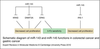
Several important targets of miR-143 or miR-145 have been discovered subsequently. Through a luciferase reporter assay, Chen et al. (Ref. Reference Chen21) identified KRAS as a key target of miR-143, and further showed that miR-143 suppressed CRC cell growth by inhibiting KRAS translation. Another important target of miR-143 is extracellular-signal-regulated kinase 5 [ERK5 (MAPK7)], which promotes cell growth in response to receptor tyrosine kinases (Refs Reference Akao, Nakagawa and Naoe22, Reference English24). Furthermore, recent studies showed that DNA methyltransferase 3A (DNMT3A), a key enzyme in DNA methylation, is a direct target of miR-143 in colon cancer cell lines: miR-143 inhibited CRC cell growth by downregulation of DNMT3A (Ref. Reference Ng23).
Borralho et al. (Ref. Reference Borralho25) investigated the roles of miR-143 in response to 5-fluorouracil (5-FU) in colon cancer cell lines. Overexpression of miR-143 resulted in a reduction of cell viability and increased cytotoxicity of 5-FU. ERK5, the transcriptional regulator NF-κB and the antiapoptotic protein Bcl-2 were also downregulated by miR-143 and subsequently reduced when exposed to 5-FU. These results suggest that miR-143 increases the sensitivity of colon cancer cells to 5-FU, possibly through ERK5/NF-κB-regulated pathways.
Additionally, two studies discovered that insulin receptor substrate 1 (IRS-1) was the downstream target of miR-145; miR-145 inhibited cell growth by downregulating IRS-1 protein (Refs Reference Shi26, Reference La Rocca27). Together, these studies indicate that miR-143 and miR-145 have potential tumour suppressor activity (Fig. 1).
Schematic diagram of miR-143 and miR-145 functions in colorectal cancer and gastric cancer. miR-143 inhibits cell proliferation by directly targeting KRAS, DNMT3A and ERK5. Downregulation of ERK5 induces sensitivity to 5-FU treatment. miR-145 also reduces the expression of IRS-1 to decrease cell proliferation. Abbreviations: DNMT3A, DNA methyltransferase 3A; ERK5, extracellular-signal-regulated kinase 5; 5-FU, 5-fluorouracil; IRS-1, insulin receptor substrate 1.

miR-126, miR-342 and miR-101
Other examples of miRNAs for which key targets are known are miR-126, miR-342 and miR-101 in CRC. miR-126, located at chromosome 9q34.3, was reported to target p85β (a regulatory subunit involved in stabilising and propagating the phosphoinositide 3-kinase signal) and to inhibit cell growth: downregulation of miR-126 and overexpression of p85β were found in primary colon cancers when compared with matched normal human colon epithelia (Ref. Reference Guo28). Reduced miR-342 expression was found in more than 50% of CRC tumour cases (Ref. Reference Grady29). Reconstitution of miR-342 in a CRC cell line induced apoptosis, suggesting that miR-342 might function as a proapoptotic gene.
Cyclooxygenase-2 (COX-2), which contributes to the growth and invasiveness of CRC, was discovered by a luciferase reporter assay to be the direct target of miR-101 (Ref. Reference Strillacci30). Subsequent analysis of miR-101 and COX-2 protein and mRNA levels in CRC and nontumour tissues confirmed that tumour tissues with higher COX-2 expression were associated with lower levels of miR-101, whereas higher levels of miR-101 were associated with lower COX-2 expression in normal samples. The authors suggested that the inverse relationship between reduced miR-101 and COX-2 overexpression and CRC malignancy could confer the tumour-suppressive role of miR-101 in CRC.
p53 and miRNAs
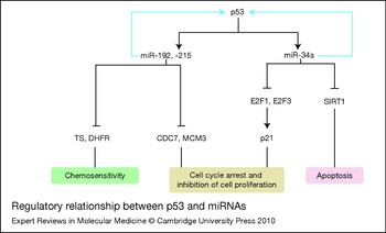
p53 (TP53) is one of the most frequently deleted and mutated tumour suppressor genes in many tumour types. Over 50% of CRC patients carry p53 mutations and deletions. A rational approach to investigate miRNAs that are potentially regulated by p53, using high-throughput expression analysis in paired HCT116 (wild-type p53) and HCT116 (p53-null) colon cancer cell lines, first indicated the impact of p53 on miRNA expression (Ref. Reference Xi31). Bioinformatic analysis revealed that over half of the 328 miRNA putative promoter regions contain p53-binding sites, including those of miR-34a, miR-192/215 and miR-26a. This is an important regulatory mechanism, because miR-34a has been shown to be regulated directly by p53 and potentially contributes to apoptosis (Refs Reference He32, Reference Chang33, Reference Raver-Shapira34). Figure 2 summarises the regulatory relationship between p53 and miRNAs.
Regulatory relationship between p53 and miRNAs. The expression of miR-192, miR-215 and miR-34 is regulated by p53 with a positive feedback mechanism (blue arrows). miR-34a suppresses the expression of E2F1, E2F3 and SIRT1 to induce cell-cycle arrest and cause apoptosis and cell growth inhibition. miR-192 and miR-215 target the cell cycle promoters CDC7 and MCM3 to induce cell cycle arrest and prohibit cell proliferation. In addition, the chemotherapeutic targets TS and DHFR are identified as targets of miR-192 and miR-215 to influence chemosensitivity. Abbreviations: CDC7, cell division cycle 7; DHFR, dihydrofolate reductase; MCM3, minichromosome maintenance complex component 3; miRNA, microRNA; p21 (CDKN1A), cyclin-dependent kinase inhibitor 1; SIRT1, silent information regulator 1; TS, thymidylate synthase.

With regard to miR-34a, transient transfection of miR-34a into colon cancer cell lines with wild-type p53 resulted in the suppression of cell proliferation and induced senescence-like phenotypes (Ref. Reference Tazawa35). miR-34a also suppressed the in vivo growth of HCT116 and RKO cells in tumours in mice. Thirty-six per cent of human colon cancer samples showed decreased expression of miR-34a compared with their counterpart normal tissues. Gene expression microarray and immunoblot analyses showed that miR-34a introduction led to downregulation of the E2F transcription factor pathway and upregulation of p53 and the cell cycle inhibitor p21 (CDKN1A) (Fig. 2). Silent information regulator 1 (SIRT1) was also a target suppressed by miR-34a (Ref. Reference Yamakuchi, Ferlito and Lowenstein36), leading to apoptosis in p53+/+ human colon cancer cells but not in p53−/− human colon cancer cells. This suggests a positive feedback loop between p53 and miR-34a (Fig. 2).
Another potential p53-mediated miRNA is miR-26a: miR-26a was shown to be downregulated as a result of the loss of p53, and might be directly regulated by p53 in colon cancer (Ref. Reference Xi31). It was recently shown that miR-26a not only influences CRC, but also plays a key role in liver cancer as both a potential therapeutic agent and a prognosis biomarker (Refs Reference Ji37, Reference Kota38). There is also direct evidence from three groups that miR-192 and its homologue miR-215 are regulated by p53 (Refs Reference Song39, Reference Sara40, Reference Christian41).
The positive feedback mechanism between p53 and these miRNAs is an important part of the regulatory networks mediated by p53. Given the broad impact of miRNAs on regulating translation rate, miRNAs exert a fine tuning upon the tumour suppressor function of p53. Furthermore, because p53 mutations and deletions are prevalent in many tumour types, we expect the modulation of p53 by miRNAs to be relevant not only in CRC but also in other cancer types.
Thymidylate synthase and dihydrofolate reductase (DHFR), two of the most important chemotherapeutic targets in cancer treatment, are regulated by miR-192 and miR-215 (Refs Reference Song39, Reference Ju, Song and Wang42, Reference Song, Wang, Titmus, Botchkina, Formentini, Kornmann and Ju43). Elevated miR-192 reduces cell proliferation and affects chemosensitivity to DHFR inhibitors. Furthermore, miR-24 regulates DHFR expression through binding to the 3′-UTR of DHFR (Ref. Reference Song, Wang, Titmus, Botchkina, Formentini, Kornmann and Ju44). A naturally occurring polymorphism (829C >T) near the miR-24-binding site in the 3′-UTR of DHFR affects DHFR expression by interfering with miR-24 function, resulting in DHFR overexpression and methotrexate resistance.
Further studies showed that miR-24 potentially acts as a tumour suppressor by reducing tumour cell proliferation in a p53-independent fashion and regulates several key cell-cycle-related genes such as p21, and the transcriptional regulators E2F and MYC (Refs Reference Mishra44, Reference Lal45).
Oncogenic miRNAs (miR-21, miR-17-92 cluster)
miR-21 and miR-17-92 have been extensively investigated in CRC, and other gastrointestinal cancer types as well, perhaps suggesting a common role of these miRNAs in gastrointestinal cancers (e.g. overexpression leading to oncogenic function).
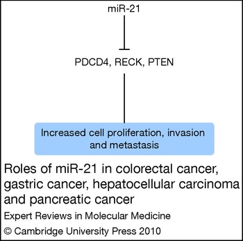
Several reports have demonstrated the upregulation of miR-21 in human CRC tissues (Refs Reference Bandrés16, Reference Schetter17, Reference Slaby18, Reference Volinia19). Increased expression of miR-21 has been associated with lymph node metastasis and the development of distant metastases in CRC patients (Ref. Reference Slaby18); another study reported increased miR-21 expression in adenomas but not in tumours with more advanced tumour, node and metastasis staging (Ref. Reference Schetter17). In situ hybridisation further supported miR-21 overexpression in CRC: the frequency and extent of miR-21 expression were found to be increased during the transition from precancerous colorectal adenoma to advanced carcinoma (Ref. Reference Yamamichi46). This study also identified tumour suppressor protein PDCD4 as a target of miR-21 (Refs Reference Frankel47, Reference Asangani48). High miR-21 expression was associated with poor survival and response to chemotherapy (Ref. Reference Asangani48). These studies suggest that miR-21 might have a clinical application as a biomarker for CRC development, poor survival and therapeutic outcome. The functional significance of miR-21 is summarised in Figure 3.
Roles of miR-21 in colorectal cancer, gastric cancer, hepatocellular carcinoma and pancreatic cancer. miR-21 is overexpressed in gastrointestinal cancers, and enhances cell proliferation, tumour invasion and metastasis by targeting several tumour suppressor genes (e.g. PDCD4, RECK, PTEN). Abbreviations: PDCD4, programmed cell death protein 4; PTEN, phosphatase and tensin homologue. RECK, reversion-inducing cysteine-rich protein with kazal motifs.

Bandrés et al. (Ref. Reference Bandrés16) identified that miR-31 expression is also significantly increased in CRC samples and correlated with the advanced clinical stage of CRC. Slaby et al. (Ref. Reference Slaby18) confirmed the higher miR-31 overexpression in CRC tissues when compared with nontumour samples, but observed the lowest levels of miR-31 in poorly differentiated tumours, suggesting the association of miR-31 expression and the degree of tumour differentiation.
The miRNAs encoded by the miR-17-92 cluster and its paralogues, the miR-106b-25 and miR-106a-363 clusters, are known to act as oncogenes. The miR-17-92 cluster (Oncomir-1) encompassing seven miRNAs (miR-17, miR-18a, miR-19a, miR-20a, miR-19b-1, miR-92a-1 and miR-106a) on chromosome 13 has previously been reported to be regulated by MYC (Ref. Reference Mendell49). One recent study, using real-time quantitative reverse-transcriptase polymerase chain reaction (qRT-PCR), showed that expression levels of the miR-17-92 cluster (all members except miR-18a) were increased in colorectal adenomas and adenocarcinomas compared with normal epithelium, in association with DNA copy number gain of the miR-17-92 locus; furthermore, there was a significant correlation between the expression of MYC mRNA and the six miRNAs (Ref. Reference Diosdado50). Three members of the miR-17-92 cluster (miR-17-5p, miR-20 and miR-106a) were also reported to be overexpressed in stage I and stage II of CRC and embryonic tissues compared with normal colonic mucosa (Ref. Reference Monzo51), and expression of its target, E2F1 protein, was inversely correlated with expression of the miR-17-92 cluster (Refs Reference O'Donnell52, Reference Woods, Thomson and Hammond53). In addition, the cluster member miR-106a was associated with poor prognosis in colon cancer (Ref. Reference Diaz54).
Some of the early translational studies of miRNA in CRC discovered several important miRNAs associated with disease prognosis. Xi et al. (Ref. Reference Xi55) evaluated ten miRNAs (let-7b, let-7 g, miR-15b, miR-181b, miR-191, miR-200c, miR-26a, miR-27a, miR-30a-5p and miR-30c) using real-time qRT-PCR analysis in 24 paired CRC and normal colorectal mucosa samples with detailed clinical follow-up information. Among the ten miRNAs, miR-15b, miR-181b, miR-191 and miR-200c were significantly overexpressed in tumours when compared with normal colorectal samples. Interestingly, miR-200c was significantly associated with patient survival, such that patients with higher miR-200c had a shorter survival time. Nakajima et al. (Ref. Reference Nakajima56) showed that let-7g, miR-181b and miR-200c were overexpressed in 46 FFPE CRC samples from 46 Japanese patients compared with corresponding normal tissues. They further showed that CRC patients who responded to S-1 treatment, a fourth generation of 5-FU, displayed a lower expression level of let-7g or miR-181b.
Circulating miRNAs in CRC
In addition to detection in primary and metastatic tumour tissues, several recent studies also showed that cell-free circulating miRNAs can be detected in plasma and serum (Refs Reference Chim57, Reference Lawrie58, Reference Wong59, Reference Mitchell60). This finding raises the possibility of using miRNAs as novel noninvasive molecular markers for cancer detection (Ref. Reference Ng61). The first report on the quantitative assessment of plasma miRNAs in CRC patients came from Ng et al. (Ref. Reference Ng61). They used real-time qRT-PCR to quantify 95 miRNAs in both plasma and tissue samples from individuals with or without CRC. Of five miRNAs (miR-135b, miR-95, miR-17-3p, miR-92 and miR-222) upregulated in both CRC plasma and tissue samples, two (miR-17-3p and miR-92) were validated in another plasma cohort. The plasma levels of miR-17-3p and miR-92 were significantly reduced after surgery. miR-92 differentiated CRC from gastric cancer, inflammatory bowel disease and normal subjects in an independent set of plasma samples with 89% sensitivity and 70% specificity. These findings suggest that miR-92 could be a potential noninvasive molecular marker for CRC screening.
Huang et al. (Ref. Reference Huang62) recently evaluated the feasibility of using plasma miRNAs as a noninvasive diagnostic test for the early detection of CRC. They measured the levels of 12 miRNAs (miR-134, miR-146a, miR-17-3p, miR-181d, miR-191, miR-221, miR-222, miR-223, miR-25, miR-29a, miR-320a and miR-92a) in plasma samples from patients with advanced colorectal neoplasia (carcinomas and advanced adenomas) and normal controls using real-time qRT-PCR. Their results showed that plasma miR-29a and miR-92a discriminated CRC and advanced adenomas from normal controls, with 83.0% sensitivity and 84.7% specificity in discriminating CRC, and 73.0% sensitivity and 79.7% specificity in discriminating advanced adenomas. The data suggest that plasma miR-29a and miR-92a are potential novel noninvasive biomarkers for the early detection of CRC.
Although studies from plasma and serum samples have offered very interesting possibilities, larger studies among several independent patient populations are still required to further validate these findings in order to determine any diagnostic potential. It will also be important to determine whether these miRNA biomarkers can predict disease recurrence after surgery, thereby providing an early detection method for tumour progression (Ref. Reference Schetter and Harris63).
miRNAs associated with gastric cancer
Gastric cancer is the second leading cause of cancer-related death in men and the third leading cause in women worldwide (Ref. Reference Forman and Burley64). In the USA in 2009, the estimated new cases and deaths of stomach cancer were 21 130 and 10 620, respectively, and the five-year relative survival rate was 25% (Ref. Reference Jemal11).
miRNA profiling analysis in gastric cancer
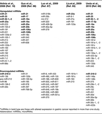
Similar to CRC, the discovery of gastric-cancer-associated miRNAs began with expression profiling by microarray analysis. Recent studies showed that miRNAs are differentially expressed in gastric cancers, as summarised in Table 2, and histological subtypes are characterised by specific miRNA signatures (Refs Reference Liu65, Reference Volinia19, Reference Guo66, Reference Luo67, Reference Ueda68).
Differentially expressed miRNAs in gastric cancera

a miRNAs in bold type are those with altered expression in gastric cancer reported in more than one study.
Abbreviation: miRNAs, microRNAs.
Differential expression of some of these miRNAs has been confirmed by real-time qRT-PCR, and their targets have been identified. miR-433 and miR-9 were confirmed to be significantly downregulated in gastric carcinoma tissues, and a growth-factor-binding protein, GRB2, and a member of the RAS oncogene family, RAB34, were identified as targets of miR-433 and miR-9, respectively (Ref. Reference Luo67). In contrast, miR-27a was confirmed to be overexpressed in gastric cancer tissues, and was shown to promote cell proliferation by directly targeting the tumour suppressor prohibitin (PHB) in a gastric adenocarcinoma cell line; thus miR-27a is a potential oncogene in gastric cancer.
Furthermore, recent miRNA microarray analysis of gastric cancer and nontumour mucosa samples from Japan showed that the observed changes in miRNAs (22 upregulated and 13 downregulated) distinguished 83% of the tumour samples in this study, and that two histological subtypes of gastric cancer showed different miRNA signatures (Ref. Reference Ueda68). Eight miRNAs (miR-105, miR-100, miR-125b, miR-199a, miR-99a, miR-143, miR-145 and miR-133a) were upregulated in diffuse type and four miRNAs (miR-373, miR-498, miR-202 and miR-494) were upregulated in intestinal type.
Functional significance, and diagnostic and prognostic potential, of miRNAs in gastric cancer
Several miRNAs (miR-451, miR-141, miR-143, miR-145 and miR-31) have been investigated further for their functional significance and clinical relevance.
Downregulated miRNAs in gastric cancer
miR-451 is reduced in gastric cancer samples and associated with worse prognosis (Ref. Reference Bandrés69). In situ hybridisation analysis indicated that miR-451 was expressed in epithelial cells of the normal mucous glands and appeared downregulated in gastric cancer. Functional studies showed that overexpression of miR-451 reduced cell proliferation and increased sensitivity to radiotherapy. The novel oncogene macrophage migration inhibitory factor was identified as a potential target of miR-451 by luciferase reporter assay. These findings indicate a role for miR-451 as a potential tumour suppressor and a target for chemo- and radiation-resistant cancers.
miR-141, which belongs to the miR-200 family, has previously been reported to inhibit epithelial to mesenchymal transition and enhance E-cadherin expression (Refs Reference Gregory70, Reference Korpal71). Real-time qRT-PCR analysis showed that miR-141 was significantly downregulated in primary gastric cancer tissues (Ref. Reference Du72). Overexpression of miR-141 significantly inhibited the proliferation of gastric cancer cells, suggesting the potential of miR-141 as a tumour suppressor. miR-143 and miR-145 were reported to function as tumour suppressor genes in CRC (Refs Reference Slaby18, Reference Michael20, Reference Chen21, Reference Akao, Nakagawa and Naoe22, Reference Ng23, Reference Borralho25, Reference Shi26, Reference La Rocca27), and reduced levels were also found in most gastric cancer tissues by real-time PCR (Ref. Reference Takagi73). The transfection of miR-143 and miR-145 suppressed cell proliferation and enhanced the sensitivity to 5-FU in human gastric cancer cell lines. These findings suggest that miR-143 and miR-145 show anti-oncogenic activity in gastrointestinal tumours in general (Fig. 1).
In contrast to the finding that miR-31 expression levels were upregulated in CRC tissues and correlated with advanced clinical stage and tumour differentiation (Refs Reference Bandrés16, Reference Slaby18), one study showed that miR-31 expression was downregulated in gastric cancer FFPE tissue specimens compared with nontumour gastric tissues. Interestingly, the study compared the serum miR-31-positive detection rate with the serum CEA-positive detection rate and found that the former was much higher than the latter (68.29% versus 21.95%). The study suggested that miR-31 might be a potential diagnostic marker for gastric cancer (Ref. Reference Zhang74).
Two large-scale microarray assays demonstrated that miR-218 expression was also reduced significantly in gastric cancer (Refs Reference Volinia19, Reference Ueda68). Gao et al. (Ref. Reference Guo130) further speculated on the role of miR-218 in gastric cancer tissues and its reaction upon Helicobacter pylori (H. pylori) infection. miR-218 expression was reduced significantly in gastric cancer tissues and in H. pylori-infected gastric mucosa. Overexpression of miR-218 inhibited cell proliferation and increased apoptosis in vitro. Epidermal growth factor receptor-coamplified and overexpressed protein (ECOP) was verified as a direct target of miR-218. Thus, miR-218 might function as a tumour suppressor and correlate with H. pylori infection.
An additional downregulated miRNA in gastric cancer, as mentioned earlier, is miR-9, which targets RAB34 (Ref. Reference Luo67). Wan et al. (Ref. Reference Wan75) also found that miR-9 expression was downregulated in gastric adenocarcinoma specimens by real-time qRT-PCR analysis. They showed that the overexpression of miR-9 inhibited growth by targeting NF-κB1 in gastric adenocarcinoma cell lines, both in vitro and in vivo. These findings suggest that miR-9 plays a tumour-suppressive role in gastric cancer pathogenesis.
Upregulated miRNAs in gastric cancer
Several studies have identified overexpressed miRNAs in gastric cancer. The miR-106b-25 cluster, the paralogue of the miR-17-92 cluster, was found to be upregulated in gastric cancer tissues (Ref. Reference Petrocca76) and might play a key role in the development of transforming growth factor β resistance and apoptosis. The expression of one member of this cluster, miR-106a, was studied by real-time qRT-PCR analysis, to explore the relationship between miR-106a levels and clinical and pathological factors (Ref. Reference Xiao77). The levels of miR-106a in gastric carcinoma tissues were significantly higher than those in nontumour tissues, and also notably associated with tumour stage, size and differentiation, and lymphatic and distant metastasis and invasion. Thus, miR-106a might have potential as a diagnostic biomarker of gastric carcinoma.
Another study (Ref. Reference Kim78) determined the levels of miR-17 from the miR-17-92 cluster, of miR-106b and miR-25 from the miR-106b-25 cluster, and of miR-222 from the miR-222-221 cluster by northern blotting. These miRNAs were upregulated in gastric tumour samples compared with their normal counterparts. This study further showed that miR-25 targeted p57 (CDKN1C), miR-106b and miR-93 controlled p21, and miR-222 and miR-221 regulated both p27 (CDKN1B) and p57. Ectopic expression of these miRNAs resulted in the activation of the cell cycle kinase CDK2 and facilitation of the G1–S phase transition.
Overexpression of miR-21 has frequently been observed in various solid tumours. Zhang et al. (Ref. Reference Zhang79) confirmed that miR-21 was significantly upregulated in human gastric cancer tissues and remarkably overexpressed in H. pylori-infected gastric mucosa. miR-21 enhanced cell proliferation and invasion in a gastric cancer cell line by targeting the tumour suppressor RECK. These findings suggest that miR-21 acts as an oncogene in gastric cancer pathogenesis and progression (Fig. 3).
miR-421, located at Xq13.2, is also overexpressed in gastric cancer tissues (Ref. Reference Jiang80). Interestingly, the positive detection rate of miR-421 was higher than that of serum CEA. Inhibition of miR-421 expression decreased growth in gastric cancer cells in vitro, by upregulating the expression of CBX7 (chromobox homologue) and RBMXL1 (RNA-binding motif protein, X-linked-like 1). Thus, miR-421 might be involved in the early stages of stomach carcinogenesis and could be used as an efficient diagnostic biomarker.
One recent report identified that miR-150 was significantly overexpressed in gastric cancer tissues and cell lines (Ref. Reference Wu81). Overexpression of miR-150 could promote the proliferation of gastric cancer cells at least partially by directly targeting the tumour suppressor EGR2, suggesting a potential strategy for the development of miRNA-based treatment of gastric cancer (Ref. Reference Wu81).
Xia et al. (Ref. Reference Xia82) investigated the possible role of miRNAs during the development of multidrug resistance in gastric cancer cells. Ten of 342 human miRNAs (let-7a, miR-15b, miR-16, miR-17-5p, miR-20a, miR-23b, miR-106a, miR-106b, miR-196a and miR-320) were downregulated by more than twofold in the vincristine (VCR)-resistant gastric cancer cell line SGC7901/VCR compared with its parental SGC7901 cell line, and miR-302b and miR-492 were upregulated by more than twofold in SGC7901/VCR cells. The downregulated miR-15b and miR-16 were further validated by qRT-PCR. The overexpression of miR-15b or miR-16 sensitised SGC7901/VCR cells treated with VCR, adriamycin (ADR), etoposide (VP-16) and cisplatin (CDDP), at least in part through increased apoptosis by suppressing Bcl-2. This suggests a potential therapeutic use of miR-15 and miR-16 in gastric cancer.
miRNAs associated with liver cancer
Cancer of the liver, including the intrahepatic bile duct, is the fifth leading cause of death from cancer in the USA. The estimated new cases and deaths were 22 620 and 18 160, respectively, in 2009; the five-year relative survival rate was about 11% (Ref. Reference Jemal11). Here, we focus on hepatocellular carcinoma (HCC) originating from the hepatocytes.
miRNA profiling analysis in HCC
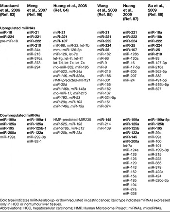
Since 2006, several studies have determined the miRNA expression profiles of liver cancer compared with adjacent nontumour tissues or benign tissues, such as cirrhotic livers and chronic hepatitis livers (Refs Reference Murakami83, Reference Huang84, Reference Wang85, Reference Ladeiro86) (Table 3). Further studies by Wang et al. (Ref. Reference Wang85) showed that miR-224 increased apoptosis as well as proliferation and targeted apoptosis inhibitor 5 (API5); miR-224 expression was inversely correlated with API-5 expression in HCC patients. Huang et al. (Ref. Reference Huang87) used bead-based microarray analysis to examine miRNA expression profiling on pairs of surgically removed HCC and adjacent nontumour tissue; 12 upregulated and 19 downregulated miRNA were found to be associated with HCC. Furthermore, miR-338 downregulation was significantly associated with clinical aggressiveness, such as tumour size, tumour–node–metastasis stage, vascular invasion and intrahepatic metastasis. This study suggests that miRNA expression could have relevance to the clinical behaviour of HCC. A recent study by Su et al. (Ref. Reference Su88) evaluated the expression of 308 miRNAs in human HCC and normal hepatic tissues by microarray and identified 29 differentially expressed miRNAs in HCC tissues.
Differentially expressed miRNAs in HCC compared with nontumour tissuesa

a Bold type indicates miRNAs also up- or downregulated in gastric cancer; italic type indicates miRNAs expressed only in HCC or nontumour liver tissues.
Abbreviations: HCC, hepatocellular carcinoma; HMP, Human Microbiome Project; miRNAs, microRNAs.
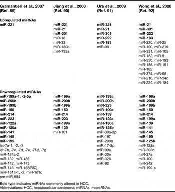
Aberrant miRNA expression profiles between HCC and benign liver tissues have also been identified. Gramantieri et al. (Ref. Reference Gramantieri89) used a genome-wide miRNA microarray, and identified 35 differentially expressed miRNAs in HCC and cirrhotic liver tissues (Table 4). Algorithms SVM and PAM correctly classified 91% and 86% of the samples, respectively. These analyses provide strong evidence that miRNA signatures are able to correctly predict most HCC and cirrhotic tissues. Jiang et al. (Ref. Reference Jiang90) compared the expression of 196 mature miRNAs in HCC and adjacent benign tissues and normal liver tissues using real-time PCR. Sixteen miRNAs were differentially expressed when the tumour data were compared with adjacent benign tissue (Table 4). A set of 19 miRNAs (let-7c, let-7 g, miR-26b, miR-29c, miR-30e-3p, miR-31, miR-99a, miR-99b, miR-100, miR-125b, miR-139, miR-148a, miR-150, miR-200c, miR-220, miR-221, miR-345, miR-372 and miR-377) significantly correlated with disease outcome, such that individuals with predominantly higher miRNA expression displayed better survival. They also identified that 50 mature and precursor miRNAs were overexpressed in both cirrhotic and hepatitis-positive liver specimens compared with the uninfected, noncirrhotic specimens. Ura et al. (Ref. Reference Ura91) used real-time qRT-PCR to measure the expression of 188 miRNAs in human HCC liver tissues linked with hepatitis B and C viral infection, and in hepatitis B and C liver tissues and normal liver tissues. Thirty-one miRNAs can differentiate the stage of liver disease (normal, chronic hepatitis and HCC), and out of the 31 miRNAs, 17 were downregulated in HCC and six were upregulated in HCC (Table 4).
Differentially expressed miRNAs in HCC compared with benign liver tissues

Bold type indicates miRNAs commonly altered in HCC.
Abbreviations: HCC, hepatocellular carcinoma; miRNAs, microRNAs.
Wong et al. (Ref. Reference Wong92) compared the expression of 156 miRNAs in HCC to matching adjacent cirrhotic livers and identified a 40-miRNA signature that could discriminate tumours from adjacent cirrhotic liver tissue (Table 4). They further confirmed that miR-222 is the most significantly upregulated candidate identified in a larger cohort of tumours. Increased miR-222 expression correlated significantly with advanced stages of HCC and with shorter disease-free survival of patients. Inhibition of miR-222 significantly retarded cell motility. miR-222 could confer metastatic potential in HCC cells possibly by targeting protein phosphatase 2A subunit B (PPP2R2A) and subsequently activating signalling by the protein kinase AKT (Ref. Reference Wong92).
Functional significance, and diagnostic and prognostic potential, of miRNAs in liver cancer
miR-122
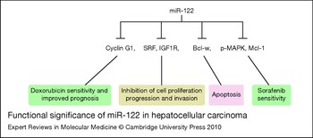
miR-122 is one of the most important and well-investigated miRNAs in HCC because of its crucial function. miR-122 is a liver-specific miRNA that is expressed in the developing liver and at high levels in the adult liver, and mainly regulates lipid and cholesterol metabolism (Refs Reference Chang93, Reference Lagos-Quintana94, Reference Esau95). In human HCC, studies have shown a downregulation of miR-122 (Refs Reference Huang87, Reference Gramantieri89, Reference Ura91, Reference Meng96, Reference Kutay97, Reference Coulouarn98), suggesting that its function is associated with hepatocarcinogenesis. An in situ hybridisation demonstrated that miR-122 was abundantly expressed in hepatocytes, but barely detectable in primary human HCC (Ref. Reference Bai99). Furthermore, in both in vitro and in vivo studies, miR-122 was reported to inhibit liver cancer cell growth, progression and invasion by directly targeting cyclin G1 (CCNG1), ADAM10 (a disintegrin and metalloprotease family 10), serum response factor and insulin-like growth factor 1 receptor (Refs Reference Gramantieri89, Reference Coulouarn98, Reference Bai99). miR-122 also downregulated Bcl-w (an antiapoptotic Bcl-2 family member) and induced apoptosis in HCC cell lines (Refs Reference Bai99, Reference Lin100). miR-122 can also sensitise HCC cells to sorafenib, which is the only oral multikinase inhibitor that enhanced overall survival in patients with advanced HCC (Ref. Reference Llovet101). The level of phospho-MAPK (p-MAPK), a target of sorafenib, was significantly reduced in miR-122-expressing cells compared with control cells. Similarly, the expression of antiapoptotic Mcl-1 gradually decreased with increasing concentrations of sorafenib in miR-122-expressing cells. This suggests that a miR-122 mimetic alone or in combination with anticancer drugs might be a promising therapeutic regimen against liver cancer.
HCC patients with higher miR-122 expression have been found to have an overall significantly longer survival time (Ref. Reference Coulouarn98). Thus, the loss of miR-122 is a marker of poor prognosis. Lower miR-122 levels have also been associated with a shorter HCC recurrence time, and higher levels of cyclin G1, verified as a direct target of miR-122, were related to a lower survival (Ref. Reference Fornari102); furthermore, miR-122 overexpression and cyclin G1 silencing increased doxorubicin sensitivity in HCC cell lines. Thus, miR-122 acts as a tumour suppressor gene and might be an indicator of prognosis and a therapeutic target in HCC (Fig. 4).
Functional significance of miR-122 in hepatocellular carcinoma. miR-122, a liver-specific miRNA, downregulates the expression of SRF and IGF1R to inhibit cell proliferation, tumour progression and invasion. miR-122 induces apoptosis by suppressing Bcl-w. miR-122 increases sensitivity to sorafenib through decreased p-MARK and Mcl-1. Cyclin G1 is also a direct target of miR-122; overexpression of miR-122 causes increased sensitivity to doxorubicin and is associated with better prognosis in HCC patients. Abbreviations: HCC, hepatocellular carcinoma; IGF1R, insulin-like growth factor 1 receptor; miRNA, microRNA; SRF, serum response factor.

Other HCC-associated miRNAs (miR-223, miR-101, miR-34a, miR-23b, miR-195 and miR-26)
Some of the other important and well-investigated miRNAs in HCC are miR-223, miR-222, miR-101, miR-23b, miR-195 and miR-26. miR-223 levels were repressed and miR-222 levels increased in HCC tumours in comparison with adjacent nontumour liver tissues (Ref. Reference Wong92), and highly deregulated miR-223 and miR-222 could unequivocally distinguish HCC from adjacent nontumour liver, irrespective of viral associations. miR-223 showed a consistent inhibitory effect on cell viability through its direct target, stathmin 1 (STMN1). The oncogenic function of miR-222 is discussed in detail in the next section.
Another miRNA that functions as a potential tumour suppressor in HCC is miR-34a. Human HCC tissues showed reduced miR-34a expression in comparison with adjacent nontumour tissues, and were further found to be associated with tumour metastasis and invasion. In addition, miR-34a directly targeted MET and reduced both the mRNA and protein levels of MET. These results indicate the suppressive role of miR-34a in tumour migration and invasion through modulation of the MET signalling pathway (Ref. Reference Li103).
miR-101 was shown to be downregulated in HCC tissues compared with their nontumour counterparts by microarray and northern blotting (Ref. Reference Su88). Furthermore, an in vivo study showed that miR-101 suppressed the formation of hepatoma cells colonies, which would result in tumours. miR-101 enhanced apoptosis in hepatoma cell lines triggered by serum starvation or chemotherapeutic drugs through the suppression of Mcl-1. This suggests that miR-101 might have potential significance in cancer therapy. Downregulation of miR-101 in HCC was confirmed by another study (Ref. Reference Li104). miR-101 expression inhibited the invasion and migration of cultured HCC cells by targeting FOS, a key component of the activator protein-1 transcription factor. These findings continue to suggest that miR-101 might play an important role in HCC.
miR-23b was downregulated in HCC specimens compared with peritumoural tissues (including cirrhosis, hepatitis, steatosis and haemosiderosis) (Ref. Reference Salvi105). miR-23b translationally repressed the expression of urokinase-type plasminogen activator and MET in HCC cells, causing a decrease in cancer cell proliferation and migration capabilities. Microarray analysis showed that miR-195, a member of the miR-15/16/195 family, was downregulated in HCC tissues compared with normal and benign liver tissues (Refs Reference Murakami83, Reference Su88, Reference Gramantieri89, Reference Wong106), and this was confirmed in paired HCC and adjacent nontumour liver tissues by northern blotting (Ref. Reference Xu107). The introduction of miR-195 suppressed the ability of HCC and CRC cells to form colonies in vitro as well as their ability to develop tumours in nude mice. Furthermore, miR-195 blocked the G1–S transition by repressing Rb–E2F signalling through targeting several crucial proteins, including cyclin D1, CDK6 and E2F3. These data highlight the important role of miR-195 in cell cycle control as well as in the molecular aetiology of HCC. The studies also suggest the potential application of miR-195 in cancer therapy.
Recent analysis of the roles of miR-26 in three independent cohorts, including a total of 455 patients with HCC who had undergone radical tumour resection, found that the expression of miR-26a and miR-26b in nontumour liver tissue was higher in women than in men (Ref. Reference Ji37). Moreover, patients whose tumours had low miR-26 expression had shorter overall survival but were more responsive to interferon therapy than patients whose tumours had a high expression of the miRNA. Therefore, the expression patterns of miRNAs in liver tissue differ between men and women with HCC, and the miR-26 expression status of such patients is associated with survival and response to adjuvant therapy with interferon α.
Oncogenic miRNAs in HCC
miR-21, an oncogenic miRNA identified in CRC, was also found to be overexpressed in human HCC tissues (Refs Reference Huang84, Reference Wang85, Reference Jiang90, Reference Ura91, Reference Meng96, Reference Wong106). Another study confirmed the upregulation of miR-21 in human HCC samples by northern blotting (Ref. Reference Connolly108). PTEN was shown to be a direct target of miR-21 in cultured HCC cells; miR-21 altered the downstream mediators of PTEN, such as focal adhesion kinase phosphorylation and matrix metalloproteases 2 and 9, to contribute to increased cell growth, migration and invasion (Ref. Reference Wu81) (Fig. 3).
miR-221 was shown to directly target both p27 and p57 in HCC-derived cells (Ref. Reference Fornari109). miR-221 was upregulated in HCC cases compared with matched cirrhosis samples, and protein levels of p27 and p57 were downregulated in HCC. Thus, miR-221 has an oncogenic function in hepatocarcinogenesis by targeting p27 and p57, thereby promoting cell proliferation by controlling cell cycle inhibitors. Besides p27 and p57, the apoptotic activator Bmf was confirmed as a target of miR-221 in HCC cell lines, wherein miR-221 inhibited apoptosis through downregulation of Bmf (Ref. Reference Gramantieri110). Overexpression of miR-221 and decreased Bmf were found in a set of HCC cell lines when compared with cirrhotic tissues. Moreover, high miR-221 levels were associated with tumour multifocality and reduced recurrence time after surgery. miR-221 levels can thus potentially predict the prognosis of HCC.
Profiling of miRNA expression in HCC, adjacent cirrhotic livers and normal livers linked a set of 12 miRNAs (including overexpression of miR-106b, miR-21, miR-210, miR-221/222, miR-224, miR-34a, miR-425, miR-519a, miR-93 and miR-96, and downregulation of let-7c) to disease progression from normal liver through cirrhosis to full-blown HCC (Ref. Reference Pineau111). In addition, miR-221 overexpression was shown to enhance tumourigenesis in vivo and DNA damage-inducible transcript 4 was identified as a bona fide target of miR-221.
Another recent report showed that miR-221 and miR-222 were overexpressed in aggressive non-small-cell lung cancer and HCC, in comparison with less invasive and/or normal lung and liver cells (Ref. Reference Garofalo112). miR-221 and miR-222 were shown to induce resistance to the apoptosis inducer TRAIL (TNFSF10) by targeting phosphatase and tensin homologue (PTEN) and TIMP3 tumour suppressors, and enhanced cellular migration through the activation of the AKT pathway and metallopeptidases. Evidence was also provided for the involvement of the oncogenic receptor MET in miR-221 and miR-222 activation through the JUN transcription factor.
miR-155 was reported to target tumour suppressor CCAAT/enhancer binding protein beta (C/EBPβ), thereby promoting cell growth in HCC cells; miR-155 was significantly upregulated in primary human HCC cells with a concomitant decrease in C/EBPβ levels compared with matching liver tissues (Ref. Reference Wang113).
The miR-17-92 cluster and its paralogue, the miR-106b-25 cluster, are differentially expressed in some human cancers. At least 50% of human HCC samples showed a significant increase in the expression of both miR-18 and miR-106b-25 when compared with corresponding paired nontumour samples (Ref. Reference Li114), and the transcription factor E2F1 was identified as a target gene for miR-106b and miR-93. Thus, the miR-17-92 cluster and the miR-106b-25 cluster have oncogenic potential in HCC.
Yamamoto et al. (Ref. Reference Yamamoto115) showed that miR-500 was abundantly expressed in human HCC tissues compared with adjacent nontumour tissues. More importantly, an increased amount of miR-500 was found in the sera of three out of ten HCC patients, and after surgical treatment the levels of miR-500 in these three patients returned to normal. These findings show that miR-500 is an oncofetal miRNA relevant to the diagnosis of human HCC.
Li et al. (Ref. Reference Li116) identified 69 miRNAs that were differentially expressed in HCC from the analysis of 78 matched cancer and noncancerous liver tissues from HCC patients and ten normal liver tissues; among the miRNAs validated by real-time qRT-PCR, miR-18a, miR-106b, miR-15b, miR-221/222 and miR-224 were upregulated and miR-125b and miR-101 were downregulated. The set of differentially expressed miRNAs could distinctly classify HCC, nontumour tissue and normal liver tissue. Most importantly, higher miR-125b correlated with good survival in HCC patients. miR-125b could evidently suppress cell growth and phosphorylation of AKT in HCC cell lines. Thus, for the first time, miR-125b was shown to have prognostic significance for HCC.
Budhu et al. (Ref. Reference Budhu117) examined miRNA expression profiles in 482 cancerous and nontumour specimens from a radical resection of 241 patients with HCC. Using a supervised algorithm and a clinically well-defined cohort of 131 cases, they built a unique 20-miRNA metastasis signature that could significantly predict primary HCC tissues with venous metastases from metastasis-free solitary tumours with a tenfold crossvalidation. A survival risk prediction analysis showed that most of the metastasis-related miRNAs were associated with poor survival. Furthermore, the 20-miRNA tumour signature was validated in 110 additional cases as a significant independent predictor of survival. It has shown significant association to both survival and relapse in 89 cases of early-stage HCC. These 20 miRNAs might provide a simple profiling approach to assist in identifying patients with HCC who are likely to develop metastases or recurrence.
miRNAs associated with pancreatic cancer
Pancreatic cancer is the fourth leading cause of cancer-related death in the USA, with an approximate five-year relative survival rate of 5% (Ref. Reference Jemal11). The poor prognosis of pancreatic cancer patients is a result of late clinical presentation, early and high metastatic potential, and resistance to chemotherapy and radiation (Refs Reference Bloomston118, Reference Greither119). Eighty-five per cent of pancreatic tumours are ductal adenocarcinomas (PDACs) (Ref. Reference Garcea120). In the past few years, increasing efforts have been made to identify miRNAs that can be used for the early detection, diagnosis and prognosis of pancreatic cancer based on human PDAC tissues.
Expression profiling analysis of miRNAs associated with pancreatic cancer
Recently, several studies have found aberrant miRNA expression profiles in pancreatic cancer tissues (Refs Reference Bloomston118, Reference Lu121, Reference Lee122, Reference Szafranska123, Reference Zhang124). We have not included a summary table in this review for the overlapping miRNAs in pancreatic cancer because this topic has been recently reviewed by Seux et al. (Ref. Reference Seux125). It is important to note that some of the important miRNAs in pancreatic cancer (e.g. miR-21, miR-122, miR-221 and miR-222) are shared among different gastrointestinal cancer types.
Lu et al. (Ref. Reference Lu121) first used a bead-based hybridisation method to screen the expression of 217 miRNAs in nine samples of pancreatic adenocarcinoma. They found a nearly universal downregulation of miRNA (129 of 131 miRNAs) in pancreatic adenocarcinomas compared with normal pancreas tissues. However, several other profiling studies reported that most of the miRNAs were increased in PDAC. Schmittgen and colleagues (Ref. Reference Lee122) analysed the expression of 222 miRNA precursors in human PDAC and adjacent benign tissues, chronic pancreatitis tissues, normal pancreas tissues and pancreatic cancer cell lines using real-time qRT-PCR. One hundred miRNA precursors were overexpressed in pancreatic cancer, including miR-155, miR-21, miR-221, miR-222, miR-100, miR-376a and miR-301. A few miRNAs were shown to have reduced expression in pancreatic cancer, such as miR-375, which was expressed in mouse pancreatic islet cells, and whose expression is possibly specific to islet cells (Ref. Reference Poy126). The upregulation of miR-155 and miR-100 and the downregulation of miR-375 were further validated by real-time qRT-PCR and northern blotting. In situ hybridisation showed that miR-221, miR-376a and miR-301 were located only to tumours. Furthermore, miRNA expression profiling was able to differentiate tumours from normal pancreas, pancreatitis and cell lines. These findings suggest that aberrant miRNAs play an important role in pancreatic tumourigenesis, and a miRNA expression profiling signature might potentially be used as a diagnostic tool.
Croce and colleagues (Ref. Reference Bloomston118) investigated the miRNA expression profiles in 65 microdissected PDACs, their adjacent noncancerous pancreatic tissues and 42 chronic pancreatitis tissues, and found that 30 miRNAs (including miR-21, miR-155, miR-221 and miR-222) were upregulated and three miRNAs (miR-148a, miR-148b and miR-375) were downregulated in pancreatic cancers compared with normal pancreatic tissue. Further analysis showed that 21 miRNAs with increased expression and four miRNAs with decreased expression correctly distinguished pancreatic cancer from benign pancreatic tissue in 90% of samples by crossvalidation; 15 overexpressed and eight underexpressed miRNAs differentiated pancreatic cancer from chronic pancreatitis with 93% accuracy; and a subgroup of six miRNAs was able to distinguish long-term survivors with node-positive disease from those dying within 24 months. High levels of miR-196a-2 significantly predicted a short duration of survival. Hahn and colleagues (Ref. Reference Szafranska123) compared miRNA expression within PDAC cell lines, normal pancreas, macrodissected chronic pancreatitis tissues and PDAC tissues using 377 featured miRNA arrays. They found that 32 miRNAs were upregulated and 41 were downregulated by at least twofold in PDAC. They selected an additional 26 miRNAs as potential biomarker candidates for PDAC. miR-155 and miR-221 were also identified by the other two groups as mentioned above. The downregulated miRNAs included miR-216, miR-217 and miR-375. Apart from islet-cell-specific miR-375, the authors suggested that very low to absent expression of miR-216 and miR-217 in PDAC samples and cell lines indicate that these two miRNAs are primarily expressed in acinar cells.
Zhang et al. (Ref. Reference Zhang124) used real-time qRT-PCR to detect the expression levels of 95 cancer-related miRNAs in pancreatic cancer specimens and cell lines as well as pancreatitis tissues. Among the 95 miRNAs, eight miRNAs (miR-196a, miR-190, miR-186, miR-221, miR-222, miR-200b, miR-15b and miR-95) were identified to be significantly upregulated in most pancreatic cancer tissue samples and cell lines. A recent study by Szafranska et al. (Ref. Reference Szafranska127) used real-time qRT-PCR analysis to quantify the levels of a panel of 13 miRNAs (miR-196a, miR-217, miR-148a, miR-130b, miR-375, miR-210, miR-155, miR-143, miR-145, miR-223, miR-96, miR-31 and miR-205) in 16 fine-needle aspirate (FNA) biopsy samples of pancreatic tissue. They also quantified miRNA levels in a training set of frozen samples from six nonpathological pancreas tissues, six patients with chronic pancreatitis and eight PDAC patients. They found that the differences in miRNAs between the nonpathological and cancer FNA samples were nearly identical to those betwen the corresponding frozen pancreas samples. Further data analysis showed that the combination of miR-196a and miR-217 clearly separated malignant from benign tissues for both the frozen and FNA sets of samples. Interestingly, the concentration of miR-217 was high in only healthy pancreatic tissues, and miR-196a was detected above the background level. These findings indicate that miRNA analysis of pancreatic FNA biopsy samples can aid in the pathological evaluation of suspicious cases and might provide a new strategy for improving the diagnosis of pancreatic diseases.
Functional significance, and diagnostic and prognostic potential, of miRNAs in pancreatic cancer
Investigation of the roles of miRNAs in pancreatic cancer is still at an early stage. The expression profiling efforts have provided miRNA candidates for functional studies. Torrisani et al. (Ref. Reference Torrisani128) discovered that let-7 expression was strongly reduced in surgery-resected PDAC samples and in endoscopic ultrasound-guided FNA material from patients with PDAC as compared with adjacent tissues. In situ RT-PCR showed that let-7 was expressed in normal acinar pancreatic cells and absent in poorly differentiated cancer samples. Functional experiments indicated that let-7 inhibited cell proliferation, KRAS expression and MAPK activation in vitro, but failed to impede tumour growth progression in vivo.
Some of the important miRNAs described earlier have also been demonstrated to influence pancreatic cancer. Oncogenic miR-21 was highly expressed in pancreatic cancers compared with benign pancreas and chronic pancreatitis (Ref. Reference Dillhoff129). Furthermore, high levels of miR-21 predicted a poorer outcome compared with absent or faint/focal miR-21 expression in patients with node-negative disease. This suggests that miR-21 might be an important prognostic biomarker (Fig. 3).
Greither et al. (Ref. Reference Greither119) recently discovered the differential expression levels of miR-155, miR-203, miR-210 and miR-222 in PDAC compared with normal pancreatic tissue. Other pancreas-specific miRNAs are miR-216 and miR-217 (Ref. Reference Szafranska123). Exploring the potential association with PDAC survival, four out of six analysed miRNAs (miR-155, miR-203, miR-210 and miR-222) showed a significant correlation with the prognosis of PDAC patients. Higher expression of miR-155, miR-203, miR-210 and miR-222 was significantly correlated with a worse outcome in PDAC patients; a lower expression of miR-217 correlated with a worse outcome in PDAC patients; and miR-216 expression showed no association with patient survival. Furthermore, the combined overexpression of all four miRNAs increased the relative risk of tumour-related death from a range between 2.20-fold and 2.50-fold to 5.24-fold.
Conclusion and future perspectives
From many miRNA studies, several miRNAs have been identified to be important in gastrointestinal cancer. Some of the key miRNAs (e.g. miR-143, miR-145, miR-17-92 cluster, miR-21, miR-26, miR-192 and miR-34) are relevant to several gastrointestinal tumour types. Furthermore, given their critical functions, some of these miRNAs might also have significant effects on many major cancer types.
There is no doubt that miRNAs will be important biomarkers in cancer diagnosis and prognosis based on some of these promising reports. An improved detection and quantification technology for miRNA expression analysis should make it possible to use various types of clinical specimens (e.g. circulating tumour cells) for cancer biomarker discovery. Collaborative efforts are essential to fully validate and realise the potential clinical utilities of these miRNA biomarkers using large patient cohorts.
miRNAs also have foreseeable potential as novel therapeutic targets for treating cancer, because modulating the expression of one particular miRNA mediates many targets and pathways in a reversible and fine-tuning manner. With clinically friendly and cost-effective miRNA expression technologies, miRNAs might soon have a direct impact on cancer treatment.
Acknowledgements and funding
We appreciate the critical reviews from the peer reviewers, and editing by Ms Cecilia Zhang. We apologise to our colleagues whose research was not cited in this review because of space limitations and timing. This study was supported in part by Stony Brook University Translational Research Laboratory Start-up funding (J.J.) and MH075020 (J.J.).