I. INTRODUCTION
Titania is known to be a very useful nontoxic, environmentally friendly photocatalyst. Reference Li, Cao, Liu, Su and Dong1–Reference Inagakia, Kondoa, Nonakaa, Itob, Toyodac, Sogabec and Tsumura4 It is a stable, photo-durable, inexpensive, and natural aid in the efficient realization of many applications, such as bio-application, dye-sensitized solar cells, photoelectrochemical cells, gas sensors, and hydrogen production. Reference Senna, Myers, Aimable, Laporte, Pulgarin, Baghriche and Bowen5–Reference Hanaor and Sorrell9 Titania has three crystal structures: anatase, rutile, and brookite. Reference Chuangchote, Jitputti, Sagawa and Yoshikawa10–Reference Liu, Wang, Yang, Cheng and Lu12 In titania phase systems transformation, amorphous titania crystallizes into the most common titania phases, namely anatase, rutile, or a mixture of them, by way of thermal treatment. Reference Albetran, Haroosh, Dong, Prida, O'Connor and Low13,Reference Albetran, Haroosh, Dong, O'Connor and Low14 The amorphous-to-anatase and subsequent anatase-to-rutile phase transformation behaviors depend on the impurities, morphology, preparation method, dopants, heating rate, calcining temperature, calcining time, and atmospheres. Reference Ghicov, Tsuchiya, Macak and Schmuki15–Reference Shannon and Pask19
One-dimensional (1-D) titania nanostructures, for example, nanowires, nanorods, nanobelts, nanofibers, and nanotubes provide unique electronic characteristics, such as a high surface area-to-volume ratio, high electron mobility, or quantum confinement effects, and high mechanical strength. Reference Li, Cao, Liu, Su and Dong1,Reference Varghese, Gong, Paulose, Grimes and Dickey3,Reference Xiong, Slater, Balasubramanian, Johnson and Rajh7,Reference Yanga, Parka, Choa, Kima and Choi8 These characteristics increase the decomposition rate of organic pollutants significantly because photocatalytic reactions occur rapidly and intensely on the catalyst surface. Reference Li, Zhang and Pan20,Reference Lee, Ha, Hong and Park21 Various techniques have been used to synthesize 1-D nanostructured titania, including sol–gel, Reference Zainal and Lee22 microwaves, Reference Monti, Ponrouch, Estruga, Palacin, Ayllon and Roig23 electrochemical anodization, Reference Macak, Tsuchiya, Ghicov, Yasuda, Hahn, Bauer and Schmuki6,Reference Arunchandran, Ramya, George and Mudali24–Reference Liao, Lin, Pan, Li, Li and Li27 hydrothermal methods, Reference Inagakia, Kondoa, Nonakaa, Itob, Toyodac, Sogabec and Tsumura4,Reference Sanchez-Tovar, Lee, Garcia-Anton and Schmuki28–Reference Bavykin, Lapkin, Plucinski, Friedrich and Walsh30 and electrospinning. Reference Tan, Pingguan-Murphy, Ahmad and Akbar31 Thus, there is a great deal of interest in synthesizing 1-D titania nanostructures using inexpensive and simple methods.
Among fabrication methods, anodization is currently one of the most commonly used method for the fabrication of 1-D titania nanostructures, Reference Li, Cao, Liu, Su and Dong1,Reference Macak, Tsuchiya, Ghicov, Yasuda, Hahn, Bauer and Schmuki6,Reference Tan, Pingguan-Murphy, Ahmad and Akbar31 and the general nanostructure obtained through this process is nanotubes. Reference Antony, Mathews, Ajikumar, Krishna, Dash and Tyagi17,Reference Zainal and Lee22 Nanotubes are 1-D nanometer scale tubes like nanowires, nanorods, and nanofiber structures. In the case of nanotubes, there is a hollow center as compared to the other 1-D structures. The anodized titania nanotube arrays are synthesized by electrochemical anodization of titanium-based alloy in an electrolyte with Pt grid used as an anode for a short period at room temperature (RT). Reference Macak, Tsuchiya, Ghicov, Yasuda, Hahn, Bauer and Schmuki6,Reference Arunchandran, Ramya, George and Mudali24–Reference Liao, Lin, Pan, Li, Li and Li27
In this paper, titania nanotube arrays were synthesized electrochemically by anodization of titanium foils, and the synthesized titania nanotubes were then implanted with indium ions. The effect of In-ions implantation on the crystallization kinetics and phase transformations of anodized titania nanotubes were investigated using in-situ high-temperature x-ray diffraction (XRD) and synchrotron radiation diffraction (SRD) from RT to 1000 °C, and then were characterized by field emission scanning electron microscopy (FESEM), and associated energy dispersive X-ray spectroscopy (EDS).
II. EXPERIMENTAL METHODS
A. Sample preparation
Titanium foils (99.6% purity) with dimensions of 10 × 10 × 0.1 mm3 were anodized to synthesize the titania nanotube arrays. The titanium foils were cleaned ultrasonically in a mixed solution of ethanol, isopropanol, and acetone for five minutes, followed by rinsing with deionized water, and were then dried using nitrogen stream. In the electrochemical anodization cell, the Ti foil acted as a cathode. Platinum was used as an anode in an electrolyte of 2.04 mL water, 100 mL ethylene glycol, and 0.34 g ammonium fluoride (NH4F). The anodization experiment was performed in a sealed environmental chamber at a constant temperature of 25 °C, and at a pH of 6. The voltage was controlled to 60 V for 20 h using a voltage power supply between the Ti foil and the platinum located several centimeters apart. The as-anodized titania arrays were rinsed/washed after being anodized with ethanol, immersed in dimethyl sulfoxide, and dried in air.
B. Field emission scanning electron microscopy
The surface morphologies of the anodized titania nanotubes before and following the in-situ high-temperature measurement were investigated using field emission scanning electron microscope (FESEM; Zeiss, Neon, 40EsB, Oberkochen, Germany). Prior to the microstructure observations, the samples were sputter-coated with a 3 nm layer of platinum to avoid charging, and then the FESEM images were taken using secondary electron with an accelerating voltage of 5 kV. EDS was used to analyze the elemental compositions of the SEM-image materials.
C. In ion implantation
Details of ion implantation are given in the preliminary studies by the authors with Cr-ions implanted titania nanotube arrays and V-ions implanted titania nanofibers. Reference Low, Albetran, Prida, Vega, Manurung and Ionescu32,Reference Albetran, O'Connor and Low33 In summary, the as-anodized samples were implanted with In ions using the MEVVA ion implanter (Lawrence Berkeley Laboratory, Berkeley, California) at the Australian Nuclear Science and Technology Organization (ANSTO). The titania nanotubes were implanted by exposing a flux of indium ions perpendicular to the sample surface. The average implantation energy of indium ions was 25 keV, and the peak penetration depth of indium in the titania nanotubes was close to the 70 nm stopping and range of ions in matter prediction based on the implantation parameters. The near-surface composition depth profiling of In ion-implanted titania nanotubes was measured by Rutherford backscattering spectrometry using He+1 ions at 2 MeV, and the implanted dose was ∼4 × 10¹⁵ ions/cm2.
D. In-situ high-temperature XRD and SRD
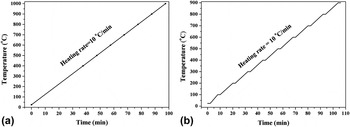
In-situ crystallization behavior of as-anodized titania nanotubes was evaluated in an air atmosphere using XRD at temperature ranging from RT to 1000 °C using an Anton Parr HTK16 hot stage (Graz, Austria). The sample was mounted and heated at a rate of 10 °C/min, from ambient temperature to 1000 °C [See Fig. 1(a)]. The XRD patterns were collected at ambient temperature and then in steps of 100 °C from 400 to 1000 °C using Cu Kα radiation with a wavelength of 0.15419 nm, settings at 40 kV, 30 mA, and each pattern was measured over the angular range of 2θ = 20°–125°.
Non-isothermal in-situ (a) XRD and (b) SRD heating protocol. Plateaus indicate the 2 min during which SRD data were acquired.

For the In-ions implanted titania nanotubes, the measurements of in-situ high-temperature SRD were conducted at the powder diffraction beamline at the Australian Synchrotron, Melbourne, Australia. The specimens were mounted and heated using an Anton Parr HTK16 hot platinum stage in conjunction with a Mythen II microstrip detector. The SRD patterns were acquired at ambient temperature and then in steps of 100 °C from 100 °C to 900 °C in air according to the heating protocol shown in Fig. 1(b). The SRD data were collected at an incident angle of 3° and a wave length of 0.1126 nm. The wavelength was determined accurately from a NIST 660a pattern with zero error from 0.007368283 to 0.0001243282° and sample displacement from 0.002781912 to 0.0008831296 mm. Each pattern was measured over the angular range of 2θ = 5°–84°.
E. Quantitative analysis
The XRD and SRD patterns were analyzed using Rietveld pattern-fitting with TOPAS (Bruker AXS, version 4.2) software, with the goodness-of-fit (GOF) being gauged from the values of the expected R-factor (R exp), the weighted pattern R-factor (R wp), and the derived Bragg R-factors (R P). The Rietveld method is expected to be more accurate than the Spurr and Myers method, which uses only the anatase (101) and rutile (110) peak intensities. Reference Albetran, Haroosh, Dong, Prida, O'Connor and Low13 The use of the Rietveld method minimizes issues relating to preferred orientation, grain size, and/or degree of crystallinity. To compute the relative crystalline phase abundances, and the lattice parameters of titanium, anatase, and rutile as a function of temperature, the parameters optimized in the Rietveld refinements were the pattern background, 2θ0, sample displacement and, for each phase, the scale factor, lattice parameters, preferred orientation, and peak shape parameters. Rietveld refinements were conducted with the crystal structures of titaniumα (ICSD 44872), anatase (ICSD 202242), and rutile (ICSD 64987). These phases were identified using the DIFFRAC. EVA software (released 2012, version 3.1) to search the International Center for Diffraction Data powder diffraction file database (PDF-4+2013 software version: 4.13.0.6, Database version: 4.1302).
III. RESULTS AND DISCUSSION
A. Microstructural imaging
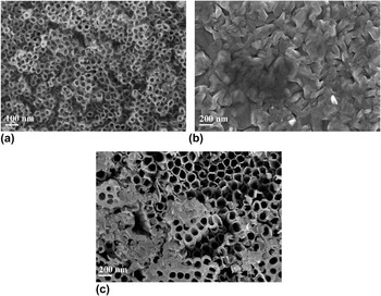
A homogeneous white layer formed on the Ti foil after the anodizing process in the ethylene glycol/NH4F electrolyte at 25 °C for 20 h (optical image not shown). Figure 2(a) shows a typical secondary electron FESEM image of as-anodized titania nanotubes, which were amorphous. The as-anodized nanotube arrays were more uniform and highly ordered than the titanium foil. The corresponding mean inner diameter and standard deviation for 100 nanotubes was 26 ± 4 nm.
FESEM micrographs of (a) as-anodized titania nanotube arrays prior to heating; (b) after thermal treatment for the XRD experiment at 1000 °C and cooling to RT; and (c) after In-ions implantation and thermal treatment for SRD experiments at 900 °C and cooling to RT.

After the in-situ high-temperature XRD at 1000 °C for as-anodized titania nanotube arrays, and for SRD at 900 °C for In-ions implanted titania nanotube arrays, the morphologies were re-examined at RT using the same FESEM conditions. Figures 2(b) and 2(c) illustrate the FESEM images of non-implanted and In-ions implanted materials, respectively. For the non-implanted sample, the nanotube array structures were destroyed and covered with rutile elongated grains. However, for In-ions implanted titania nanotubes, some grains were randomly distributed among the titania nanotubes, which indicates that implantation had inhibited or slowed the grain growth of titania nanotube arrays. These grains are crystalline rutile because metastable anatase transformed into stable crystalline rutile at elevated temperatures (see Sec. III. B).
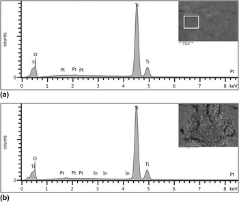
Figure 3(a) shows the EDS spectrum for the as-anodized titania nanotube arrays in Spectrum 1, showing that strong signatures for Ti, O, and Pt, with the Pt content begin due to the platinum coating. Assuming that the Pt peak from the platinum coating is negligible, the quantitative EDS analysis indicated 66.18% O Kα and 33.82% Ti Kα, thus confirming the presence of titania. It should be noted that quantitative EDS is performed for flat samples, which is approximated for the nanotubes. The EDS for the nanotubes was collected from a small square to minimize the nonflat sample issue, using EDS software (AZtec Software, Oxford). The qualitative EDS spectrum for non-implanted and In-ions implanted nanotubes after cooling was essentially the same for the as-anodized nanotubes, whereas the quantitative EDS analysis indicated approximately 33.82% Ti Kα and 66.18% O Kα, thus confirming and providing evidence of titania with a TiO2 chemical formula structure. The existence of In ions in-implanted titania nanotubes was verified by the quantitative EDS analysis in Spectrum 2 as shown in Fig. 3(b) that indicates 0.8(4)% In. This occurs because the surface composition is detected to a greater depth by SEM.
EDS spectra of (a) anodized TiO2 nanotubes prior to heating; and (b) after thermal treatment and In-ions implantation and cooling to RT.

B. Crystallization behavior
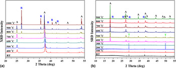
Figures 4(a) and 4(b) show the stacked XRD and SRD plots during thermal annealing in air from RT to 1000 °C for non-implanted titania nanotubes and In-ions implanted titania nanotubes, respectively. Diffraction results showed that both titania nanotube arrays were initially amorphous, but crystalline anatase formed at 400 °C for both samples. The anatase-to-rutile transformation started at 600 °C by a small crystalline rutile peak observed for non-implanted material, compared with 700 °C for In-ions implanted material. The reason for the 100 °C delay in the initial crystallization of rutile for the In-ions implanted titania nanotubes was that the In-ions implant provided greater structural rigidity and prevented relaxation in the Ti bonding environment. This behavior of severe/difficult rearrangement of the atoms in the anatase and rutile lattices could be due to the presence of In-ions interstitially or substitutionally. The presence of interstitial ions can inhibit the anatase-to-rutile transformation, whereas substitutional ions can also inhibit phase transformation if the substitutional ion is larger and does not differ significantly from Ti4+. Reference Shannon and Pask19,Reference Okada, Yamamoto, Kameshima, Yasumori and MacKenzie34 The In3+ radius (0.080 nm) is relatively larger than the Ti radius (0.061 nm). (For a discussion of whether In-ions enter the titania interstitially or substitutionally, see Sec. III. C)
Stacked XRD and SRD plots for the temperature range of RT-1000 °C of (a) non-implanted material, and (b) after In-ions implantation, respectively. [Legend: anatase (A), rutile (R), titanium peaks (T), and RT].

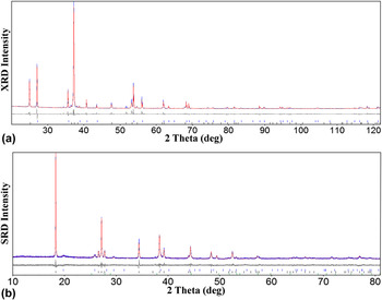
The relative phase levels of crystalline titanium, anatase, and rutile in the anodized nanotubes were calculated using the Rietveld method. Figures 5(a) and 5(b) show typical XRD and SRD Rietveld residual plots at 900 °C for data measured in non-implanted and In-ions implanted titania nanotube arrays, respectively. In nonimplanted material, R wp = 13.23, R exp = 3.75, R B (anatase) = 4.791, and R B (rutile) = 6.416%, and for the In-ions implanted material, R wp = 12.14, R exp = 9.94, R B (titanium α) = 2.817, R B (anatase) = 2.908 and R B (rutile) = 2.951%. The GOF values are 3.53 for non-implanted nanotube arrays and 1.22 for In-ions implanted nanotube arrays. The ideal value for the GOF is 1.0, but 4 or higher can be accepted for abundance data analysis. Reference Kisi35
XRD and SRD Rietveld difference plots for data measured in (a) non-implanted and (b) In-ions implanted titania nanotubes at 900 °C, respectively. Measured patterns are indicated by black crosses and calculated patterns by solid red lines. The Gray residual plot shows the difference between calculated and measured patterns. Peak positions for titanium, anatase, and rutile are indicated by green, black, and blue bars, respectively.

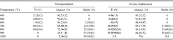
Table I shows the variation of relative phase concentrations of crystalline titanium, amorphous anatase, and rutile in the temperature range of 400–1000 °C for non-implanted and In-ions implanted material. At 600 °C, the relative phase concentrations were 1.40(12)% Ti, 96.55(64)% anatase, and 2.05(63)% for non-implanted nanotubes, whereas they were 1.56(47)% Ti, and 98.44(47)% anatase without any crystalline rutile formed for In-ions implanted titania nanotubes. The metastable anatase crystallizes from amorphous titania, which formed from the anodized Ti foils at a relatively low temperature, and the anatase subsequently transforms into stable crystalline rutile at a higher temperature. For the non-implanted sample, it is evident that the relative phase concentration of the crystalline rutile increased dramatically as the temperature increased from 600 to 1000 °C. However, the titania transformations were observed to increase more slowly in In-implantation than in nonimplantation samples. At 900 °C, the relative phase concentration was 0.2194(68)% Ti, 84.16(32)%, and just 15.66(31)% rutile for the In-ions implanted nanotubes, whereas all of the crystalline Ti, and more than 53% of the crystalline anatase transformed into crystalline rutile for the non-implanted sample. The relatively larger In3+ than Ti4+ radius would inhibit the transformation of titania, since it involves greater structural rigidity and prevents relaxation in the Ti bonding environment.
The effect of In-ions implantation on the relative weight percentages of anatase and rutile as a function of temperature, using the Rietveld method.

C. Influence of In-ions implantation on lattice parameters
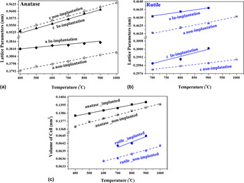
Figure 6 shows the temperature variations with the anatase and rutile lattice parameters and cell volumes for non-implanted and In-ions implanted nanotubes. The unit-cell parameters and cell volumes for both anatase and rutile in both non-implanted and In-ions implanted titania nanotube arrays increased linearly with temperature, but the lattice parameters and cell volumes showed an increase in the In-ions implanted sample relative to the non-implanted sample. For example, at 900 °C, the anatase lattice parameters a , and c for the non-implanted titania nanotube arrays were 0.380523(18) and 0.961642(26) nm, whereas they were 0.381530(75), and 0.960651(81) nm for the In-ions implanted samples, respectively. Moreover, the rutile lattice parameters a, and c at the same temperature, were they 0.4626490 (98) and 0.2986160 (75) nm for the non-implanted sample, whereas they were 0.46360 (13) and 0.300084 (79) nm for In-ions implanted samples, respectively. Thus, the cell volumes of anatase and rutile were 0.139243(13) and 0.0639170(31) nm3 for non-implanted samples, and 0.139838(27), 0.064495(41) nm3 for In-ions implantation at 900 °C, respectively. As can be seen, the volume of cells for the anatase and rutile were higher for the In-ions implanted samples than the non-implanted samples. The current lattice parameter increase in the In-ions implantation suggests that added ionic In substitutes for Ti4+ rather than occupying interstitial sites, which causes the ions to be incorporated as dopants in the titania lattice. Reference Shannon and Pask19,Reference Okada, Yamamoto, Kameshima, Yasumori and MacKenzie34 The lattice deformation of both titania phases caused by the incorporation of foreign Indium dopants affects the lattice parameter of In-ions implanted samples with temperature. The relatively difficult anatase-to-rutile transformation in the In-implanted titania nanotubes appears from an In-ions radius occupying the Ti sublattice substitutionally.
Variation of the lattice parameters as a function of temperature for (a) anatase, and (b) rutile; and (c) the cell volume in non-implanted and In-ions implanted anodized titania nanotube arrays.

IV. CONCLUSIONS
The effect of In-ions implantation on the dynamic crystallization behavior of anodized titania nanotubes was investigated using in-situ high-temperature XRD and SRD in the range from RT to 1000 °C. Diffraction results show that the diffraction peaks of the Ti foils were only observed before calcination until 300 °C, which indicated the amorphous titania nanotubes. The amorphous-to-anatase transformation started at 400 °C for non-implanted and In-implanted materials, including the characteristic crystalline anatase peaks. The crystalline rutile temperature increased from 600 to 700 °C for In-implantation, with the anatase-to-rutile transformation being inhibited relative to the non-implanted material. It was concluded that In-ions occupy the Ti sublattice substitutionally and then inhibit the anatase-to-rutile transformation. The relatively difficult anatase-to-rutile transformation in the In-implanted material appears to result from the relatively larger In3+ than Ti4+ radius, which provides greater structural rigidity and prevents relaxation in the Ti bonding environment.
ACKNOWLEDGMENTS
This research was supported by the funding from the Australian Institute of Nuclear Science and Engineering (AINGRA-11134), and the Australian Synchrotron (PD-3611). The authors would like to thank Prof. V.M. Prida from Department of Physics, University of Oviedo for XRD collection. Dr M. Ionescu of ANSTO assisted with work on ion-implantation.