Introduction to virosphere of Earth
Studies suggest that there are at least 1031 viruses on Earth (Bergh et al. Reference Bergh, Borsheim, Bratbak and Heldal1989; Hendrix et al. Reference Hendrix, Smith, Burns, Ford and Hatfull1999). They are a major factor in carbon cycling in the oceans (Borsheim et al. Reference Borsheim, Bratbak and Heldal1990; Santelli et al. Reference Santelli, Orcutt, Banning, Bach, Moyer, Sogin, Staudigel and Edwards2008) and they might induce the highest selective pressure on microbe evolution (Comeau & Krisch Reference Comeau and Krisch2005; Weitz et al. Reference Weitz, Hartman and Levin2005). Viruses also mediate horizontal gene transfer between organisms (Kidambi et al. Reference Kidambi, Ripp and Miller1994; Canchaya et al. Reference Canchaya, Fournous, Chibani-Chennoufi, Dillmann and Brussow2003) and they can induce major evolutionary changes due to their genome editing competence (Witzany Reference Witzany2006). All viruses share a few basic principles: the genome is enclosed inside a protective protein capsid and virus reproduction is dependent on cellular resources. Differing from all discovered cellular organisms, virus genomes can be RNA in addition to DNA. Capsids, however, are always made of proteins, although some viruses also contain lipids as structural components (see, for example, Cota-Robles et al. (Reference Cota-Robles, Espejo and Haywood1968), Nagy et al. (Reference Nagy, Pragai and Ivanovics1976) and Laurinavicius et al. (Reference Laurinavicius, Kakela, Somerharju and Bamford2004)).
Certain viruses have been observed to have similar folds in their major capsid proteins despite the fact that these viruses infect cells of different domains of life (Benson et al. Reference Benson, Bamford, Bamford and Burnett1999). Therefore, it has been suggested that a common ancestor virus existed before the last universal common ancestor (LUCA) of cells separated into Bacteria, Archaea and Eukarya (Bamford et al. Reference Bamford, Burnett and Stuart2002; Benson et al. Reference Benson, Bamford, Bamford and Burnett2004). This view has received support from other similar observations (Akita et al. Reference Akita2007) and from genomics (Koonin et al. Reference Koonin, Senkevich and Dolja2006), which thus might indicate that most prokaryal viruses have originated before the LUCA.
Panspermia of endospores
The term panspermia refers to the possibility that life might be able to travel from one planet to another. Meteorite or comet impacts can elevate rocks to space and subsequently these interplanetary missiles can land on other planets. The rocks might seed sterile environments with resistant micro-organisms that have survived the journey within the protective mineral layers. Several Martian meteorites have been found from Earth, thus lithopanspermia (the panspermia via rocks) seems to be a reasonable event (Davies Reference Davies1996; Melosh Reference Melosh2003).
Previously it has been tested that Bacillus subtilis endospores are able to survive in space for long periods if the spores are covered with a layer providing decent protection against ultraviolet (UV) irradiation (Horneck Reference Horneck1993; Dose & Klein Reference Dose and Klein1996; Horneck et al. Reference Horneck, Rettberg, Reitz, Wehner, Eschweiler, Strauch, Panitz, Starke and Baumstark-Khan2001). Experiments also suggest that bacteria might be able to handle the shock of an asteroid impact and remain viable (Burchell et al. Reference Burchell, Galloway, Bunch and Brandao2003). However, studies rarely try to determine the type of ecosystem these interplanetary travellers might establish. Such knowledge is not trivial as, for example, the hypothesized life on Mars might be a result of panspermia and therefore successful detection of life partly depends on the correct prediction of the agents that missions should be looking for. Because of this we estimated whether viruses may be present in an alien ecosystem of lithopanspermial origin.
Are there viruses of endospore forming bacteria on extraterrestrial bodies?
Is it probable to find viruses from an ecosystem that was founded by one or a few bacterial endospores? Several studies have explored the possibility of whether bacterial virus (i.e. bacteriophage) genomes are introduced into the forming endospores. Interestingly, some bacteriophages are able to convert spore-negative Bacillus mutants (incapable of forming endospores) into sporulating bacteria (Bramucci et al. Reference Bramucci, Keggins and Lovett1977; Keggins et al. Reference Keggins, Nauman and Lovett1978; Perlak et al. Reference Perlak, Mendelsohn and Thorne1979). Several phages seem to have genetic elements that either directly affect the sporulation of their hosts or alter the phage life cycle in accordance with the sporulation of their host (Silver-Mysliwiec & Bramucci Reference Silver-Mysliwiec and Bramucci1990; Lewis et al. Reference Lewis, Brannigan, Offen, Smith and Wilkinson1998; Zimmer et al. Reference Zimmer, Scherer and Loessner2002; Meijer et al. Reference Meijer, Castilla-Llorente, Villar, Murray, Errington and Salas2005). Therefore, sporulation itself does not render post-panspermia ecosystems virus-free.
However, in all the previous studies only tailed phages were found to be able to mediate their own inclusion into the forming endospore. Nothing is known about the other bacterial viruses, although in a paper by Keggings et al. in 1978 the conversion of spore-negative mutants to sporulating bacteria was screened from environmental samples and thus all viruses capable of inducing spore formation were given the opportunity to become isolated. In this study, it was tested whether the conserved, icosahedral, temperate tectivirus Bam35 (Ravantti et al. Reference Ravantti, Gaidelyte, Bamford and Bamford2003; Benson et al. Reference Benson, Bamford, Bamford and Burnett2004; Laurinmaki et al. Reference Laurinmaki, Huiskonen, Bamford and Butcher2005), which is genetically and structurally distinct from tailed phages, can introduce itself to the forming endospores of Bacillus thuringiensis. Only 23 cells of the 83 revived spores of HER1410_L7 (which hosts a lysogenized Bam35) contained the prophage after sporulation (the inclusion frequency being 28%). It seems, therefore, that either these prophages only randomly get into spores or that most phage-containing spores are destroyed during revival. However, as some of the reviving colonies produced lysogenized viruses, it is evident that even a single bacterial endospore is capable of establishing a biosphere in which viable viruses exist.
Most bacteriophages have been observed to be tailed viruses (Ackermann Reference Ackermann2007). Tailed phages consist of an icosahedral head and a tail. This order of Caudovirales is divided into families Podoviridae, Myoviridae and Siphoviridae according to their differing tail structures (Ackermann Reference Ackermann1998). Despite the generally accepted polyphyletic origin of viruses, tailed phages are considered to be monophyletic. Viruses of endospore forming bacteria are currently grouped as follows: Myoviridae 40%, Siphoviridae 50%, Podoviridae 8% and non-tailed phages 2% (Ackermann Reference Ackermann2003); however, this division might be somewhat biased. Yet, it seems logical to assume that lithopanspermia of endospores is most likely to yield a virosphere where tailed phages, if any, are present.
How the life may change after lithopanspermia?
It is possible that successful lithopanspermia establishes a virus-free ecosystem as reviving spores might not host viruses or virus-hosting spores face lysis right after their revival. Since viruses are known to affect the evolution of cellular organisms in many ways (as reviewed and discussed by Villareal (Reference Villareal2006) ), the subsequent evolution in such an ecosystem might become highly distorted. Viruses can be responsible for such important evolutionary transitions as the origin of the nucleus, the emergence of secondary membranes over Gram-negative bacteria, the evolution of adaptive immunity system and the speciation of some organisms (Bell Reference Bell2001; Takemura Reference Takemura2001; Villareal Reference Villareal2006; Jalasvuori & Bamford Reference Jalasvuori and Bamford2008). Viruses are also known to mediate horizontal gene transfer, maintain microbial diversity and cause major evolutionary changes due to their genome editing competence (Canchaya et al. Reference Canchaya, Fournous, Chibani-Chennoufi, Dillmann and Brussow2003; Weinbauer & Rassoulzadegan Reference Weinbauer and Rassoulzadegan2004; Witzany Reference Witzany2006). Absence of viruses can thus be a highly striking feature of an ecosystem, for which the life on Earth cannot be used as a credible analogue.
Bacteria and Archaea are capable of only minor differentiation between homologous cellular populations, whereas nucleus-containing Eukaryotes have the potential to form complex multi-cellular organisms. Emergence of the nucleus can be seen as one of the most important steps in the evolution of modern complexity and, interestingly, the absence of viruses may limit this step (Villareal Reference Villareal2006). If panspermia is a common cause of the origin of life on any given planet in the Universe and the type of life seen on Earth is a usual one, then complexity may be less likely to emerge if viruses are absent. Of course it is possible that the viruses of prokaryotes can emerge later on, but this does not seem to be the case on Earth (Koonin et al. Reference Koonin, Senkevich and Dolja2006).
However, the situation might also be totally reversed. Certain bacteria have been noted to evolve faster into spatial niche specialists in the absence of viruses (Buckling & Rainey Reference Buckling and Rainey2002). Viruses are thought to be the major force in driving microbial evolution because they control which mutations and which characteristics are favoured (Weitz et al. Reference Weitz, Hartman and Levin2005). Therefore, a virus-free environment might allow the overall complexity of cells to increase faster as the progress is not limited by viral selection (Jalasvuori & Bamford Reference Jalasvuori and Bamford2009).
Could low diversity of viruses indicate panspermial origin of life?
The majority of bacterial viruses seem to have retained their morphology for the time life has existed on Earth. Viruses might not spread from one planet to another within endospores and only a limited amount of different viruses can in any case spread along a limited number of endospores. Therefore, it is possible that life of lithopanspermial origin has, in a sense, a distorted virosphere. Indeed, it could be hypothesized that only a few viral morphotypes can be found from such a planet. To evaluate this idea, the virosphere of Earth was considered.
Viruses of thermophilic Crenarcheaota are highly diverse (Rachel et al. Reference Rachel, Bettstetter, Hedlund, Haring, Kessler, Stetter and Prangishvili2002; Prangishvili & Garrett Reference Prangishvili and Garrett2005), but most of the euryarchaeal and bacterial viruses share only a few different morphologies (Dyall-Smith et al. Reference Dyall-Smith, Tang and Bath2003; Ackermann Reference Ackermann2007). The hypothesized early origin of creanarchaeal viruses finds support from the notion that these viruses share very few genes with each other (Prangishvili et al. Reference Prangishvili, Garrett and Koonin2006). If the assumption that most viral morphotypes of prokaryotes formed simultaneously with the early evolution of life is accepted, then the diversity of crenarchaeal viruses seems to indicate that crenarchaeal cells are either formed on Earth or that the panspermial event bringing the cells to Earth was big enough to sustain a high number of viable cells. As the bacterial viruses are nowadays mostly tailed-phages, it is hypothetical but yet possible that in the past some bacteria that hosted only viruses with tailed morphology were ejected to space. Perhaps tailed phages began to dominate the bacterial domain when bacteria reseeded Earth, as most of the other morphotypes were already eliminated by, for example, meteorite-induced extinction events. Crenarchaeal viruses may have retained their diversity in hot environments such as the boiling oceans, and the pouring acid rains did not affect their thermo- and acidophilic hosts (Jalasvuori & Bamford Reference Jalasvuori and Bamford2009). Planetary thermal habitats might thus be likely to sustain divergent viral forms anywhere in the Universe and this concept could be used to bring some insights to clarify the history of life in extraterrestrial environments. Moreover, warm vents and similar environments could be targeted for the search for remnants of existing life that has emerged independently on alien planets. Within these habitats it may also be possible to find virus-like life forms that have emerged during the early steps of evolution of life, as virus-like selfish entities have been hypothesized to precede the origin of free-living cells (Koonin et al. Reference Koonin, Senkevich and Dolja2006; Jalasvuori & Bamford Reference Jalasvuori and Bamford2008).
The diversity of the virosphere can be measured by enriching environmental samples for viruses and analyzing different virus morphologies with an electron microscopy (as has been done here on Earth). If metagenomic studies are ever performed on extraterrestrial locations, then virus sequences related to viruses of Earth might be recognizable due to their sequence similarity. To perform these studies on extraterrestrial locations, however, massive, manned or sample returning missions are required. Several samples from different regions should be studied in any case.
Detection of life on other planets
Detection of possible extant or extinct life on Mars (or elsewhere) is dependent on recognizing features that most likely indicate the present or past existence of living beings in the studied environment. Many features are conserved among cellular organisms on Earth and have thus been suggested to serve as possible biomarkers for recognizing extraterrestrial life that shares a common ancestry with life on Earth. Among the proposed molecular targets for the immunoassay experiments of ESA's Aurora ExoMars Life Marker Chip are ATP synthases, membrane or cell wall components, agents of metabolism and even complete cells (Parnell et al. Reference Parnell2007). An immunological approach for the search of alien life was tested to some extent by Schweitzer et al. in 2005 and, while they concluded that it is a viable method, they also suggested that hemes pose an ideal choice for the detectable element (Schweitzer et al. Reference Schweitzer, Wittmeyer, Avci and Pincus2005). Ribosomal RNA genes have been proposed to serve as universal templates for polymerase chain reaction (PCR) assays (for a review and critique, see Poole & Willerslev (Reference Poole and Willerslev2007)). However, viruses do not have any of the components mentioned above (with the exception of lipids as structural components in some viruses).
Viruses are the most abundant genetically reproducing elements on Earth (Bergh et al. Reference Bergh, Borsheim, Bratbak and Heldal1989). If there was a way to trap single genetic entities, the chances to catch a virus would be at least tenfold higher than other living beings (Wommack & Colwell Reference Wommack and Colwell2000; Brüssow & Kutter Reference Brüssow, Kutter, Kutter and Sulakvelidze2005). This notion raises the concern that extraterrestrial life might go unnoticed just because the possibility that only viruses managed to be included (in required quantities) in the studied sample is being ignored. However, viruses evolve quickly and possibly all the earthly molecular determinants have been lost from the putative viruses on Mars or elsewhere. Yet, certain virus characteristics are highly conserved and not susceptible to rapid changes.
Viruses as possible biomarkers
The presence of viruses in the environment indicates the presence of cells due to the obligatoriness of viruses on cellular machinery and resources for reproduction. One bacterial cell can release several hundreds of viral particles upon lysis (Delbruck Reference Delbruck1945; Wang Reference Wang2006). Viruses have been observed to travel airborne between cell populations (Snyder et al. Reference Snyder, Wiedenheft, Lavin, Roberto, Spuhler, Ortmann, Douglas and Young2007) and therefore viruses could extent the area from which a cell can be indirectly detected. Samplings, however, should be directed to regions from which cellular life is also expected to be found.
A notable share of the mass of all viruses comes from the conserved major capsid proteins (MCPs). For instance, a tectiviral Bacillus anthracis phage can produce 300 virions upon lysis of a single cell (Sozhamannan et al. Reference Sozhamannan, McKinstry, Lentz, Jalasvuori, McAfee, Smith, Dabbs, Ackermann, Bamford, Mateczun and Read2008) and each of the virions has 720 capsid sub-units that are made of three identical double-beta barrel MCPs. In other words, a single virus can execute an infection in a cell releasing about 105 conserved proteins to the surrounding environment. A very local virus epidemic occurring within a few cubic centimetres of densely populated cells can yield a billionfold more proteins. MCPs are usually conserved (Benson et al. Reference Benson, Bamford, Bamford and Burnett2004), thus it might be possible to detect these proteins on an alien planet. This approach, however, assumes a common ancestry with the alien and terrestrial viruses. Nevertheless, panspermia seems to be a possible scenario (Horneck et al. Reference Horneck, Stöffler, Ott, Hornemann, Cockell, Moeller, Meyer, de Vera, Fritz, Schade and Artemieva2008); therefore the possibility of using MCPs as biomarkers should be considered.
Structural conservation of MCPs may produce the means for virus recognition with antibodies
Bacteriophage PRD1 and human Adenovirus were noticed to possess similar folding in their MCPs regardless of their sequence differences (Benson et al. Reference Benson, Bamford, Bamford and Burnett1999). Eventually almost identical double beta-barrel folds were found from the MCPs of viruses that infected other eukaryal and archaeal organisms (Nandhagopal et al. Reference Nandhagopal, Simpson, Gurnon, Yan, Baker, Graves, Van Etten and Rossmann2002; Khayat et al. Reference Khayat, Tang, Larson, Lawrence, Young and Johnson2005). Tailed viruses show similar relationships with each other (Agirrezabala et al. Reference Agirrezabala, Velazquez-Muriel, Gomez-Puertas, Scheres, Carazo and Carrascosa2007). If the MCP fold had retained its composition for the last three to four billion years, then it is possible that a similar MCP fold can be found from the viruses that infect hypothetical Earth-originated organisms on Mars (or elsewhere). The wet period on Mars ended approximately three billion years ago, thus any marker candidates should be expected to have retained their identities from that period of time.
Antibodies are able to bind both three-dimensional structures and linear amino acid sequences and therefore they might pose a good probe for recognizing biomolecules (Schweitzer et al. Reference Schweitzer, Wittmeyer, Avci and Pincus2005). Antibodies are known to cross-react with other proteins and sometimes, but not always, sequence similarity can be used to predict the cross-reaction (Michaud et al. Reference Michaud, Salcius, Zhou, Bangham, Bonin, Guo, Snyder, Predki and Schweitzer2003). Thus the common features in the MCPs of the viruses of PRD1-Adenovirus lineage were identified.
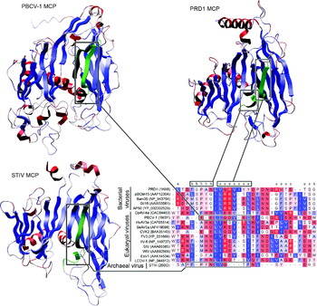
Multiple amino acids in the primary sequences of MCPs are conserved and the three-dimensional structure also matches some of these conserved sites in those proteins for which X-ray crystallography structure is available (Fig. 1). Development of antibodies recognizing this quaternary structure along with some of the conserved amino acids might be possible. In order to test this idea a polyclonal antibody raised against PRD1 MCP was used to detect MCPs of AP50 and Bam35 as described in the Materials and methods section. Bam35 and AP50 are ancient relatives of PRD1 and they all belong to the Tectiviridae family. The Enzyme-Linked ImmunoSorbent Assay (ELISA) failed to produce a positive signal with these antigens (even at high protein concentrations) while it functioned effectively on PRD1 (data not shown), thus suggesting that immunoassays are not spontaneously effective in detecting very distantly related but still conserved virus proteins. It is possible that a set of antibodies raised against different but homologous virus capsid proteins, if any, can be used to maximize the chances of detecting viruses. Exhaustive experimental studies are required to identify the possibility of detecting viruses via an immunological approach.
Structural and sequence comparison of double-beta barrel major capsid proteins of archaeal, bacterial and eukaryal viruses. Only a short region of the protein alignment is shown. The amino acids in the alignment are coloured according to their hydrophobicity (blue for low hydrophobicity, red for high hydrophobicity). H, helix; e, beta-sheet.

Materials and methods
Bacterial strains and bacteriophages
The Bacillus thuringiensis strains used in this study were HER1410 (Ravantti et al. Reference Ravantti, Gaidelyte, Bamford and Bamford2003) and HER1410_L7 (Gaidelyte et al. Reference Gaidelyte, Jaatinen, Daugelavicius, Bamford and Bamford2005). The latter strain hosts a lysogenized bacteriophage Bam35 and releases virions spontaneously. Bacteriophage AP50 (Nagy and Ivanovics Reference Nagy and Ivanovics1977) genomic DNA was kindly obtained from Dr. Shanmuga Sozhamannan, Naval Medical Research Institute, Silver Spring, USA.
General methods for growth, sporulation and plaque assays
For routine growth of B. thuringiensis, a Luria Broth (LB), medium was used (Sambrook & Russel Reference Sambrook and Russel2001). To sporulate HER1410_L7 strains, bacteria grown overnight were dissolved in 1:20 water diluted LB medium and then grown for an additional 16 hours. Sporulated bacteria were washed twice with distilled water in order to remove spontaneously released virions from the solution. Non-sporulated bacteria were eliminated from the solution by heating it to 80°C for 15 mins. 200 μl of the spore containing solution was plated on LB-Agar. Reviving spores formed detectable colonies after two days of cultivation at room temperature. Cells from these colonies were transferred into fresh LB-medium. Spontaneously released virions were detected using plaque assay (as described in Gaidelyte et al. (Reference Gaidelyte, Jaatinen, Daugelavicius, Bamford and Bamford2005)). If the lysogenized Bam35 genome was introduced into the forming spore, then plaques were observed on the bacterial lawn. The absence of virus genome in the bacterial cell was confirmed for several revived spores with the PCR using Bam35 specific primers
Bam35_ORF28F (CGTAGGATCCATGACGAAGGATAATACATGGCGT)
and
Bam35_ORF28R (CGTAGTCGACTTACACCCCCAAAATACGGACAGT).
Immunologic assays
ELISA was used to test the cross-reactivity of bacteriophage PRD1 major coat protein specific antibodies against major coat proteins of bacteriophages Bam35 and AP50. Polyclonal antiserum against PRD1 P3 was raised by immunizing a rabbit (Hänninen et al. Reference Hänninen, Bamford and Bamford1997). PRD1 and Bam35 virus stocks of 1011 pfu ml−1 heated to 70°C for 15 min were used for the antigen solutions. Heating disrupts virions but keeps capsid proteins intact. The AP50 major coat protein encoding gene was cloned into expression vector pMG60 (Strömsten et al. Reference Strömsten, Bamford and Bamford2005) and was produced in Escherichia coli HMS174 cells. Expression of a correctly sized protein was confirmed in SDS-PAGE. Protein extract (heated to 70°C for 15 min) was used as an antigen. ELISA was performed following Ausubel et al. (Reference Ausubel, Brent, Kingston, Moore, Seidman, Smith and Struhl2002) with appropriate controls. ELISA-plates (Nunc-Immuno™ plate with MaxiSorp™ Surface) were coated with protein antigens. Optimal antigen concentration was determined to be 10 μg ml−1 for PRD1 P3. However, for AP50 and Bam35 protein concentrations of 20 μg ml−1 and 100 μg ml−1 were also tested. The primary antibody, anti-P3 (in phosphate buffered saline containing 0.05% NaN3), was used to bind the antigen. The secondary antibody (alkaline phosphatase-conjugated goat-anti-rabbit, Bio-Rad), 500 ng ml−1 in a blocking buffer (0.017 M sodium borate, 0.12 NaCl, 0.05% Tween 20, 1 mM ethylenediamine tetraacetic acid (ETDA), 0.25% bovine serum albumin, 0.05% NaN3, pH 8.5) was added and the signal with the substrate p-nitrophenyl phosphate disodium hexahydrate (1 mg ml−1 in 1 M diethanolamine, 0.5 mM MgCl2, pH 9.0. Sigma-Aldrich) was detected by measuring the absorbance units of each well at wavelength 405 nm.
Amino acid sequence analyses
The MCP GenBank ids or PDB files used in the alignment are shown in Fig. 1. The visualizations of Protein Data Bank files and the alignment of MCPs were done using Bodil (Lehtonen et al. Reference Lehtonen2004).
Acknowledgements
This study was supported by the Finnish Centre of Excellence Program of Academy of Finland (1213467, 2006–2011). We thank Alice Pawlowski for technical support.