




No CrossRef data available.
Article contents
Surgery for mitral valvar disease in childhood: should we choose reconstruction or repair?
Published online by Cambridge University Press: 24 May 2005
Abstract
Objective: Modern surgical techniques now offer the potential for reconstructing rather than replacing the mitral valve in those patients who require an operation during childhood. The purpose of this study was to assess the outcome in our centre following mitral valvar surgery in children. Methods: Between 1992 and 2002, 51 consecutive children, with a mean age of 12.5 ± 3.5 years, underwent surgical treatment of mitral valvar stenosis and/or insufficiency in the Cardiovascular Surgical Department of Istanbul University. Of the children, 20 underwent repair, whilst we replaced the valve in 31 patients. In one, we inserted a pulmonary valvar autograft in mitral position. Results: There were neither early nor late deaths in those who underwent mitral valvar repair. Similarly, there were no early deaths in those in whom the valve was replaced, but two patients died late, while six outgrew their prosthetic valves and required insertion of a further valve within 10 years of their first operations. Conclusion: Our experience shows that procedures designed to repair the mitral valve are effective and reliable when performed in children, with low mortality, and a lower incidence of reoperation when compared with those having replacement of the mitral valve.
Information
- Type
- Original Article
- Information
- Copyright
- © 2003 Cambridge University Press
In developed countries, diseases afflicting the mitral valve during infancy and childhood are generally congenital rather than acquired, although rheumatic disease probably still predominates in countries of the third world. They are rare and complex malformations, with isolated congenital stenosis or insufficiency involving the mitral valve affecting less than 1% of patients with congenital cardiac defects.1 The surgical treatment of these diseases has now been transformed by advanced techniques of valvar reconstruction that offer a better alternative to replacement with a prosthetic valve. Repair, however, is often technically challenging, while replacement with a prosthetic valve also presents inherent difficulties when undertaken in childhood, particularly because of the diameter of the prosthesis in relation to the growing child, and doubts concerning the efficacy of anticoagulant therapy. Reconstructive surgery, therefore, is gaining in popularity when intervention is needed in children.2 Repair offers the advantages of avoidance of thromboembolism, preservation of cordal function, and potentially a reduced need for reoperation.3 With all these considerations in mind, we have assessed our own experience with the surgical treatment of mitral valvar disease in children.
Methods
From January 1992 through July 2002, 51 children, ranging in age from 3 years to 18 years of age, with an average of 12.5 ± 3.5 years, underwent surgery to treat mitral incompetence and/or stenosis at the Department of Cardiovascular Surgery of the Medical Faculty of Istanbul University. Their characteristics are summarized in Table 1. Of the children, 12 were under 6 years of age, 21 were aged from 6 to 12 years, and the remaining 18 were between 12 and 18 years old. In 27 of the children, rheumatic disease was believed to be the cause of the mitral valvar abnormality, one was known to have had endocarditis, while the remaining 23 children all had congenital malformations. In 30 patients, there was predominant mitral stenosis (Table 2), while the other 21 patients had predominant mitral incompetence (Table 3). The mitral valve was replaced in isolation in 25 patients, and was repaired in 20 patients. Concurrent replacement of the aortic valve was performed in five patients, while insertion of an autogenous pulmonary valve was performed as an alternative to placing a prosthetic mitral valve in one patient. We excluded from the study all those patients with atrioventricular septal defect and common atrioventricular junction, discordant atrioventricular connections, and those with functionally univentricular hearts.
Table 1. Mitral valvar lesions and operative techniques in patients.

Table 2. Classifications according to Carpentier.

Table 3. Classifications according to Carpentier.

Preoperative symptoms were chronic congestive heart failure, dyspnea, repeated pulmonary infections, and retarded growth. Associated cardiac anomalies were present in 12 patients. Preoperative cardiac catheterization was performed in all patients.
We employed cardiopulmonary bypass and moderate systemic hypothermia for all procedures, routinely using cold potassium cardioplegia. After aortic cross-clamping, the mitral valve was exposed through a left atriotomy, and the entirety of the mitral valvar apparatus was carefully inspected. Cold saline solution was routinely injected into the left ventricular cavity to test the competence of the valve, and to assess the motion of the leaflets.
Replacement
We replaced the mitral valve in 31 patients, having deemed the valvar deformities unsuitable for repair (Table 1). Rheumatic heart disease was believed to be the cause of the valvar abnormality in 55% of these patients. Of those requiring valvar replacement, six were aged from 3 to 6 years, nine from 6 to 12 years, and 16 were between the ages of 12 and 18 years. We implanted 18 Carbomedics valve, 10 St Jude Medical, and 2 Björk-Shiley. The mechanical valve was placed in the native annulus in all patients. An autogenous pulmonary valve, having a diameter of 21 mm, was used in the mitral position in one patient with endocarditis. The valve was seated in a dacron tube of 22 mm diameter, which was then secured in the mitral annulus. The right ventricular outflow tract was reconstructed using a homograft.
Of these patients, six required insertion of a second valve within 10 years of the original operation. Anticoagulation was maintained in all children using warfarin sodium (Coumadin). Anticoagulation was judged to be adequate when values of 15% to 30% were obtained using the thrombotest twice monthly.
Repair
We repaired the valve in 20 children aged from 3 to 18 years, most frequently because of annular dilation, followed by commissural fusion, prolapsing leaflets, and elongated tendinous cords. The methods of repair included annuloplasty in 8 children, splitting with resection in 5, cordal shortening in 3, triangular resection in 2, and repair of a cleft leaflet in 2 (Table 4). We used the De Vega annuloplasty to reduce the annular diameters. The cleft in the aortic leaflet was closed with interrupted sutures placed on the edge of the leaflet. Prolapsing aortic leaflets secondary to cordal elongation were corrected by plication of the cords and triangular resection of the leaflet. Restricted motion was treated by resection of secondary cords and splitting of the papillary muscles.
Table 4. Type of lesions and type of repair techniques in mitral valvar diseases.

Results
There were neither hospital deaths nor late deaths in those who underwent valvar repair. Cardiac catheterization was performed in all the patients after discharge from the hospital. Left ventriculography revealed mitral incompetence at the second grade in 4, and of the first grade in 8 patients. In only one of the patients was it necessary subsequently to replace the valve over a period of ten years. This patient developed mitral stenosis after annuloplasty combined with triangular resection of the aortic leaflet. The valve was replaced 5 years after the first operation.
There were no early deaths in those in whom the valve was replaced, but two patients died later, one as a result of prosthetic valvar dysfunction, and the other with sepsis. Among the 30 patients who underwent valvar replacement, 6 outgrew their prosthesis and needed insertion of a second valve within a period of ten years. These patients had all undergone the initial replacement when aged less than 12 years, a 19 mm Bjork-Shiley prosthesis being inserted in 2, and a 19 mm St Jude Medical prosthesis in 4. Hemorrhage or thrombosis of the leaflets did not occurr in any of these patients. Echocardiography confirmed the excellent functioning of the pulmonary autograft placed in mitral position up to 18 months postoperatively.
Discussion
Surgery for mitral valvar disease in children carries both technical and clinical difficulties due to the morphologic abnormalities.4, 5 The lesions of the mitral valve are frequently associated with other congenital malformations that may be obscured by the valvar malformations. Surgical treatment, nonetheless, has been transformed by modern techniques of valvar reconstruction that offer a better alternative to replacement with a prosthetic valve.6 Fortunately, these malformations are relatively rare, with isolated congenital mitral stenosis or insufficiency affecting less than 1% of patients with congenital cardiac defects.1
The mitral valve is complex in both its structure and function. The mitral apparatus consists of an annulus, leaflets, tendinous cords, and papillary muscles. Limitation to left ventricular inflow may be caused by a supravalvar mitral ring, hypoplasia of the mitral annulus, or malformation of the leaflets, cords, or the papillary muscles themselves. Commonly, obstruction is due to abnormalities at multiple levels within the valvar apparatus.
Congenital mitral stenosis often is quite severe. If untreated, death usually occurs within the first five years of life.7 Isolated mitral insufficiency commonly causes symptoms later in childhood, and this may be medically controlled, delaying surgical intervention. Because of associated cardiac lesions, a mortality of up to 30% has been observed during infancy in children with less severe mitral stenosis.8 Those who survived, however, have not exhibited a progression in the severity of stenosis when followed over time. Thus, medical management is prudent, followed by valvar repair when symptoms become severe or uncontrollable. Valvar replacement should be undertaken only in those patients in whom repair has failed to make the symptoms medically manageable.9
There is no truly satisfactory substitute to replace mitral valve in a patient of any age, and lack of such a suitable replacement is a real problem for infants and small children. In infants born with congenital mitral valvar disease, there is a tendency to carry the patients longer on medical management, since the longterm risk of persistent pulmonary hypertension due to elevated left atrial pressure is not well established. Surgical intervention is considered when symptoms become severe, or when limitations of exercise are unacceptable. Medical management, followed by valvar repair, is the optimal course of action. At the present time, only mechanical valves are suitable to replace the valve in an infant or a child.10 All patients with mechanical valves, however, require life-long anticoagulation. Despite the use of anticoagulation, thromboembolic events may still occur, running the risk of late valvar entrapment by formation of pannus.11 Smaller patients will eventually outgrow the prothesis, and require reoperation. Annular enlargement is not possible when the mitral valve is replaced, since there is no analogue to the Konno procedure for the mitral valve. Porcine heterograft valves were utilized in the mitral position in children for a number of years but, due to early calcific degeneration, these are now considered to be contraindicated in infants and children.12
As an alternative to replacement with a prosthetic valve, it is possible to use the pulmonary autograft, used in the Ross operation, which has now been performed worldwide since 1967, by inserting the autograft in aortic position, particularly in younger patients. The advantages of this procedure are the use of the patient's own valve, a low risk of endocarditis, low thrombogenicity, avoidance of anticoagulant therapy, and an alleged potential for growth of the autograft in children.13 This operation is also indicated for severe valvar insufficiency. Having used the native pulmonary valve to replace the diseased valve, the right ventricular outflow tract is reconstructed with a homograft or xenograft. Although the Ross operation has become the accepted technique for replacement of the aortic valve in children,14 the long term fate of the pulmonary valve in mitral position remains unknown. Kabbani et al.15 reported their short and medium term experience with use of the pulmonary autograft in mitral position, opining that the autograft is a worthwhile alternative to use of mechanical prostheses in developing countries, but cautioning that full endorsement of the procedure will have to wait until long-term results are available.
Replacement of the mitral valve carries a higher mortality and poorer prognosis than does repair. In infants, the operative mortality has been reported to range from 32% to 50%.16, 17 In recent years, due to improved techniques, and the use of low-profile valves, mortality has reduced to the range of 10%. Although prosthetic valvar replacement was not associated with any operative deaths in our series, the major problem encountered was the need for a second valvar replacement. The results in the long term of mechanical valvar replacement, therefore, do not compare favorably with repair.5, 18, 19 Furthermore, the functional results after repair are usually quite satisfactory.20, 21 In our experience, only one patient undergoing repair required reoperation during a period of follow-up that ranged from 6 months to ten years. In contrast, 6 of the patients undergoing valvar replacement have required reoperation during this period.
We conclude, therefore, that in chidlren replacement of the mitral valve should be avoided if at all possible. The techniques of repair have advanced. The current attitude to disease of the mitral valve, therefore, should be an aggressive approach to repair.
References
Davachi F, Moller JH, Edwards JE. Diseases of the mitral valve in infancy. Circulation 1971; 43: 565.Google Scholar
Carpentier A, Branchini B, Cour JC, et al. Congenital malformations of the mitral valve in children: pathology and surgical treatment. J Thorac Cardiovasc Surg 1976; 72: 854–866.Google Scholar
Aharon AS, Laks H, Drinkwater DC, et al. Early and late results of mitral valve repair in children. J Thorac Cardiovasc Surg 1994; 107: 1262–1271.Google Scholar
Oosthoek PW, Wennink ACG, Wisse LJ, Gittenberger-de Groot AC. Development of the papillary muscles of the mitral valve: morphogenetic background of parachute like asymmetric mitral valves and other mitral valve anomalies. J Thorac Cardiovasc Surg 1998; 116: 36–46.Google Scholar
Okita Y, Miki S, Kusuhara K, et al. Early and late results of reconstructive operation for congenital mitral regurgitation in pediatric age group. J Thorac Cardiovasc Surg 1988; 96: 294–298.Google Scholar
Stellin G, Bortolotti U, Mazzucco A, et al. Repair of congenitally malformed mitral valve in children. J Thorac Cardiovasc Surg 1988; 95: 480–485.Google Scholar
Collins Nakai RL, Rosenthal A, Castaneda AR, Bernhard NF, Nadeo AS. Congenital mitral stenosis. A review of 20 years' experience. Circulation 1977; 56: 1039–1047.Google Scholar
Moore P, Adatia I, Spevak PJ, et al. Severe congenital mitral stenosis in infants. Circulation 1994; 89: 2099–2106.Google Scholar
Embrey RP, Behrendt DM. Congenital abnormalities of the mitral valve. In: Baue AE (ed.). Glenn's Thoracic and Cardiovascular Surgery, 6th edn, Volume II, Appleton & Lange, Stamford, Connecticut, 1996, p 1468.
Lamberti JJ, Mainwaring RD. Congenital anomalies of the mitral valve. In: Constantine Mavroidis (ed.). Pediatric Cardiac Surgery, 2nd edn. Mosby, St. Louis, Missouri, 1994, pp 463–464.
Alexiou J, Galogavrou M, Chen O, et al. Mitral valve replacement with mechanical prostheses in children: improved operative risk and survival. Eur J Cardiothorac Surg 2001; 20: 105–113.Google Scholar
Ross DN. Replacement of aortic and mitral valves with a pulmonary autograft. Lancet 1967; 2: 956–958.Google Scholar
Elkins RC, Lane MM, Mc Cue C. Ross operation in children: late results. J Heart Valve Dis 2001; 10: 736–741.Google Scholar
Kabbani SS, Jamil H, Hammoud A, et al. Use of the pulmonary autograft for mitral replacement: short- and medium-term experience. Eur J Cardiothorac Surg 2001; 20: 257–261.Google Scholar
Zweng TN, Bluett MK, Mosca R, Callow LB, Bove EL. Mitral valve replacement in the first 5 years of life. Ann Thorac Surg 1989; 47: 720–724.Google Scholar
Kadoba K, Jonas RA, Mayer JE, Castaneda AR. Mitral valve replacement in the first year of life. J Thorac Cardiocasc Surg 1990; 100: 762–768.Google Scholar
Guyton SW, Paull DL, Anderson RP. Mitral valve reconstruction. Am J Surg 1992; 163: 497–501.Google Scholar
Lamberti JJ, Jensen TS, Grehl TM, et al. Late reoperation for systemic atrioventricular valve regurgitation after repair of congenital heart defects. Ann Thorac Surg 1989; 47: 517–522.Google Scholar
Yoshimura N, Yamaguchi M, Oshima Y, et al. Surgery for mitral valve disease in the pediatric age group. J Thorac Cardiovasc Surg 1999; 118: 99–106.Google Scholar
Lorier G, Kalil RA, Barcellos C, et al. Valve repair in children with congenital mitral lesions: late clinical results. Pediatr Cardiol 2001; 22: 44–52.Google Scholar
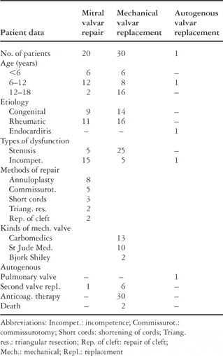
Table 1.
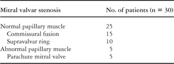
Table 2.
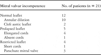
Table 3.
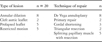
Table 4.
 You have
Access
- 4
- Cited by