Introduction
The over-underlay tympanoplasty technique is traditionally followed for residency training in our centre. In this operative technique, the graft is typically placed medial to the annulus but lateral to the handle of the malleus.Reference Sarkar1 The handle of the malleus serves as an excellent medial support, ensuring adequate middle-ear space. However, complete removal of the epithelial remnant from the malleus remains an essential requirement in this technique. Epithelial remnants over the handle of the malleus and under the graft inevitably carry an increased risk of future epithelial pearl formation or cholesteatoma.Reference Rizer2
Quite often, residents, and sometimes even senior surgeons, find themselves unconvinced of the successful completion of de-epithelisation of the malleus, particularly over the umbo. Indeed, the first author prefers performing malleus exteriorisation (medialisation of the graft to the handle of the malleus) in all cases, irrespective of the de-epithelisation status, as it gives better post-operative hearing results.Reference Vijayendra, Mahadeviah, Surendran and Sangeetha3
Intra-operative assessment by the surgeon is considered adequate for assuring proper clearance of disease in middle-ear surgical procedures.Reference Ryan and Briggs4 However, it is unclear how accurate we are at detecting epithelial remnants over the malleus in tympanoplasty, especially given the limited magnification of an operating microscope, and without the aid of any staining or fluorescent techniques. This study therefore aimed to determine the accuracy of the surgeon's assessment in detecting epithelial remnants after de-epithelisation in type 1 tympanoplasty and to realistically assess the need for malleus exteriorisation in residency training.
Materials and methods
This study was planned as a diagnostic test evaluation. It was performed in the ENT Department of the Government Medical College, Kottayam, India.
We selected 42 patients who voluntarily underwent primary tympanoplasty (for dry central perforations with the umbo touching the promontory) at our centre during the study period. Cases with active infection, retraction pockets or cholesteatomas were intentionally excluded.
The majority of the operations were completed under local anaesthesia. All surgical procedures were performed by the first author, who employed a retro-auricular approach and underlay technique. Freshening of the perforation margin and de-epithelisation of the malleus were conducted with proper attention, using a sickle knife and an angled pick.
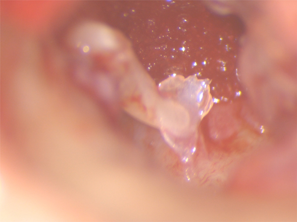
After elevation of the tympanomeatal flap, the umbo was carefully examined under various levels of magnification using an operating microscope (Figure 1). As there were no guidelines available, if any part convincingly demonstrated a change of texture over the umbo or attached connective tissue when compared to the surroundings, or if any tissue remnant retained an appearance of the elevated tympanic membrane remnant, it was taken as positive for epithelium. In contrast, when the surfaces of the umbo and adherent connective tissue were unvarying, it was recorded as negative for epithelium. Observations were also made regarding the presence of the lamina propria, tympanosclerotic plaques, relations between the perforation and the handle of the malleus, and so on.
Intra-operative examination image of the umbo, obtained using a Leica M320 ENT microscope (×25 magnification).

The umbo was resected only in cases where it was touching the promontory, in order to adequately establish space medial to the handle of the malleus;Reference Sanna, Sunose, Mancini, Russo and Taibah5 in practice, this does not affect hearing results.Reference Mahadevaiah and Parikh6 Any case with adequate space medially was excluded from the study. Malleus exteriorisation was performed in all cases, as is routinely conducted by the first author.
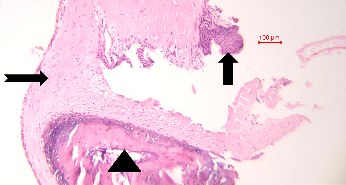
The resected specimens were placed in 10 per cent buffered formaldehyde and promptly sent for histopathological examination, which is considered the ‘gold standard’ for accurately determining the presence or absence of epithelium. The pathologist was not given any details regarding the surgeon's opinion, to avoid bias. Chosen specimens were fixed in 10 per cent buffered formalin, decalcified with 10 per cent formic acid and embedded in paraffin blocks. Consecutive 4–5 µm sections were taken and deparaffinised. After staining with routine haematoxylin and eosin, the appropriate sections were examined with a binocular microscope. The confirmed presence of even a minuscule amount of epithelium was reported as positive (Figure 2).
Histopathology image of a resected umbo specimen obtained using a Labomed Lx400 microscope (H&E; ×100). The upwards arrow indicates an epithelial remnant attached to the lamina propria. The notched right-pointing arrow shows the lamina propria and the triangle indicates the malleus.

Results
A total of 42 patients who underwent primary tympanoplasty were selected for the study. The group included 16 males (aged 18–56 years; mean, 33.68 years) and 26 females (aged 18–47 years; mean, 33.61 years). The operation was undertaken on the left side in 26 patients (16 females, 10 males) and on the right side in 16 patients (10 females, 6 males).
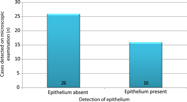
In 31 of the 42 cases, the umbo was in direct relation to the perforation before de-epithelisation, as observed on microscopic examination. The microscopic assessment indicated findings suggestive of epithelium in 16 cases (side of surgery – right 5, left 11); the remaining 26 cases (right 11, left 15) did not show any changes (Figure 3).
Results of the intra-operative microscopic assessment.

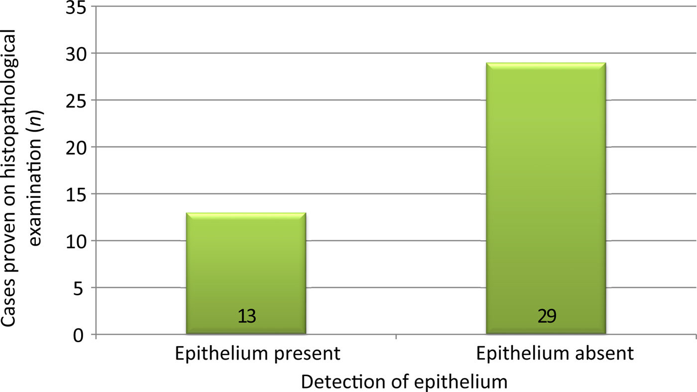
Histopathological examination revealed the presence of epithelium in 13 specimens (7 specimens from the right ear and 6 from the left ear). That is, 30.95 per cent of cases had residual epithelium over the umbo even after careful de-epithelisation (Figure 4). Twelve of these 13 specimens were resected from patients in whom the umbo was involved in the perforation.
Results of the histopathological examination.

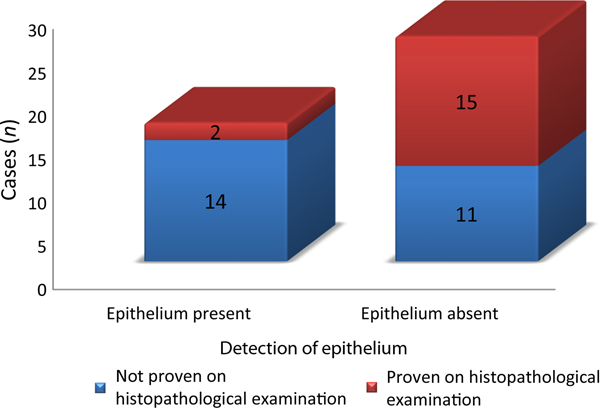
The intra-operative microscopic detection of epithelium was correct in only two cases, with a sensitivity of 15.38 per cent. False-negative reporting occurred in 11 cases. Epithelial remnants were absent in 29 specimens on histopathological examination. Of these, 14 specimens were reasonably suspected to have epithelium on microscopic examination (specificity of 51.72 per cent).
Microscopic examination (diagnostic test) reported epithelium in 16 cases, of which only 2 were correct. Of the 26 cases with a negative test result, these findings were correct in only 15 cases (Figure 5); the remaining 11 cases had epithelium. Therefore, the accuracy or ability of microscopic examination to correctly identify the presence or absence of epithelium was low (40.47 per cent). Further evaluation revealed a positive predictive value of 12.5 per cent and a negative predictive value of 57.69 per cent. These findings indicate that microscopic examination is less useful in detecting epithelial remnants. Based on the abovementioned calculations, the identification of the two correct cases could have been a coincidence. A positive likelihood ratio of 0.31 and a negative likelihood ratio of 1.63 were also derived from the available data.
Results of the diagnostic test evaluation of the intra-operative microscopic examination compared to the histopathological examination.

Discussion
The first documented attempt at repairing a tympanic membrane perforation was performed in 1640, by Banzer, using a pig's bladder.Reference Benzer7 Similar documented attempts were made by Toynbee in 1853 using a rubber disc with silver wire,Reference Toynbee8 and by Blake in 1887 using a paper patch.Reference Blake9 However, Berthold is credited with performing the first true tympanoplasty, as he applied plaster over the tympanic membrane remnant for de-epithelisation and then applied a skin graft.Reference Berthold10 All tympanoplasty techniques are based on the classic concept of the middle-ear transformer mechanism put forward by Helmholtz in 1868.Reference Helmholtz11
Tympanoplasty is broadly divided into overlay and underlay techniques, with each having its own advantages and disadvantages. The over-underlay tympanoplasty technique was introduced as a means of incorporating the advantages of both overlay and underlay techniques, whilst avoiding the disadvantages.Reference Kulduk, Dundar, Soy, Guler, Yukkaldiran and Iynen12 It is relatively simple and straightforward, with a good success rate.Reference Sarkar1 The handle of the malleus remains an asset in this technique and prevents atelectasis. According to Kartush et al., one vital requirement in this technique, like the overlay method, is the complete removal of epithelium from the handle of the malleus.Reference Kartush, Michaelides, Becvarovski and Larouere13 Many consider this impossible to achieve surgically.Reference Hough14
Nejadkazem and colleagues’ study on intratympanic cholesteatoma after tympanoplasty stated that the typical site of these cholesteatomas is near the umbo. They suggested that incomplete removal of epithelium from the umbo is the cause.Reference Nejadkazem, Totonchi, Naderpour and Lenarz15 This is not exclusively the result of a lapse by the surgeon, as normal tympanic membrane anatomy plays a crucial role too. The tympanic membrane is intimately connected to the handle of the malleus, especially the umbo, and serves as one of the main supports of the malleus.Reference Gulya16 According to Tos, the umbo represents the only site where the handle of the malleus attaches to the tympanic membrane.Reference Tos17 Graham et al. also reported that the umbo maintains a uniform distribution of fibrous tissue on its medial and lateral surfaces.Reference Graham, Reams and Perkins18 According to the authors, these fibres are intimately attached to the perichondrium surrounding the manubrium over the lower third. Furthermore, optical coherence tomography has also shown the tympanic membrane to have naturally increased thickness (100–120 µm) near the umbo.Reference Van der Jeught, Dirckx, Aerts, Bradu, Podoleanu and Buytaert19
Studies have demonstrated that the experience and skill set of the surgeon determines the ultimate outcome in tympanoplasty, irrespective of other clinical variables.Reference El-Seifi and Fouad20 According to Emir et al., the outcomes of tympanoplasty are more unsatisfactory when performed by trainees, and practical guidance from a senior surgeon is essential for better post-operative results.Reference Emir, Ceylan, Kizilkaya, Gocmen and Uzunkulaoglu21 It is our opinion that residents, with their limited experience and skills, are more likely to leave residual epithelium over the handle of the malleus in tympanoplasty.
Intentional placement of graft material medial to the handle of the malleus can reduce the chances of epithelial entrapment in tympanoplasty in such situations and avoid unwanted ossicular mobilisation. During the course of this study, the utilisation of other methods were suggested, including using lasers for the removal of tissues over the umbo, to ensure complete de-epithelisation.Reference Kartush, Michaelides, Becvarovski and Larouere13 This circumvents the excessive ossicular mobilisation involved in de-epithelisation that may affect ossicular chain stability.Reference Mahadevaiah and Parikh6 However, the apparent lack of proper techniques for epithelium identification and the cost factor dissuaded us from it.
When the likelihood of residual epithelium over the umbo is significant, the surgeon can resect the umboReference Mahadevaiah and Parikh6,Reference Kutluhan, Yalçıner, Güler, Kösemehmetoğlu, Bozdemir and Bilgen22 and conveniently place the graft medial to the handle of the malleus.Reference Sanna, Sunose, Mancini, Russo and Taibah5 In our study, 13 specimens (30.95 per cent) had residual epithelium on histopathological examination. Selective removal of the umbo and exteriorisation of the malleus should have sufficiently reduced the likelihood of cholesteatoma in these cases. However, intra-operative assessment was accurate in only two cases. Thus, if the graft was resting over the handle of the malleus in the remaining 11 cases, it could have led to epithelial entrapment. In this study, 29 specimens were negative for epithelium on histopathological examination. However, intra-operative findings were misleading in 14 of these cases, naturally leading to inaccurate positive reporting for epithelium. Therefore, according to the first author, the two cases of correct identification could have occurred unintentionally.
• Over-underlay tympanoplasty is a common technique; complete epithelium removal from the umbo is essential
• This study aimed to critically assess the surgeon's accuracy in identifying epithelial remnants over the umbo
• Intra-operative assessment of epithelial remnants over the umbo was conducted after de-epithelisation
• Resected umbo specimens were sent for histopathological assessment
• Calculated sensitivity, positive predictive values and likelihood ratios showed intra-operative assessment to be poor
• Malleus exteriorisation or umbo resection should be considered in residency training programmes, to prevent epithelial entrapment
However, our study has some inadequacies. First, the viability of the detected epithelium is unascertained; in other words, whether epithelial pearl or cholesteatoma would have developed in these cases is unknown. There are published studies utilising over-underlay tympanoplasty that did not show any epithelial pearl formation on follow up.Reference Khalifa and Khalifa23 Because of the comparatively brief duration of our study, the incidence of epithelial pearl or cholesteatoma in our study group remains unascertained. The second and most significant limitation is the inadequate sample size. The required sample size was difficult for a lone surgeon to realistically achieve in the comparatively short study period. A multicentre meta-analysis is required.
Conclusion
It is essential for residents and their mentors to be aware of the practical possibility of residual epithelium over the handle of the malleus, despite performing meticulous de-epithelisation. Direct assessment based on intra-operative microscopic examination does not guarantee the presence or absence of epithelium, as observed findings can be misleading. It is our opinion, based on this study, that malleus exteriorisation or resection of the umbo should be incorporated in tympanoplasty training for residents, with the ultimate aim of preventing epithelial entrapment in the middle ear, especially if the surgeon is unconvinced of adequate epithelium clearance from the handle of the malleus.
Acknowledgements
The authors would sincerely like to thank Dr H Vijayendra, Vijaya ENT Clinic, Bangalore, India, for his moral support and necessary guidance in meticulously preparing this academic paper, and Dr Sangeetha Merrin Varghese (Community Medicine), Government Medical College, Kottayam, India, for the statistical analysis of observed data. We would also like to humbly acknowledge the proper guidance and support of Dr K M Krishnan (Director) and Mrs Nisha Sidhique (guest faculty member), School of Letters, Mahatma Gandhi University, Kottayam, in editing this paper.
Competing interests
None declared