The idea that early experience, including the environment and quality of maternal care, affects developmental outcomes is not new. It is well documented that experiences that occur early in development, both positive and negative, can affect an individual for their entire lifetime. Behavioral and cognitive development seems especially sensitive to early life experiences, including quality of maternal care in mammalian species. Maternal responsiveness and competence are positively correlated with measures of infant mental development (Clarke-Stewart, Reference Clarke-Stewart1973), and with long-term cognitive and social functioning (Connell & Prinz, Reference Connell and Prinz2002). Adverse early experiences such as infant and child maltreatment, including both physical abuse and neglect, lead to poor developmental outcomes including alterations in social behavior, affective disorders, and other psychopathologies (Cicchetti & Valentino, Reference Cicchetti, Valentino, Cicchetti and Cohen2006). Although the biological mechanisms through which these adverse experiences have such long-lasting effects are not fully understood, they are thought to involve the deleterious effects of repeated stress activations, leading to “allostatic load” (cost) on behavioral/cognitive systems (Grassi-Oliveira, Ashy, & Stein, Reference Grassi-Oliveira, Ashy and Stein2008). However, in this paper we review findings suggesting that early adverse care results in developmental psychopathology via additional nonstress, nonglucocorticoid mechanisms that include alterations in neurotrophic factors mediated by lack of maternal tactile stimulation, as well as alterations in social learning. Understanding these mechanisms will increase our ability to improve and develop prevention and intervention strategies and would open doors for novel therapeutic treatments.
Stress, Homeostasis, Allostasis, and the Reactive Scope Model
As evident from the elegant dialogue between Romero, Dickens, and Cyr (Reference Romero, Dickens and Cyr2009) and McEwen and Wingfield (Reference McEwen and Wingfield2010), there are still active discussions regarding the exact definitions of the terms stress, homeostasis, and allostasis. The term stress can be vague, as it has been used to describe several things: the activation of the stress response, the stressful stimuli themselves, and the consequences of the stressful experience. To clarify this ambiguity the general consensus is that the term stressor refers to the stimulus, “stress response” refers to the activation of the bodily systems that respond to potential threats (stressors), and chronic stress refers to prolonged stress response activations that can lead to pathological consequences. As pointed out in Romero et al. (Reference Romero, Dickens and Cyr2009), the most commonly used definition of a stressor is “any unpredictable or uncontrollable stimulus that could—potentially—pose a threat to the organism” (Levine & Ursin, Reference Levine, Ursin, Brown, Koob and Rivier1991). This distinction is important as it does not include stimuli that vary in a predictable manner, such as seasonal shifts that are associated with elevated stress axis activity, which result in elevated levels of physiological stress mediators such as cortisol (outcome hormone of hypothalamic–pituitary–adrenal [HPA], stress neuroendocrine axis). The levels of physiological mediators such as cortisol that vary seasonally are maintained in equilibrium via a dynamic process referred to as homeostasis, which was expanded by Sterling and Eyer (Reference Sterling, Eyer, Fisher and Reason1988) into the concept of allostasis, and more recently redefined in other articles (e.g., McEwen & Wingfield, Reference McEwen and Wingfield2003).
The basic ideas that led to the concept of homeostasis were first proposed in the mid-19th century by Claude Bernard (Reference Bernard1957), a French physiologist, although he referred to it as the milieu interieur. Walter Cannon (Reference Cannon1932) would later coin the term “homeostasis” in the early 20th century defined as “the coordinated physiological processes which maintain most of the steady states in the organism are so complex and so peculiar to living beings—involving, as they may, the brain and nerves, the heart, lungs, kidneys and spleen, all working cooperatively—that I have suggested a special designation for these states, homeostasis.” Homeostasis in the context of this review is the ability of an organism to maintain appropriate levels of physiological or behavioral “mediators” to sustain optimal function (e.g., in the case of stress response systems, a main “mediator” would be cortisol (or corticosterone) glucocorticoid released in response to HPA axis stress activation and that, in turn, affects a myriad of other bodily systems such as metabolism, growth, immune, and reproductive function). These appropriate levels of mediators are not rigid, but determined by several factors including time of year, time of day, age, and life history. A clear-cut example of homeostatic process in the stress response is the glucocorticoid negative feedback loops that maintain cortisol at appropriate levels; however, as mentioned above, appropriate levels of cortisol can be different at different times of the day, year, and life cycle. Thus, the idea that biological systems are set up to defend just one appropriate physiological level was considered too strict and simplistic to explain all these observations, and thus the concept of allostasis was developed.
Allostasis is the ability of an organism to adjust the range of the levels of these mediators (e.g., cortisol) depending on the body's needs in a specific context, in order to maintain stability in other physiological systems that have narrow ranges for survival (e.g., electrolyte balance, blood glucose). Thus, allostasis was defined as “maintaining stability through change” (McEwen & Wingfield, Reference McEwen and Wingfield2003). These authors describe homeostasis as the phenomenon involved in the maintenance of life-supporting processes, whereas allostatic processes are not critical for life and are employed in response to potential threats to homeostasis (McEwen & Wingfield, Reference McEwen and Wingfield2003). Critics of the allostasis concept contest that the traditional idea of homeostasis does, indeed, include these responses to predictable seasonal and life cycle changes and that allostasis is redundant, and in fact, can lead to confusion (Dallman, Reference Dallman2003). Dallman contends that the concept of allostasis is a restatement of basic concepts of homeostasis, because “stability through change” is in fact built into homeostatic processes (e.g., those that take place to adapt to seasonal changes or migratory phases) and are absolutely necessary to support life. However, in a recent paper McEwen and Wingfield (Reference McEwen and Wingfield2010) emphasize that allostasis builds on the idea of homeostasis by describing the active processes involved in maintaining homeostasis when adapting to new environments and challenges. It is because of this “stability through change” that organisms are able to cope with their environments, both predictable and unpredictable. McEwen and Wingfield (Reference McEwen and Wingfield2003) also point out that one of the most important differences between the concepts of homeostasis and allostasis is that the operating range of mediators is much larger during adaptation to new situations, and that chronic exposure to the extremes of this range can lead to pathology through the “wear and tear” of systems, which they termed “allostatic load.” The reactive scope model (Romero et al., Reference Romero, Dickens and Cyr2009) has been proposed more recently as a more comprehensive model built on the allostasis concept but addressing some of its criticisms. As such, the goal is to be more generalized to different species, contexts, and systems, and the focus is not so much on energy expenditure as on understanding the individual's acute responses to threats (fitted for survival), how these adaptive acute responses become pathological if chronically sustained, and the underlying mechanisms. As an example, an animal freezing in response to an acute threat may actually conserve energy; however, if this type of response is exhibited in a sustained form and at high levels, it may become a pathological fear response that impairs reproductive success. In this regard, the reactive scope model is more inclusive because it proposes that physiological (and behavioral) systems have normal ranges of mediators (e.g., cortisol as an HPA axis mediator; cytokines and antibodies as immune function mediators) that include responses to predictable internal and external changes (e.g., circadian and seasonal oscillations, pregnancy and lactancy, migration), called “predictive homeostasis”; however, they can also move above or below these predictive ranges in response to threat or to unpredictable challenges (called “reactive homeostasis”). Levels maintained above the reactive or below the predictive homeostasis ranges lead to pathology and are called homeostatic overload (similar concept to the “wear and tear” concept in the allostatic load model) or failure, respectively. The advantage of the reactive scope model is that it can be generalized to systems beyond those directly involved in energy balance used by the allostatic load model.
Stress-Induced Psychopathology Explained by the Allostatic Load and Reactive Scope Models
As briefly described above, two theoretical frameworks have been developed to help explain how stress exposure leads to deleterious outcomes. The first is the allostatic load model, as proposed by McEwen and Wingfield (Reference McEwen and Wingfield2003). In this model, allostasis, as described above, is achieved at a cost to the organism. In other words, the organism is subject to wear and tear because it must adjust the operating range of physiological mediators of the stress response in a manner that increases work load on the organism and leads to pathology in other systems. This cost is referred to as allostatic load. This model examines these costs in the very global context of energy requirements and available energy resources. Within this framework McEwen and Wingfield (Reference McEwen and Wingfield2003) describe two situations in which allostatic load is sustained, resulting in what they call allostatic overload, which leads to adverse outcomes. Type 1 allostatic overload results when energy demands increase above available resources and is often seen in natural environments during unpredictable environmental changes such as storms or unseasonable conditions. Type 2 allostatic overload occurs when access to resources is not a limiting factor and organisms continue to gather and store energy when energy demands are not exceeded (e.g., leading to diabetes, obesity), which is more pertinent to developed societies. The biological mechanisms through which allostatic overload leads to adverse outcomes are proposed to involve exposure to chronic high levels of physiological mediators of the stress response, mainly glucocorticoids (GCs). GCs (such as cortisol or corticosterone) are released in response to stress-induced activation of the HPA axis and can be damaging if exposure to high levels is sustained. This wear and tear due to exposure to increased levels of cortisol includes pathological outcomes such as hypertension, hyperlipidemia, insulin resistance (diabetes), weight gain, amenorrhea, impotence, and alterations in the immune system and brain regions such as the hippocampus (McEwen, Reference McEwen1998). In this model, allostatic overload is thought to be the primary mechanism through which stress can lead to psychopathological outcomes such as depression and anxiety disorders because chronically elevated levels of stress hormones can impact behavioral and cognitive processes through the ability of GCs to access the brain and bind to region-specific corticosteroid receptors (Sanchez, Reference Sanchez2006; Sanchez, Young, Plotsky, & Insel, Reference Sanchez, Young, Plotsky and Insel2000). As an example, stress-induced elevated levels of GCs (or direct local GC administration) mediate increased expression of corticotropin-releasing hormone (CRH) in the central nucleus of the amygdala (CeA) via binding to glucocorticoid receptors (GRs) in this nucleus (e.g., Kolber et al., Reference Kolber, Roberts, Howell, Wozniak, Sands and Muglia2008; Shepard, Barron, & Myers, Reference Shepard, Barron and Myers2000). The amygdala (in particular, the CeA) is a key limbic region involved in the regulation of fear and anxiety responses and in fear learning, and studies have shown that GCs can lead to long-lasting increases in fear and anxiety responses through increased CRH expression in CeA. Increased activity of these amygdala (and extended amygdala, i.e., the bed nucleus of the stria terminalis) CRH efferent pathways to hypothalamus and brain stem regions have been proposed to mediate the increased reactivity to potential threats (increased fear, anxiety, vigilance, sympathetic, and HPA responses) in individuals that experience chronic/repeated stress, particularly early in life (Sanchez, Ladd, & Plotsky, Reference Sanchez, Ladd and Plotsky2001), and are interpreted as behavioral and physiological states of allostatic load (Schulkin, Gold, & McEwen, Reference Schulkin, Gold and McEwen1998). Stress-induced psychopathology not only results in increased fear, anxiety, and reactivity to stress, but also in alterations in reward, motivation, and mood regulatory circuits (Nestler & Carlezon, Reference Nestler and Carlezon2006; Southwick, Vythilingam, & Charney, Reference Southwick, Vythilingam and Charney2005), leading to much of the symptomatology observed in anxiety and mood disorders, which have been considered stress-related disorders, linked to chronic stress or early life stress experiences (e.g., Heim & Nemeroff, Reference Heim and Nemeroff2001).
However, it is difficult to apply the allostatic load model to other behavioral and cognitive domains or to some developmental questions because many of these processes do not necessarily seem mediated by increases in stress mediators (such as GCs) nor by alterations in energy consumption, or if they do, the results do not always fit the model or there is not a clear way of measuring allostatic load. One example of the complexity of these questions is the relationship between social dominance and allostatic load. Social animals take advantage of cooperation and support from others, but this also involves costs due to social competition and conflict. Goymann and Wingfield (Reference Goymann and Wingfeld2004) examined the association of allostatic load of social status with GC levels in free-ranging social animals with strong hierarchies, from hawks and sparrows to elephants, wolves, and nonhuman primates. Although the cost (or allostatic load) of social conflict has been typically considered to be higher in subordinate animals and paralleled by higher levels of GCs (von Holst, Reference von Holst1998), other studies have reported just the opposite, that allostatic load and GC levels are higher in dominants (Creel, Reference Creel2001; Creel, Creel, & Monfort, Reference Creel, Creel and Monfort1996; Rohwer & Wingfield, Reference Rohwer and Wingfield1981). Goymann and Wingfield's (Reference Goymann and Wingfeld2004) analysis of data in the field suggests that the allostatic load related to social status depends on the way hierarchy is reached and maintained by each species, which is a very logical concept. Thus, if dominance status is inherited and accepted by others without challenges (e.g., in some species of hyenas), the cost of acquiring and maintaining higher ranks would be low. However, if dominance is acquired and maintained by displaying aggression against other animals (e.g., in some nonhuman primate species), the allostatic load of the alpha status would be high (Sapolsky, Reference Sapolsky, Becker, Breedlove, Crews and McCarthy2002). In the case of subordinates, allostatic load would depend on how much aggression they receive from dominants, as well as their coping strategies (e.g., social support from family members, possibilities of escape, etc.). Of course, all this is also affected by other environmental factors, such as access to resources. The results from these analyses suggest that in species where cost (allostatic load) is higher in dominants than in subordinates, GCs levels are usually higher in the dominant animals (e.g., in marmosets; Saltzman, Schultz-Darken, Scheffler, Wenger, & Abbott, Reference Saltzman, Schultz-Darken, Scheffler, Wegner and Abbott1994; Saltzman, Schultz-Darken, Wegner, Wittwer, & Abbott, Reference Saltzman, Schultz-Darken, Wegner, Wittwer and Abbott1998); but, when allostatic load was higher in subordinates than dominants, the relationship with GCs was not clear. Thus, the relationship between allostatic load (cost) of social status and GC levels seems extremely complex, suggesting that there are other important factors that modulate GC secretion, not clearly related to the allostatic load related to social status.
In an attempt to address some of the limitations of the allostatic load model, Romero and colleagues developed the reactive scope model, which takes a more ecological and general approach to the same situation. Thus, although the allostatic load model greatly emphasizes the role of the stress response in regulating energy expenditure in response to a stressor and is especially focused on the ecological impact of the availability of energetic resources, the reactive scope model takes a closer look at the underlying mechanisms and other potential mediators. This does not mean that they are mutually exclusive concepts, nor that one is necessarily overall better than the other, but rather that they are different perspectives focused on the same problem. Both consider the same physiological mediators, and both acknowledge that too high levels of mediators are detrimental to the organism. However, it is difficult to apply the allostatic load model when many proposed mediators of allostatic load do not necessarily have a direct interaction with changes in energy demand. The reactive scope model, in contrast, can be expanded beyond traditional physiological mediators of stress to include others, such as behavioral mediators.
For the remainder of this review we will discuss the idea that behavior is homeostatically regulated and subjected to allostatic processes. We will then apply the models discussed above to understand how dysregulation of behavioral homeostasis can lead to developmental psychopathology.
Behavioral Development as an Allostatic Process
It is important to step back and consider whether behavioral development is in fact an allostatic process, that is, is it necessary for behaviors to be maintained within dynamic ranges that change depending on age, current environment, and past experiences to ensure survival? In other words, do behavioral manifestations change over time (development) to adapt to, or maintain the individual's balance with the environment? There is abundant evidence that behavioral development is indeed an allostatic process, and is described as such in both models proposed by Romero et al. (Reference Romero, Dickens and Cyr2009) and McEwen and Wingfield (Reference McEwen and Wingfield2003). Development is not an entirely deterministic process, but rather interacts with the environment to produce an organism that is able to survive within the current context. Throughout development behaviors are expressed at age appropriate levels that are not often deviated from without adverse consequences.
An example of behavioral change (allostatic process) during development in rhesus monkeys, a nonhuman primate species commonly used in research, is aggression. Infant monkeys begin to rough and tumble play around the fourth month of life (Mears & Harlow, Reference Mears and Harlow1975), which involves rolling around on the ground, chasing, and gentle biting. Aggression, considered here to be any action with clear intent to harm or that elicits fearful or painful behavior in the victim, does not really begin to appear until around 6–7 months of age (Suomi, Reference Suomi, Fox and Davidson1984). If this behavior is displayed too soon the infant would have a hard time finding playmates, thus affecting social development. If aggression is expressed at too low or high levels once it does become appropriate the infant would be unable to defend itself or could become the target of increased aggression from other members of the social group, respectively, thus making it important for the infant to maintain an appropriate level of aggression according to age and context. These examples are evidence that behaviors have to be maintained within certain ranges to facilitate normative function, but that these undergo drastic changes with age.
An additional example that behavioral development is an allostatic process is the striking developmental switch between approach and avoidance in infants. The view that the infant brain is an immature version of the adult's has shifted and we now understand that it is simply different and uniquely adapted to survive early in life. In mammals, one of these unique brain functions is to bond with the mother—or whatever caregiver is available—at any cost. Bowlby (Reference Bowlby1965) proposed that infants quickly form attachment to caregivers after birth and that the health of this relationship determines later social relationships. The elegant work of Regina Sullivan's lab has provided us with a new view on the neurobiological attachment systems that help mammalian infants identify and strongly bond with the caregiver (Moriceau & Sullivan, Reference Moriceau and Sullivan2005; Sullivan & Lasley, Reference Sullivan and Lasley2010). Sullivan and colleagues' studies in rat pups have shown that they are similar to human and nonhuman primate infants in the way their brains are wired to form and maintain the bond with the mother, although rat pups do so through the olfactory system. Their initial studies suggested that the neonatal rat uses a different learning circuit than adults, which includes the locus coeruleus (LC) noradrenergic (NE) system. This LC–NE system is hyperfunctional during the sensitive period for formation of attachment to the mother, when the pups are confined to the nest (postnatal days 1–9 in the rat put), releasing high amounts of NE during interactions with the mother, which helps the pup learn a rapid and robust preference for her. In addition, the brain circuits responsible for fear and avoidance learning (i.e., amygdala) are turned “off” during this sensitive period. These findings explain why this strong bond is not broken to avoid pain when the caregiver is physically abusive, as has been shown in humans (Helfer, Kempe, & Krugman, Reference Helfer, Kempe and Krugman1997), nonhuman primates (Harlow & Harlow, Reference Harlow, Harlow, Shrier, Harlow and Stollnitz1965; Sanchez, McCormack, & Maestripieri, Reference Sanchez, McCormack, Maestripieri, Plotsky, Schechter and Cummings2010), or even dog puppies (Fisher, Reference Fisher1955), because avoidance and fear learning circuits are “offline” during the sensitive period for attachment formation. However, even more of interest, Sullivan's lab has also demonstrated that when the pups start exploring outside the nest (after postnatal day 10), the sensitive period of enhanced approach and attenuated aversion learning ends (i.e., the LC becomes less active and the amygdala-fear system is turned “on,” as in the adult), although the mother's presence still acts as a switch for the pups amygdala during associative learning. Thus, during the pairing of a stimulus with pain, the pups still exhibit approach learning toward that stimulus if the mother is present because it turns off the activity of the amygdala in the pup; but, if the mother is not present, the amygdala is “on” and the pup develops avoidance of the stimulus paired to pain. The development of these infant brain–behavioral strategies that involve turning “on” attachment/approach neurobiological systems and turning “off” fear learning and avoidance systems in parallel is very adaptive for survival in altricial species where the infant depends on parental care early in life. Thus, early on proximity to the mother provides with nutrition, protection, comfort, and physiological regulation, at the same time that the suppression of the fear system protects the bond and gives the infant the boldness to explore in the presence of the mother as she serves as an external regulator and secure base. However, when the infant grows older, the fear switch is turned on when the pup is exploring, which is a great evolutionary strategy to stir youngsters from danger and back to the nest.
A similar developmental switch has also been reported in human children, known as stranger anxiety, a behavior characterized by increased fear behaviors such as freezing and gaze aversion, which appears around 1 year of age when a child is learning to move independently and is faced with more possibly dangerous interactions with unknown people (Sroufe, Reference Sroufe1977). Thus, if an infant does not exhibit a particular level of wariness toward strangers they risk making themselves vulnerable to harm. Just as in other systems, development of a specific behavior does not always occur at the same age or in the exact same way for all individuals, yet the actual rates and manifestations of behaviors is strikingly similar across individuals (Bijou & Baer, Reference Bijou and Baer1961).
Is behavioral development affected by early life history and experiences like other physiological systems under strong homeostatic/allostatic control are?
Early Life Stress and Developmental Psychopathology
There is abundant evidence that behavior is sensitive to early experience, making it particularly vulnerable to stress early in life. Long-term behavioral outcomes associated with early life stress in different species include enhanced emotional and stress reactivity that lead to increased fear and anxiety, as well as high levels of aggression and social withdrawal and to conduct, drug abuse, and attention-deficit hyperactivity, and mood disorders in humans (e.g., for reviews, see Gunnar & Vazquez, Reference Gunnar, Vazquez, Cicchetti and Cohen2006; Heim & Nemeroff, Reference Heim and Nemeroff2001; Sanchez et al, Reference Sanchez, Ladd and Plotsky2001; Teicher, Andersen, Polcari, Anderson, & Navalta, Reference Teicher, Andersen, Polcari, Anderson and Navalta2002). The allostatic load model explains this developmental psychopathology as resulting from repeated stress activations and sustained exposure to high levels of GCs, which act on brain regions such as the amygdala. However, there are other pathways to developmental psychopathology. Early life stress can also interfere with the normative allostatic processes that modulate behavioral development (such as those described in the previous section for the development of aggression, approach, and avoidance), changing the set point for future behaviors that depend on earlier “expected” behavioral patterns and performance, and in this sense, an early balanced relationship with the caregiver is crucial.
As stated above, early life stress is strongly linked to developmental psychopathology which in humans includes anxiety and mood disorders, aggressive, impulsive, and antisocial behavior, as well as conduct disorder, substance abuse, and attention-deficit/hyperactivity disorder (Cicchetti & Rogosch, Reference Cicchetti and Rogosch2001; Cicchetti, Rogosch, Gunnar, & Toth, Reference Cicchetti, Rogosch, Gunnar and Toth2010; Gunnar & Vazquez, Reference Gunnar, Vazquez, Cicchetti and Cohen2006; Heim & Nemeroff, Reference Heim and Nemeroff2001; Teicher et al., Reference Teicher, Andersen, Polcari, Anderson and Navalta2002, Reference Teicher, Andersen, Polcari, Anderson, Navalta and Kim2003). Clinically these disorders are diagnosed based on alterations in normative behaviors. Development does not occur in a vacuum, and thus an organism must adjust to its environment, not only physiologically, but also behaviorally; however, these adjustments are not always adaptive in all contexts, especially when the context under which the original adjustment was made ends. For example, physically abused children tend to develop high levels of aggressive and antisocial behavior, many times ending up in arrests for violent offenses, which has been interpreted as an overreaction to social stimuli resulting from a mix of problems, including poor emotional regulation and attention bias toward threat (for a review, see Sanchez & Pollak, Reference Sanchez, Pollak, Haan and Gunnar2009). Therefore, a child that is physically abused is exposed to negative emotions exhibited by the parent(s) and subsequently learns to pay more attention to facial expressions communicating negative emotions (i.e., angry faces become more salient to the child; Pollak, Cicchetti, Hornung, & Reed, Reference Pollak, Cicchetti, Hornung and Reed2000) and to overread threat and overreact with elevated aggression and violence toward his/her social environment (Kimonis, Frick, Munoz, & Aucoin, Reference Kimonis, Frick, Munoz and Aucoin2008; Teisl & Cicchetti, 2008). These findings are consistent with the view that infants/children learn to “tune” their early perceptual skills to detect salient aspects of their social environment so that they become exceptionally good at discriminating potential threat cues (anger expressions) that are adaptive for survival in an environment where threat signals may predict an abuse event. Although adaptive in an abusive environment, these perceptual skills have a cost for the child in terms of attentional flexibility, filtering of irrelevant information, emotional regulatory ability and, consequently, for establishing healthy social relationships (Sanchez & Pollak, Reference Sanchez, Pollak, Haan and Gunnar2009). In contrast, neglected children do not show attention bias toward threat, but a clear difficulty discriminating between emotional facial expressions and responding to them (Hildyard & Wolfe Reference Hildyard and Wolfe2002; Pollak et al., Reference Pollak, Cicchetti, Hornung and Reed2000; Shackman, Shackman, & Pollak, Reference Shackman, Shackman and Pollak2007). These differences in outcomes support the view that different kinds of early adverse experiences (e.g., two types of maltreatment: physical abuse vs. neglect) have differential effects on behavioral development via social and emotional learning processes, in addition to potential common effects resulting from stress-induced elevations in GCs or catecholamines.
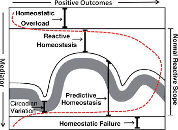
Once the child is removed from the situation their predisposition to recognize negative emotion or inability to recognize positive and neutral emotional cues leads to social incompetence as he/she is unable to recognize and respond appropriately to emotions of peers. It is through adaptations like these that develop in response to an early stressful environment, that children's behavior becomes maladaptive in “normal” (nonthreatening) situations, and psychopathology emerges. These psychopathological consequences could be explained from the reactive scope model perspective as happening due to the high vulnerability to stress exposure of these behaviors during development, so that if the behavioral response to the stressful situation (childhood maltreatment) goes higher than the reactive homeostasis levels into homeostatic overload, the threshold of the later can be lowered so that future threatening situations can push the animal to immediately exhibit behavioral responses into the homeostatic overload level, that is, overreacting with psychopathology (e.g., extreme aggression or anxiety, pathological fear, social withdrawal). If children are not removed from these highly stressful situations, the risk for this threshold to be permanently lowered is much higher. In physically abused children, we can think of attention to angry faces as a behavioral mediator that increases beyond reactive homeostasis levels, and reaches levels of homeostatic overload, leading to psychopathology (see Figure 1). In neglected children, discrimination of emotion can be considered as another behavioral mediator, but in this case the levels of this mediator drop below the reactive homeostasis range and into homeostatic failure range, leading to a different psychopathological manifestation than in abused individuals (see Figure 1). It is important to note that these conditions are so detrimental in the long term because they are occurring during development, when organisms are extremely vulnerable to social experiences (Andersen, Reference Andersen2003).
Reactive scope model could be interpreted as an inverted U-shaped curve. The dashed line represents the levels of mediators (including physiological and experiential such as maternal care) that can affect developmental outcomes of early life experiences, including stress reactivity and emotionality. Exposure to levels of mediators that fall within the “normal reactive scope” results in positive outcomes, whereas exposure to levels of mediators in either the “too high” range (“homeostatic overload”) or “too low” range (“homeostatic failure”) results in negative developmental outcomes. Adapted from “The Reactive Scope Model—A New Model Integrating Homeostasis, Allostatis, and Stress,” by L. M. Romero, M. J. Dickens, and N. E. Cyr, 2009, Hormones and Behavior, 55. Copyright 2009 by Elsevier. Adapted with permission. [A color version of this figure can be viewed online at journals.cambridge.org/dpp]

From the developmental psychopathology field point of view, these early adverse experiences can alter not only brain circuits that modulate behavioral function, but the behavioral alterations themselves can change the interactions with the environment, provoking additional adverse experiences that can further cause deviations in brain–behavioral development (Cicchetti & Toth, Reference Cicchetti and Toth2009).
The allostatic load model focuses more on energy balance, and states that an organism is subjected to wear and tear as a result of having to adjust to their environment, leading to increased energy demand. Although there have been no studies into energy expenditure associated with the behavioral adaptations expressed by physically abused or neglected children, the idea that there is an energetic cost associated with the adaptation is a useful one. Both are valid models, but we are applying the reactive scope model because it seems to be more amenable to mediators beyond the HPA axis, and to explain behavioral development.
Mediators of the Effects of Early Life Stress
As described above, a critical mediator of the pathological consequences (wear and tear) of sustained stress in different systems, such as elevated blood pressure, diabetes, cardiovascular disease, or pathological fear and anxiety, is elevated levels of GCs. However, it is helpful to consider how other systems and mediators beyond the stress axis and GCs support behavioral homeostasis, and how dysregulation of these systems caused by early life stress lead to developmental psychopathology. In their review, Romero and colleagues (Reference Romero, Dickens and Cyr2009) claim that the reactive scope model is better fitted than the allostatic load model to explain the way early life experiences related to maternal care affect neurochemical and neurotrophic factors that impact brain and behavioral development. Their main argument is that, although the allostatic load model produces a very useful theoretical framework to predict that early life stress results in stress-induced elevations in glucocorticoids that can lead to allostatic overload and, therefore, developmental psychopathology by, for example, increasing CRH expression in the central amygdala in the offspring, it cannot explain why in turn in some cases mild early life stress can result just in the opposite; that is, positive developmental outcomes such as increased resiliency to allostatic overload. We are going to elaborate on this point brought up by Romero and colleagues (Reference Romero, Dickens and Cyr2009). This body of evidence suggests that, when early life stress poses a mild (but not overwhelming) challenge to the infant, the outcomes are actually beneficial for his/her development, including decreased stress reactivity, less anxiety and fear, increased exploratory traits, improved social behavior and reproductive success. For example, handling rat pups for 15 min (considered a mild stressor) leads to decreased stress reactivity in adulthood (in terms of both reduced HPA axis activations and fear/anxiety levels) than those exhibited by pups that are either not handled at all or handled for 180 min (Meaney et al., Reference Meaney, Mitchell, Aitken, Bhatnagar, Bodnoff and Iny1991). These results suggest that a little bit of stress early in life in the form of brief maternal separations is beneficial for rat pups because the individual develops as less stress reactive (better fitted) toward changes in the environment. However, in order to understand the adaptive implications of these findings, we have to emphasize the ethological relevance of these short separations (20–30 min), which are typical in the wild, where the dams are away from the nest and litter for short periods of time to forage (Rosenblatt, Reference Rosenblatt1975).
A similar phenomenon has been described in detail by David Lyons' group in nonhuman primates, labeled as “stress inoculation” (Lyons & Parker, Reference Lyons and Parker2007; Lyons, Parker, Katz, & Schatzberg, Reference Lyons, Parker, Katz and Schatzberg2009), as well as in humans (Rutter, Reference Rutter1981). The notion is that moderately stressful experiences early in life lead to a “stress inoculation” that help develop arousal regulation and resiliency. This concept is embedded in different cultures, sometimes called “toughening” or “character building” and reflected in expressions such as “what doesn't kill you makes you stronger.” The “stress inoculation” notion fits well with the physiological concept of “habituation,” although the former involves persistent and broader implications, so that the reduced reactivity is long term and not stressor specific. The long-term implications of early life stress inoculation involve better functioning in multiple domains, including reduced stress reactivity (i.e., lower HPA axis and sympathetic activations), reduced levels of fear and anxiety, increased exploration in novel environments, as well as enhanced prefrontal-dependent cognitive function. These improved developmental outcomes involve underlying neurodevelopmental changes (mostly an enlarged ventromedial prefrontal cortex and increased myelination in the prefrontal cortex), all of which leads to individuals with more adaptive strategies to a changing world. In those studies, brief intermittent separations at 3–6 months of age (1 hr separation per week, for 10 weeks) initially elicit distress calls and increase plasma levels of cortisol in infant squirrel monkeys, with partial habituation of these measures of arousal observed over repeated separations (Coe, Glass, Wiener, & Levine, Reference Coe, Glass, Wiener and Levine1983; Hennessy, Reference Hennessy1986). Later in life, monkeys exposed to repeated intermittent separations show less anxiety, blunted cortisol stress responses, and increased sensitivity to glucocorticoid feedback regulation of the HPA axis compared to monkeys not exposed to separations (Levine & Mody, Reference Levine and Mody2003; Lyons, Lopez, Yang, & Schatzberg, Reference Lyons, Lopez, Yang and Schatzberg2000; Lyons, Martel, Levine, Risch, & Schatzberg, Reference Lyons, Martel, Levine, Risch and Schatzberg1999). The “inoculated” animals have other long-term positive effects, including higher levels of exploration in a novel environment and improved performance in a cognitive test that required behavioral inhibition and cognitive flexibility, functions regulated by the prefrontal cortex. As discussed in the rat studies described above, we have to understand the ethological implications of these experimentally induced separations for squirrel monkey infants, because in this species mothers and other group members periodically leave newly weaned offspring on their own, beginning at 3–6 months of age, to forage for food (Boinski & Fragaszy, Reference Boinski and Fragaszy1989; Lyons, Kim, Schatzberg, & Levine, Reference Lyons, Kim, Schatzberg and Levine1998). So, these separation stress experiences are built into the normative developmental experiences of this nonhuman primate species.
Thus, as many other biological systems do, it seems like the effects of early life stress on the offspring development follow an inverted U-shaped curve, with exposure to moderate stress during early development resulting in positive/beneficial outcomes such as resilience, and either too little or too much stress exposure (or intensity) leading to negative outcomes. This inverted U-shaped response could be thought of as turning the reactive scope model on its side, with extremes of exposure to stress at either end (“homeostatic overload” or “failure”) resulting in adverse developmental outcomes (see Figure 1). In the following sections we will discuss evidence supporting the role of nonglucocorticoid mechanisms, such as maternal care-related cues, underlying these developmental effects.
Additional (Nonglucocorticoid) Mediators of Early Life Stress Effects on Behavioral Development
Maternal care
Several lines of research provide evidence that maternal care is a potent regulator of the infant's homeostasis, particularly during early development. Dam–pup separations have been used in rats to examine the specific maternal behaviors that affect the development of specific behavioral and physiological systems in the offspring (Hofer, Reference Hofer1984; Kuhn, Butler, & Schanberg, Reference Kuhn, Butler and Schanberg1978), demonstrating that the mother works as an external regulator of the infant's physiological and behavioral states, and affects their development. For example, (a) feeding regulates heart rate in pups via interaction of milk with gastrointestinal receptors, whereas (b) the nonnutritional aspect of sucking, in particular, the rhythmicity of milk ejection, regulates sleep patterns in the pups, and (c) tactile stimulation via licking, nursing, and retrieving regulates pup behavioral activity (Hofer, Reference Hofer1984). Additional recent evidence suggests that maternal care is also a major factor in both rodent and primate brain development and is, therefore, important for behavioral development.
In rats, specific maternal behaviors such as licking and grooming during the first week of life strongly affect the development of brain regions like the prefrontal cortex and hippocampus (Bredy, Grant, Champagne, & Meaney, Reference Bredy, Grant, Champagne and Meaney2003; Liu et al., Reference Liu, Diorio, Tannenbaum, Caldji, Francis and Freedman1997) and stress reactivity of the rat pups (Caldji, Diorio, & Meaney, Reference Caldji, Diorio and Meaney2000), demonstrating the ability of specific maternal behaviors that involve physical contact to affect brain structure and function in the offspring. Alterations in amount and timing of tactile stimulation provided by the mother could therefore cause alterations in development of neural systems, leading to alterations in behavior.
How does maternal care regulate behavioral development? One of the most thoroughly characterized mechanisms is the effect of maternal care on gene expression in brain regions like hippocampus, via epigenetic modifications such as DNA methylation of gene promoter regions. For example, prolonged and repeated maternal separations (180 min) affects rat pups' HPA axis and emotional development, so that these animals have elevated levels of anxiety and fear and stress reactivity in adulthood. Some of these alterations have been linked to decreases in GR expression in the hippocampus (Caldji et al., Reference Caldji, Diorio and Meaney2000), due to epigenetic changes in DNA methylation in the GR gene (Szyf et al., Reference Szyf, Weaver, Champagne, Diorio and Meaney2005). These changes and parallel epigenetic modifications detected in the estrogen receptor gene associated with alterations in reproductive and maternal behavior of the offspring were mediated by alterations in rat maternal care after the prolonged separations (i.e., reduced levels of licking and grooming and arched-back nursing; Cameron et al., Reference Cameron, Champagne, Parent, Fish, Ozaki-Kuroda and Meaney2005), and not by increased levels of GCs in the pups, emphasizing that GCs are not the only “mediator” (or mechanism) involved. These regulatory properties of maternal care on offspring development have been termed the “maternal mediation hypothesis” and they not only apply to extreme changes in maternal behavior, but to natural variations in maternal care, which have a tremendous impact on the development of defensive and reproductive behavior, maternal behavior, stress reactivity, and neural function of rats, and gets transmitted from generation to generation (Champagne et al., Reference Champagne, Bagot, van Hasselt, Ramakers, Meaney and de Kloet2008). Of interest, this intergenerational transmission of behavioral individual differences has also been reported in primates, including vervet and rhesus monkeys (Fairbanks, Reference Fairbanks1996; Maestripieri, Reference Maestripieri2005a) as well as in humans (Miller, Kramer, Warner, Wickramaratne, & Weissman, Reference Miller, Kramer, Warner, Wickramaratne and Weissman1997), at least for individual differences in maternal care. An example is poor maternal care in the form of physical abuse, which has been reported in nonhuman primate species including macaques, marmosets, and baboons (Brent, Koban, & Ramirez, Reference Brent, Koban and Ramirez2002; Johnson, Kamilaris, Calogeron, Gold, & Chrousos, Reference Johnson, Kamilaris, Calogero, Gold and Chrousos1996; Maestripieri, Reference Maestripieri1999), operationalized as violent behaviors directed toward the infant that cause pain and distress. Of interest, abusive parenting seems to be a stable maternal trait, so that abusive mothers tend to abuse all their infants with very similar patterns and rates (Maestripieri, Reference Maestripieri1999). These maternal behaviors are also transmitted across generations along the maternal line (for review, see Sanchez et al., Reference Sanchez, McCormack, Maestripieri, Plotsky, Schechter and Cummings2010), and studies of females that were cross-fostered at birth have shown that intergenerational transmission of abusive as well as neglectful and inconsistent parenting is due to differences in early maternal care that were experienced (Maestripieri, Reference Maestripieri2005a, Reference Maestripieri2005b). The problem with these primate studies, in comparison to those in rats, is that we have very limited experimental evidence of the underlying mechanisms mediating the changes, as well as the maternal behaviors that most significantly regulate infant development or the critical developmental windows.
Despite these limitations, some of the epigenetic effects of early maternal care (e.g., higher levels of DNA methylation in the promoter region of the GR gene in rats that received low levels of maternal care) that affect GR expression in the rat hippocampus (e.g., Weaver et al., Reference Weaver, Cervoni, Champagne, D'Alessio, Sharma and Seckl2004) have also been reported in hippocampi of humans with histories of childhood abuse (McGowan et al., Reference McGowan, Sasaki, D'Alessio, Dymov, Labonte and Szyf2009), suggesting the possibility of similar cross-species epigenetic mechanisms mediating the effects of poor maternal care. Reduced GR expression in the hippocampus would result in reduced GC negative feedback, and, therefore, more prolonged stress-induced elevations of GCs.
However, GR is not the only gene whose expression is affected by maternal care. There are hundreds of other hippocampal genes whose expression in adulthood are affected by maternal care via epigenetic mechanisms and are related to anxiety, at least in rats (Weaver, Meaney, & Szyf, Reference Weaver, Meaney and Szyf2006). A recent study supports very extensive epigenetic changes associated with natural variation in maternal care, suggesting long-term genome programming effects (McGowan et al., Reference McGowan, Suderman, Sasaki, Huang, Hallett and Meaney2011). Although extensive, these epigenetic modifications do not seem to be generalized, but specific to big groups of functionally related genes, suggesting that these epigenetic modifications are part of a coordinated biological response. Thus, in addition to the epigenetic modifications to the hippocampal GR gene, these authors reported extensive effects of rat maternal care on gene expression of protocadherins, neural proteins that regulate synaptic development (Junghans, Haas, & Kemler, Reference Junghans, Haas and Kemler2005). Differential levels of protocadherins during development could potentially affect hippocampal development in a significant way because, for example, these proteins are crucial for serotonergic projections to appropriately innervate the hippocampus (Katori et al., Reference Katori, Hamada, Noguchi, Fukuda, Yamamoto and Yamamoto2009). Consequently, the effects of differential maternal care on hippocampal protocadherin expression could underlie the reported effects of maternal care on hippocampal synaptic density, long-term potentiation, fear conditioning, and spatial learning previously reported in adult rats (Champagne et al., Reference Champagne, Bagot, van Hasselt, Ramakers, Meaney and de Kloet2008; Liu, Diorio, Day, Francis, & Meaney, Reference Liu, Diorio, Day, Francis and Meaney2000). Therefore, although the epigenetic mechanisms underlying the long-term effects of early maternal care on brain, behavior, stress responses, and cognitive function has been mostly studied for GR expression in the rat hippocampus, it could potentially mediate effects on the development of other brain regions, neurochemical systems, and in other species.
Female offspring of high licking and grooming rat mothers also show increased expression of estrogen receptor-alpha (ER-a) in the medial preoptic area (MPOA), mediated by lower levels of DNA methylation than those seen in daughters of low licking and grooming dams (Champagne et al., Reference Champagne, Weaver, Diorio, Dymov, Szyf and Meaney2006). Expression of maternal behavior in the rat is dependent upon the activation of this ER-a in the MPOA and downstream effects on oxytocin receptors (OTRs; Numan & Sheehan, Reference Numan and Sheehan1997; Pedersen, Caldwell, Wlaker, Ayers, & Mason, Reference Pedersen, Caldwell, Walker, Ayers and Mason1994). Because the MPOA levels of ER-a control expression of OTR in that region (Young, Wang, Donaldson, & Rissman, Reference Young, Wang, Donaldson and Rissman1998), hypomethylation of the ER-a promoter of female offspring of high licking and grooming mothers would lead to increased expression of OTR in response to increased levels of estrogen during pregnancy, which in turn, would upregulate positive maternal behaviors, including elevation in rates of licking and grooming (Champagne et al., Reference Champagne, Chretien, Stevenson, Zhang, Gratton and Meaney2004; Numan & Sheehan, Reference Numan and Sheehan1997).
As it relates to other species, although there is less supporting evidence and mechanistic studies available in primates, this “maternal mediation hypothesis” has also been proposed to explain how extreme alterations in early caregiving, or just normal variation in parental care, results in long-term changes in behavior, neuroendocrine, and neurobiological function in humans and nonhuman primates humans (Gunnar & Donzella, Reference Gunnar and Donzella2002). Although much research is still needed to understand the underlying mechanisms of early caregiving regulation on the development of brain and behavior in primates, as well as the maternal behaviors that most significantly regulate development and the sensitive periods, the existing evidence suggest that parental sensitivity might be a component of caregiving that strongly affects offspring development in primates. This view is supported by several reports that low responsive/sensitive parenting in humans is associated with larger stress responses in toddlers (Gunnar, Brodersen, Nachmias, Buss, & Rigatuso, Reference Gunnar, Brodersen, Nachmias, Buss and Rigatuso1996) and enhanced fearfulness in infants, which is linked to more right frontal EEG asymmetry (Hane & Fox, Reference Hane and Fox2006) and to greater behavioral inhibition, anger, aggressive play, and internalizing problems later in development, during the toddler years (Hane, Henderson, Reeb-Sutherland, & Fox, Reference Hane, Henderson, Reeb-Sutherland and Fox2010). Similar effects of poor maternal care have been reported in rhesus monkeys, where infant maltreatment leads to increased levels of anxiety/fear in the offspring, behavioral alterations that are associated with reduced levels of brain serotonergic function (Maestripieri, McCormack, Higley, Lindell, & Sanchez, Reference Sanchez2006; McCormack, Newman, Higley, Maestripiere, & Sanchez, Reference McCormack, Newman, Higley, Maestripieri and Sanchez2009). When different components of maternal care were dissected in these nonhuman primate studies to examine their specific effect on infant development, maternal rejection (and not so much physical abuse) was the best predictor of poor developmental outcomes (Maestripieri, Lindell, et al., Reference Maestripieri, Lindell, Higley, Newman, McCormack and Sanchez2006; Maestripieri, McCormack, et al. Reference Maestripieri, McCormack, Higley, Lindell and Sanchez2006), supporting the view of the human studies above that low maternal sensitivity/responsivity maybe a particularly important caregiving dimension for proper development of primates.
In summary, maternal care regulates the behavior developmental trajectory of the offspring. Alterations in normative maternal care cause behavioral alterations that can lead to developmental psychopathology. There has to be an evolutionary explanation for why behavioral development is so sensitive to variations in maternal care. If the environmental adversity faced early on persists throughout life, it could be very adaptive to incorporate some neurodevelopmental programming that involves behavioral, cognitive, and physiological strategies that increase survival of the adult organism in high pressure environments. For example, the high levels of vigilance, anxiety, fear, aggression, and stress reactivity reported in individuals that experience early adversity in the form of poor maternal care in different species, including maternal separation in rats (above), childhood maltreatment in humans and nonhuman primates (e.g., Heim et al, Reference Heim, Newport, Heit, Graham, Wilcox and Bonsall2000; Sanchez & Pollak, Reference Sanchez, Pollak, Haan and Gunnar2009), as well as other nonhuman primate models of adverse early caregiving such as peer rearing (Barr et al., Reference Barr, Newman, Becker, Parker, Champoux and Lesch2003) and variable foraging demand (Coplan, Rosenblum, & Gorman, Reference Coplan, Rosenblum and Gorman1995), could potentially be a protective trait for offspring that continue to develop under stressful environments due to limited resources, high social pressure, or high levels of predation. In the case of rat pups, for example, the increased behavioral reactivity could attract higher levels of maternal care by, for example, increasing ultrasonic vocalizations. Thus, from an evolutionary point of view postnatal maternal care could potentially program the offspring's brain to be more or less reactive to stress depending on the early environmental pressure in order to be better fitted to survive in such an environment later in life. In the context of the developmental psychopathology field, although this developmental plasticity allows for neurodevelopmental adaptations that improve behavioral functioning in specific contexts, it also causes malfunction in other contexts, leading to psychopathology (e.g., see Cicchetti & Gunnar, Reference Cicchetti and Gunnar2008).
Neurochemical mediators (brain serotonin [5-HT] systems, neurotrophic factors)
Brain 5-HT systems
The brain 5-HT systems play an important role in emotional regulation, stress physiology, mood, and behavioral inhibition, and their development is sensitive to alterations in the early environment. This is due to the high density of 5-HT innervation found in limbic structures such as amygdala, prefrontal cortex, hippocampus, hypothalamus, and striatum (Bauman & Amaral, Reference Bauman and Amaral2005; Freedman & Shi, Reference Freedman and Shi2001) This localization explains the strong role of 5-HT regulating fear, anxiety, and stress responses. Previous studies have shown that early adverse experiences impact brain 5-HT function both in humans (Kaufman et al., Reference Kaufman, Birmaher, Perel, Dahl, Stull and Brent1998) and nonhuman primates (e.g., Higley, Suomi, & Linnoila, Reference Higley, Suomi and Linnoila1996; Maestripieri, Lindell, et al., Reference Maestripieri, Lindell, Higley, Newman, McCormack and Sanchez2006; Maestripieri, McCormack, et al., Reference Maestripieri, McCormack, Higley, Lindell and Sanchez2006; Sanchez et al., Reference Sanchez, Alagbe, Felger, Zhang, Graff and Grand2007), and that early life stress alterations in brain 5-HT neurotransmission, typically reduction, are associated with different forms of psychopathology, including anxiety, increased aggressive, and impulsive behaviors and mood disorders (Manji, Drevets, & Charney, Reference Manji, Drevets and Charney2001) and related physiological alterations, including HPA axis dysregulation. Thus, in our studies we have reported that infant maltreatment impacts the development of brain 5-HT systems in rhesus monkeys, whose function was reduced and associated with increased anxiety and with the perpetuation of maltreatment across generations of females (Maestripieri, Lindell, et al., Reference Maestripieri, Lindell, Higley, Newman, McCormack and Sanchez2006; Maestripieri, McCormack, et al., Reference Maestripieri, McCormack, Higley, Lindell and Sanchez2006). Altogether these findings emphasize, once again, the important role of early maternal care for proper neurobiological development of systems involved in the control of mood, stress responses, and behavioral inhibition in the offspring.
Why are brain 5-HT systems so vulnerable to early life stress and maternal care? Because the brain 5-HT system and the stress axis are interconnected in several ways, allowing them to influence each other (Leonard, Reference Leonard2005) and to potentially mediate the psychopathological effects of early life stress. The cells that comprise the ascending 5-HT fibers are located in the dorsal raphé nucleus of the brainstem, and their cell bodies express CRH receptors (Meloni, Reedy, Cohen, & Carlezon, Reference Meloni, Reedy, Cohen and Carlezon2008), allowing for direct regulation of 5-HT synthesis and/or release by CRH and, therefore, by stress. For example, it has been proposed that a potential neurochemical pathway by which stress-induced CRH increases lead to increase in fear behaviors, such as freezing, in rats involve increased CRH release at the level of the 5-HT synthesizing neurons in the dorsal raphé nucleus, which cause increased release of 5-HT at the level of the central nucleus of the amygdala (CeA), which leads to freezing; in contrast, a delayed wave of 5-HT release detected in medial prefrontal cortex (PFC) is associated with cessation in freezing behavior, suggesting that 5-HT innervations in these two limbic regions have opposing roles in the regulation of fear behavior (Forster et al., Reference Forster, Feng, Watt, Korzan, Mouw and Summers2006).
Based on the abundant evidence that maternal care affects brain CRH and 5-HT systems in the offspring, and the cross-regulation between CRH neurocircuits and brainstem monoaminergic circuits that include 5-HT and noradrenergic ascending pathways Tallie Baram's and other groups have studied the specific brain pathways that transfer sensory cues associated with maternal behaviors such as licking, grooming, feeding, and anogenital stroking needed to induce micturition/defecation in the rat pup to limbic and hypothalamic areas that express CRH and control stress responses (Eghbal-Ahmadi, Avishai-Eliner, Hatalski, & Baram, Reference Eghbal-Ahmadi, Avishai-Eliner, Hatalski and Baram1999; for reviews, see Korosi & Baram, Reference Korosi and Baram2009; Sanchez et al., Reference Sanchez, Ladd and Plotsky2001). A crucial target of these neurocircuits is the amygdala (particularly, the central CeA, basomedial, and medial nuclei) and its connections with regions like the PFC. Thus, somatosensory cues associated with licking, grooming, and suckling behavior can reach the CeA via afferents from the pedunculopontine and lateral dorsal tegmental nuclei, which receive substantial somatosensory inputs from the spinal cord. In addition, the CeA also receives integrated sensory information from the medial amygdala (which projects to the basomedial amygdala, and thalamic paraventricular nucleus [PVT]). Viscerosensory cues related to anogenital stroking-induced micturition are relayed via the nucleus of the solitary tract to the Barrington's nucleus (micturition center with high CRH expression) and from this to the PVT via a monosynaptic pathway. The CeA and PVT can then relay sensory and micturition information, respectively, to the basomedial amygdala, where this sensory information is integrated and sent to hypothalamic and brain stem nuclei (including CRH-synthesizing neurons in the PVN) via either direct projections or through relay in the CeA. Therefore, differential activation of these multiple pathways due to different levels of maternal behaviors (e.g., licking, grooming, anogenital stroking) can alter the development of these neural circuits and therefore PVT- and amygdala-mediated stress responses including fear and anxiety, HPA axis, and autonomic responses (e.g., via amygdala and PVT projections to the PVN and locus coeruleus, Pihok, Owens, Kuhn, Schanberg, & Nemeroff, Reference Pihoker, Owens, Kuhn, Schanberg and Nemeroff1993; and to prefrontal cortex and hippocampus; for reviews, see Korosi & Baram, Reference Korosi and Baram2009; Sanchez et al., Reference Sanchez, Ladd and Plotsky2001).
Activation of the HPA axis by stress also modulates 5-HT receptor transcription via GR-mediated glucocorticoid action (Flugge, Reference Flugge1995) because the genes that code for the 5-HT1A and 5-HT2 receptor subtypes contain glucocorticoid response elements (Garlow & Ciaranello, Reference Garlow and Ciaranello1995) that allow GCs to regulate their gene expression. GCs functionally act to downregulate transcription of the 5-HT1A receptor subtype (Flugge, Reference Flugge1995). Alterations in 5-HT1A receptors have been implicated in many psychopathologies, including depression, anxiety, bipolar disorder, and schizophrenia (Mackowiak, Czyrak, & Wedzony, Reference Mackowiak, Czyrak and Wedzony2000; Savitz, Lucki, & Drevets, Reference Savitz, Lucki and Drevets2009). Therefore, via alterations of 5-HT production, release, reuptake, and/or receptor expression, repeated stress activations could contribute to the 5-HT system dysregulation observed in many developmental psychopathologies, particularly in the case of early life stress, when the brain 5-HT systems may still be developing or playing a critical neurotrophic role.
What is this neurotrophic role of 5-HT? Although the brain 5-HT system develops early in humans, being visible at 5 weeks of gestation, it increases slowly through 2 years of age, and then decreases again to reach adult levels by 5 years (Whitaker-Azmitia, Reference Whitaker-Azmitia2001). A similar developmental pattern has been reported in nonhuman primates (Goldman-Rakic & Brown, Reference Goldman-Rakic and Brown1982), although in some regions like the PFC the 5-HT adult innervation is reached by 2 weeks postnatal, ahead of other monoamines. Although some aspects of the brain 5-HT system may develop early, 5-HT has a crucial neurotrophic role throughout development, affecting the maturation of target tissues (Whitaker-Azmitia, Druse, Walker, & Lauder, Reference Whitaker-Azmitia, Druse, Walker and Lauder1996). For example, neocortex and hippocampus in rats are affected by 5-HT levels during development. Prenatal depletion of 5-HT causes a decrease in the number of neurons in the adult neocortex and hippocampus (Lauder & Krebs, Reference Lauder and Krebs1976), as well as slowing differentiation in areas known to contain 5-HT terminals in the adult (Lauder & Krebs, Reference Lauder and Krebs1978). In cell cultures of neonatal rat visual neocortex, administration of physiological levels of 5-HT results in glial proliferation, neuron differentiation, synaptogenesis, and axon myelination (Chubakov, Gromova, Konovakov, Sarkisova, & Chumasov, Reference Chubakov, Gromova, Konovalov, Sarkisova and Chumasov1986). These effects are potentially mediated through 5-HT1A receptors on astroglia, which upon 5-HT binding release neurotrophic factors (Whitaker-Azmitia & Azmitia, Reference Whitaker-Azmitia and Azmitia1994). These 5-HT receptors reach adult levels shortly after birth in humans (Whitaker-Azmitia, Reference Whitaker-Azmitia2001), and their stimulation causes maturation of the astroglia from radial glia into more stellate forms (shorter and more numerous processes; Whitaker-Azmitia et al., Reference Whitaker-Azmitia, Druse, Walker and Lauder1996). Thus, if early life stress leads to reductions in synthesis and release of 5-HT in different brain target regions, as has been reported in different early adverse experiences (see above), at specific critical developmental periods this could alter its important role in the maturation of glial cells, neurons, synapses, and myelination of axons in target regions of 5-HT innervation. This could, in turn, lead to alterations in behavioral and cognitive development.
Role of maternal care-related tactile stimulation on brain and behavioral development: Effects on 5-HT systems
As described above, maternal licking and grooming during the first week of life affects development of rat pups hippocampus (Bredy et al., Reference Bredy, Grant, Champagne and Meaney2003; Liu et al., Reference Liu, Diorio, Day, Francis and Meaney2000) and stress reactivity, including fear, anxiety, and HPA axis activity (Caldji et al., Reference Caldji, Tannenbaum, Sharma, Francis, Plotsky and Meaney1998, Reference Caldji, Diorio and Meaney2000), demonstrating the ability of maternal physical contact to alter brain structure and related functions of the offspring, independently of other maternal behaviors, such as nursing. Of interest, the first week of life in rat pups has also been reported as the critical period for maternal tactile stimulation to define barrel receptor representation in primary somatosensory cortex (Fox, Reference Fox1995). Altogether, this evidence supports a critical role of maternal tactile stimulation during the first week of life on rat pup brain development.
Meaney's group has proposed a cascade of effects by which handling-induced increases in maternal tactile stimulation of rat pups via increased licking and grooming behaviors increase neonatal brain 5-HT release and turnover, at least in hippocampus and frontal cortex of the offspring in the first week of life (Smythe, Rowe, & Meaney, Reference Smythe, Rowe and Meaney1994), which leads to long-term alterations in GR expression during adulthood (Meaney et al., Reference Meaney, Mitchell, Aitken, Bhatnagar, Bodnoff and Iny1991, Reference Meaney, Diorio, Francis, Weaver, Yau and Chapman2000; Mitchell, Betito, Rowe, Boksa, & Meaney, Reference Mitchell, Betito, Rowe, Boksa and Meaney1992; Mitchell, Iny, & Meaney, Reference Mitchell, Iny and Meaney1990). Tactile stimulation of rat pups by gentle stroking them with a paint brush mimics the benefits associated with high maternal licking/grooming, including lower anxiety/fear and stress reactivity, and improved cognitive function and maternal behavior in adulthood (for a review, see Kaffman & Meaney, Reference Kaffman and Meaney2007), demonstrating the crucial role of maternal tactile stimulation on the offspring brain and behavioral development in rodents. Although such a strong relationship has not been established in primates, there is some evidence that early physical contact with the caregiver is also necessary for proper development of primate neonates and young infants. This includes early work by Harlow showing that, in the absence of the biological mother, physical contact with an inanimate surrogate mother improved emotional development of infant rhesus monkeys otherwise receiving adequate nutrition and care (Harlow & Zimmermann, Reference Harlow and Zimmermann1959), as well as evidence that skin to skin contact of premature human babies during their 2 first weeks of life leads to better cognitive and motor outcomes at 6 months of age (Feldman, Eidelman, Sirota, & Weller, Reference Feldman, Eidelman, Sirota and Weller2002).
Thus, level and timing of tactile stimulation provided by the mother is one aspect of maternal care that strongly regulates the development of brain systems involved in behavioral and physiological stress responses (including anxiety, fear, and the HPA axis), social and maternal behavior, and cognitive function, mediated by direct effects of maternal tactile stimulation—and not necessarily GCs—on neurobiological systems (Van Oers, de Kloet, Whelan, & Levine, Reference Van Oers, de Kloet, Whelan and Levine1998). One of the brain systems strongly affected by maternal tactile stimulation is 5-HT, whose release has been demonstrated to increase in the hippocampus of rats after stroking of the fur and the tail for similar durations as those in the handling paradigms (15 min), using microdialysis probes (Cenci & Kalen, Reference Cenci and Kalen2000). However, there are other neurobiological systems directly affected by maternal tactile stimulation, such as neurotrophic and oxytocin (OT) systems, as will be described in more detail below.
Role of maternal care-related tactile stimulation on brain and behavioral development: Effects mediated by growth and neurotrophic factors and OT
Despite the great amount of progress described above, the exact molecular mechanisms underlying the effects of early adverse experiences on brain development are not completely understood. In the case of adverse experiences related to alterations in mother–infant relationship, such as those caused by repeated and prolonged (e.g., 3 hr) maternal separations in rats, it seems that the deleterious neurodevelopmental effects are not due as much to stress-induced elevations in GCs, but to alterations in maternal care after the prolonged separations that lead to low maternal tactile stimulation. This reduced tactile stimulation, via unknown mechanisms but probably mediated by the lack of secretion of growth factors and cytokines caused by skin stimulation, results in low levels of brain 5-HT and neurotrophic factors such as brain-derived neurotrophic factor (BDNF) and fibroblast growth factor-2 (Bredy et al., Reference Bredy, Grant, Champagne and Meaney2003; Chatterjee et al., Reference Chatterjee, Chatterjee-Chakraborty, Rees, Cauchi, de Medeiros and Fleming2007; Liu et al., Reference Liu, Diorio, Day, Francis and Meaney2000), which regulate neurodevelopmental processes such as cell proliferation, migration, differentiation, and synaptogenesis. A support for this view is that stroking of pups with a brush reverses the effects of 24-hr maternal separation on BDNF levels and neurodevelopmental processes, for example (Chatterjee et al., Reference Chatterjee, Chatterjee-Chakraborty, Rees, Cauchi, de Medeiros and Fleming2007). In the case of rats, maternal stimulation produced by licking and grooming increases the secretion of brain 5-HT (as described above by Meaney's group), which early in development has neurotrophic effects, including a downstream activation of BDNF mRNA expression in different brain regions, such as the hippocampus (Castren, Reference Castren2004; Zetterstrom et al., Reference Zetterstrom, Pei, Madhav, Coppell, Lewis and Grahame-Smith1999). The mechanisms by which maternal separation of rat pups causes decreased expression of BDNF in their hippocampus and PFC include epigenetic modifications (DNA methylation) in the BDNF gene (Roth, Lubin, Funk, & Sweatt, Reference Roth, Lubin, Funk and Sweatt2009).
There are additional pathways by which maternal stimulation could modulate brain and socioemotional development in the offspring, such as via tactile stimulation-induced release of OT, which has been reported both in human adults (Grewen, Girdler, Amico, & Light, Reference Grewen, Girdler, Amico and Light2005) and children (Uvnas-Moberg, Reference Uvnas-Moberg1996). This maternal effect does not seem limited to just tactile stimulation but can also be elicited in children by listening to the mother's voice (Seltzer, Ziegler, & Pollak, Reference Seltzer, Ziegler and Pollak2010). Of interest, animal and human studies have shown that OT is a neuropeptide that plays a critical role mediating affiliative behaviors (maternal behavior, attachment, and social bonding), and reduces anxiety and HPA axis responses to stress. Early adverse experiences consisting of mother–infant relationship disruption do, indeed, result in decreased function of OT neural circuits of nonhuman primates (Winslow, Noble, Lyons, Sterk, & Insel, Reference Winslow, Noble, Lyons, Sterk and Insel2003) and in humans, as shown by lower cerebral spinal fluid levels of OT in women with histories of childhood maltreatment (Heim et al., Reference Heim, Newport, Mletzko, Miller, Young and Nemeroff2006). This reduced brain OT activity could be contributing to the impaired affiliative behavior and stress vulnerability of primates with early adverse caregiving experiences. Similar effects of maltreatment have also been reported in children with severe neglect who showed lower OT salivary levels than comparison controls (Wismer-Fries, Ziegler, Kurian, Jacoris, & Pollak, Reference Wismer Fries, Ziegler, Kurian, Jacoris and Pollak2005). These OT findings are intriguing given the difficulties severely neglected children have forming secure attachments (van IJzendoorn & Juffer, Reference van IJzendoorn and Juffer2006).
Maternal tactile stimulation can also affect offspring development through the release of growth hormone and other growth promoting hormones, which stimulate anabolic processes necessary to support growth during development, including that of the brain. This tactile stimulation-induced release of growth hormone and its effect on growth during development has been demonstrated both in rat pups and human and nonhuman primate infants (Kuhn & Schanberg, Reference Kuhn and Schanberg1998). Children who experienced omission of normal environmental stimulation (e.g., institutionalization or neglect) do indeed show alterations in growth processes, including stunted physical growth (Rutter, Reference Rutter1998), that are not related to poor nutrition. These growth alterations could be explained by decreased levels of growth hormone secretion due to deprivation of tactile stimulation as well as by stress-induced increases in GC secretion and CRH, which could also affect the growth system (Albanese et al., Reference Albanese, Hamill, Jones, Skuse, Matthews and Stanhope1994; Kertes, Gunnar, Madsen, & Long, Reference Kertes, Gunnar, Madsen and Long2008).
This list of mediators is not meant to be exhaustive, but our aim was to provide with examples of potential glucocorticoid-independent mechanisms that underlie the effects of early life stress on behavioral development. We acknowledge that these are all interrelated systems, and future studies need to dissect the cycle of cause and effect to further understand the critical factors that trigger early life stress-related developmental psychopathology.
Allostatic Load: Prevention, Intervention, and Social Policy
The concepts of homeostasis, allostasis, and adverse effects of repeated exposure to inappropriate levels of homeostatic mediators, whether in the framework of the allostatic load or the reactive scope models, are useful for the field of developmental psychopathology because they bring up the advantages of using a multiple levels of analysis approach. As pointed out by Cicchetti and colleagues (Cicchetti & Dawson, Reference Cicchetti and Dawson2002; Cicchetti & Toth, Reference Cicchetti and Toth2009), it is vital that researchers come together to approach developmental psychopathology questions from different vantage points using multiple levels of analysis (i.e., behavioral, biological, and molecular). This multidisciplinary and multilevel approach is required because the field of developmental psychopathology deals with behavioral alterations potentially caused by changes in brain circuits, cellular processes, hormones, genes, environment, and so forth during development; therefore, these alterations have to be integrated and interpreted across all levels of analyses, even if it is difficult to find a common language to do so (particularly when the multidisciplinary approach involves collaborations between experts from disparate fields). The models discussed throughout this review are useful platforms from which to begin this dialogue, as they allow us to establish a common framework to integrate mediators from different systems (i.e., molecular, behavioral, and neurobiological).
This is especially relevant for the fields of prevention and intervention, where those in the developmental psychopathology community have an opportunity to utilize randomized trials to assess the efficacy of treatments not only at the behavioral but also at the biological level to simultaneously gather information about the underlying mechanisms of pathology and recovery. As deduced from the preclinical evidence provided in this review, biological factors need to be considered in addition to behavioral, psychological, and social factors when addressing the effects of preventions and interventions for many reasons, including the need to understand the pathways through which they work and to detect potential critical periods for intervention, as biological changes may precede (and even be more sensitive than) behavioral ones (Cicchetti & Gunnar, Reference Cicchetti and Gunnar2008).
There is very little work directly addressing the implications of allostatic load for intervention and prevention (or social policy) in the field of developmental psychopathology. However, given the multilevel approach of both the reactive scope and allostatic load models, they seem potentially helpful to guide the design and evaluation of intervention and prevention strategies (e.g., by providing information about normative ranges of mediators). By following multiple levels of analysis and applying the models explored here, information about underlying mechanisms can be gathered and applied to develop effective treatments using information from basic science and fostering collaborations between different disciplines in developmental psychopathology (Cicchetti & Gunnar, Reference Cicchetti and Gunnar2008).
Conclusions
In this review we applied both the allostatic load model proposed by McEwen and Wingfield (Reference McEwen and Wingfield2003) and the reactive scope model proposed by Romero and colleagues (Reference Romero, Dickens and Cyr2009) to understand the link between early life stress and developmental psychopathology. This is an important discussion as it helps in understanding the underlying mechanisms and translating findings from animal studies to human health. The reactive scope model focuses on specific mechanisms, including physiological and behavioral mediators of behavioral homeostasis, some of which are measureable in both animals and humans, and applicable to understand how maternal behavior modulates behavioral development in the offspring. The allostatic load model is more focused on energy availability and explains the effects of early life adversity as being mediated mostly by stress-induced activations that cause elevations in stress hormones such as GCs.
As has been discussed in other publications, some of the shortcomings of the Allostatic Load Model could be due to confusion involving terms. One difficulty we faced while writing this review was to accomplish the differentiation between the applicability of both theoretical models to the interpretation of data in the literature. In fact, we agree with the view that differences between the two models may be mostly semantic and that the original description of homeostasis has implicitly built the concept of allostasis developed later on (Dallman, Reference Dallman2003). The reactive scope model, however, seems more broadly applicable to systems and contexts besides those involved in energy homeostasis, such as the regulation of behavioral development by maternal care discussed in this review.
Behavioral development is very plastic in comparison to some physiological systems and involves a very complex regulation by external and internal factors. In this review, we have tried to dissect some of these regulators as well as what we (and others) think is the evolutionary purpose of this plasticity. Thus, in mammals, maternal (or paternal, or both) care, partially via tactile stimulation, seems a crucial aspect of the early environment that regulates the development of neural pathways that control affiliative, aggressive, and emotional behaviors for optimal, normative function. However, if the environmental conditions change and become challenging, maternal care changes accordingly producing striking differences in brain and behavioral development of the offspring, which are adaptive for survival in response the new environmental pressure. However, these adaptive changes may come with a cost to other aspects of the offspring functioning, some of them included within our view of developmental psychopathology. This obviously takes place in parallel to brain alterations caused by stress-induced elevations in GCs, in the case of early life stressful experiences. There also seem to be critical periods (such as the first week of life for rat pups and the neonatal period in primates), so that the development of specific neural circuits is modified in an activity-dependent manner by specific sensory input provided by maternal behaviors, as it has been previously demonstrated for the development of visual and auditory systems.