Ventricular inversion without transposition of the great arteries, i.e., isolated ventricular inversion, is a rare congenital cardiac malformation. Most patients with this defect are symptomatic in infancy with cyanosis and congestive heart failure. Survival beyond childhood is rare. This patient presented with a rare diagnosis of IVI with hypoplastic right ventricle in adolescence. Various surgical options are available for such cases, but in our case, we decided to go ahead with a single ventricular physiology since the RV was hypoplastic which could not function as the systemic ventricle in the future.
Case report
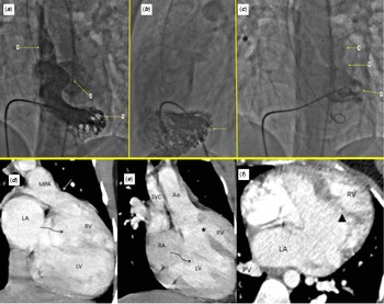
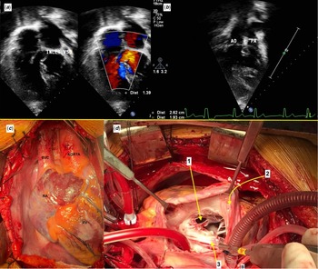
A 15-year-old girl presented to the outpatient department of our hospital with complaints of cyanosis and respiratory distress on and off since birth. On examination, she was cyanotic with a saturation of 70–75% on room air. She was further evaluated with a transthoracic echocardiography (Fig 2a–b) which revealed situs solitus, levoacardia with atrioventricular discordance and ventriculoarterial concordance. There was associated hypoplasia of the left-sided right ventricle with thickening of the pulmonary valve and severe pulmonary stenosis with a peak systolic gradient of 87 mmHg. Also, the branch pulmonary arteries were confluent and of adequate size for the body surface area. There was the presence of a large inlet ventricular septal defect (33 mm) with intact interatrial septum along with severe left atrioventricular valve regurgitation. She further underwent a catheter angiography (Fig 1a–c) and a CT angiography (Fig 1d–f) to confirm the diagnosis which revealed the exact anatomy of the right atrium emptying into the left ventricle through the mitral valve which further emptied into the right and anteriorly placed aorta. After all pre-operative investigations, she was planned for a total cavopulmonary connection with atrial septostomy and left AV valve repair. After median sternotomy and pericardial harvestation, the surface anatomy of the heart (Fig 2c–d) was assessed and the diagnosis was confirmed. The patient was heparinised and cardiopulmonary bypass was instituted with bicaval cannulation and ascending aortic cannulation. The heart was arrested in diastole with del Nido cardioplegia. After routine dissection, the superior caval vein was anastomosed to the right pulmonary artery and the inferior caval vein was anastomosed to the undersurface of the RPA using a 19 mm polytetrafluoroethylene tube graft. Also, an atrial septostomy was made to allow adequate mixing of the blood. The left AV valve was tricuspid and a commissurotomy was done which lead to adequate coaptation of the leaflets on saline injection intraoperatively. The patient was subsequently shifted to the ICU on ionotropic support and was successfully discharged on post-op day 7. She is currently doing well in the post-op period (Supplementary file).
Catheter angiography – ( a ) catheter course – IVC-RA-LV, contrast injection showing 1 – LV, 2 – VSD, 3 – Aorta, LV is identified by virtue of the fine trabeculations in a criss-cross pattern. ( b ) Lateral LV angiogram showing the same with the LV (yellow arrow). ( c ) Catheter course – IVC-RA-VSD-RV, contrast injection showing 1 – RV with coarse trabeculations, 2 – valvular pulmonary stenosis, 3 – MPA, RV is identified by virtue of its coarse trabeculations, RV appears to be small and hypoplastic. ( d – f ) CT angiography showing atrioventricular discordance, ventriculoarterial concordance with morphological right atrium draining into morphological left ventricle, and morphological left atrium draining into morphological right ventricle. Large VSD (*) and hypoplastic right ventricle are seen. There is valvular pulmonary stenosis (white arrow). (arrow head) showing the moderator band.

( a – b ) Transthoracic echocardiography showing ( a ) four- chamber view with the hypoplastic left-sided right ventricle with an inlet VSD, ( b ) relationship of the great vessels with aorta being to the right and anterior, ( c – d ) intraoperative images – ( c ) external anatomy of heart ( d ) view after opening the RA showing 1 – VSD, 2 – RA wall, 3 – posterior mitral leaflet. It can be seen that the septum is free from the attachment of the papillary muscles characteristic of the left ventricle.

Discussion
The nomenclature of this anomaly remains controversial. Reference Anderson and Willcinson1 Van Praagh and Van Praagh Reference VanPraagh and VanPraagh2 used the term isolated ventricular inversion in 1966, while Shinebourne and colleagues, Reference Shinebourne, Macartney and Anderson3 using a segmental approach, referred to the malformation as “visceroatrial situs solitus, discordant atrioventricular and concordant ventriculoarterial connections.” Echocardiography plays a vital role in diagnosing IVI while the diagnosis is usually confirmed by catheter angiography which allows visualisation of ventricular roentgenographic features and the AV valves. In ventricular inversion (discordant atrioventricular connections), the morphologically right ventricle with its septoparietal muscle bundles and atrioventricular valve closer to the cardiac apex is connected to the morphologically left atrium. Reference Murat, Aysel and Ersin4
Suitability for a biventricular repair is the main thing to decide in such cases depending on the associated lesions and anatomy. After a thorough discussion in our Heart Team meeting, we decided to go ahead with total cavopulmonary connection in our patient as the patient had a hypoplastic RV, along with other lesions such as severe valvular and infundibular pulmonary stenosis and severe left-sided AV valve regurgitation which could complicate the biventricular repair.
The morphologically left ventricle with its smooth septal surface and atrioventricular valve further from the cardiac apex
is connected to the morphologically right atrium. In our patient, the morphologically right ventricle was to the left and posterior to the left ventricle. A handful of cases of IVI have been reported in the literature till date but none with the morphology as in this case. Depending on the associated cardiac lesions, the definitive surgical management of this cyanotic malformation can vary, like associated lesions such as hypoplastic right ventricle and severe pulmonary stenosis, in this case, prompted us to enter the single ventricular repair pathway.
Conclusion
In summary, isolated ventricular inversion is a rare defect characterised by discordant connections between the atria and ventricles and normal relationships between the great arteries and the ventricle. Optimal management of such a lesion can vary according to the presence of the associated lesions.
Supplementary material
To view supplementary material for this article, please visit https://doi.org/10.1017/S1047951121003607.
Financial support
None.
Conflict of interest
None.