Introduction
The use of assisted reproductive technologies, such as artificial insemination, superovulation, ovum pick-up, in vitro embryo production and timed-embryo transfer, have the potential to enhance reproductive outcomes and disseminate selected genetics, therefore improving milk and meat production of buffalo herds (Baruselli et al., Reference Baruselli, Soares, Bayeux, Silva, Mingoti and Carvalho2018).
Oocyte maturation is the initial phase in successful in vitro embryo production and is the starting point for many biotechnological applications, such as in vitro fertilization, cloning, transgenesis and stem cell research (Kakkassery et al., Reference Kakkassery, Vijayakumaran and Sreekumaran2010). In vitro maturation of buffalo oocytes is influenced by factors including the composition of culture medium, method of oocyte recovery, oocyte quality, season and ovary status (Mahmoud and El-Naby, Reference Mahmoud and El-Naby2013). Culture conditions and choice of medium also affect embryo development in the laboratory (Demyda and Genero, Reference Demyda and Genero2011). Oocyte maturation is a complex process, during which oocytes gain the intrinsic ability to support further embryo development and activate the embryonic genome. The process includes events in both cytoplasmic and nuclear maturation. Nuclear maturation mainly involves segregation of chromosomes, and cytoplasmic maturation entails organelle reorganization, and storage of mRNA, proteins and transcription factors that are essential for fertilization and subsequent early embryonic development (Ferreira et al., Reference Ferreira, Vireque, Adona, Meirelles, Ferriani and Navarro2009).
Assisted reproductive techniques involve in vitro handling of gametes and embryos that increases the risk of exposure to levels of reactive oxygen species (ROS) that exceed typical physiological levels (Agarwal et al., Reference Agarwal, Said, Bedaiwy, Banerjee and Alvarez2006). The cellular antioxidant system balances generation and scavenging of ROS to maintain normal cellular functions. Enzymatic and non-enzymatic antioxidants, especially vitamins and minerals, are available in the ovarian follicle microenvironment and protect oocytes from the detrimental influence of ROS. However, excessive ROS production beyond antioxidative capacity may result in oxidative stress and reduce oocyte quality (Kala et al., Reference Kala, Shaikh and Nivsarkar2017). During in vitro maturation of mammalian oocytes, ROS production increases in response to factors such as incubation in high oxygen concentration (Leoni et al., Reference Leoni, Rosati, Succu, Bogliolo, Bebbere, Berlinguer, Ledda and Naitana2007), exposure to visible light (Liebel et al., Reference Liebel, Kaur, Ruvolo, Kollias and Southall2012), pollutants (Khoudja et al., Reference Khoudja, Xu, Li and Zhou2013) and some culture medium components (Agarwal et al., Reference Agarwal, Said, Bedaiwy, Banerjee and Alvarez2006).
ROS levels that exceed the physiological range destabilize maturation promoting factor and trigger programmed cell death in oocytes from several mammal species (Aghaz and Khazaei, Reference Aghaz and Khazaei2017; Tiwari et al., Reference Tiwari, Prasad, Tripathi, Pandey, Singh, Shrivastav and Chaube2016). Effects of oxidative damage can continue during the late stages of pregnancy. Furthermore, many metabolic disorders affect mitochondrial functions and DNA methylation patterns. DNA fragmentation and mutations during successive proliferation of cells exhibiting sublethal damage (Menezo et al., Reference Menezo, Silvestris, Dale and Elder2016).
Sodium selenite, as a source of selenium, protects cellular function and improves culture procedures in various ways, such as reducing production of ROS and inhibiting lipid peroxidation (Ebert et al., Reference Ebert, Ulmer, Zeck, Meissner-Weigl, Schneider, Stopper, Schupp, Kassem and Jakob2006). Zinc deficiency is associated with oxidative stress. Zinc is essential for several cellular functions and for many enzymes that participate in producing and neutralizing ROS (Kloubert and Rink, Reference Kloubert and Rink2015). Zinc can also improve cell growth during embryo culture. Zinc is therefore involved in each stage of the development of oocytes and embryos (Picco et al., Reference Picco, Anchordoquy, De Matos, Anchordoquy, Seoane, Mattioli, Errecalde and Furnus2010). Furthermore, zinc is an adjunct to proteins associated with DNA (Cathomen and Joung, Reference Cathomen and Joung2008) and is released from mitochondria in the oxidation reaction by an oxidative form of glutathione (Rice et al., Reference Rice, Zweifach and Lynes2016).
The current study investigated the influence of zinc chloride (ZnCl2) and sodium selenite (Na2SeO3) as antioxidants in maturation medium for buffalo oocytes on in vitro maturation rate, oxidative biomarkers and gene expression related to antioxidant, antiapoptotic and proapoptotic genes.
Materials and methods
Ovaries and oocytes collection
Buffalo ovaries were collected from a local slaughterhouse (El-Batanoun–Menofeya governorate) during the breeding season (autumn and winter). Ovaries were removed immediately after slaughter in a thermos containing warm saline solution (0.9%; 30°C) supplemented with antibiotics (100 IU/ml penicillin and 100 µg/ml streptomycin). In the laboratory, ovaries were washed three times with warm (30°C) saline solution supplemented with antibiotics to remove adhering tissues and clotted blood on ovary surfaces. All visible follicles on surfaces (2–8 mm in diameter) were aspirated using a 20-gauge hypodermic needle attached to a sterile disposable 10 ml syringe. After aspiration, contents of the syringe were slowly dispelled into a sterile Petri dish (30 × 60 mm) to identify COCs under a stereomicroscope. Only compact cumulus–oocytes complexes (COCs) with at least four layers of complete cumulus cells and evenly granulated dark ooplasm were used for maturation.
Experimental design
Good quality COCs were randomly allocated during IVM (22 h) to one of four treatment groups: [1] control maturation medium (basic medium), [2] basic medium supplemented with ZnCl2 (1.5 µg/ml), [3] Na2SeO3 (5 µg/l), or [4] ZnCl2 + Na2SeO3 (1.5 µg/ml + 5 µg/l). These concentrations are based on Picco et al. (Reference Picco, Anchordoquy, De Matos, Anchordoquy, Seoane, Mattioli, Errecalde and Furnus2010) and Tareq et al. (Reference Tareq, Akter, Khandoker and Tsujii2012), respectively.
In the first four replicates, oocytes were matured for 22 h of IVM, fixed, and stained to evaluate the stage of nuclear maturation. Spent media were collected for biochemical assays of total antioxidant capacity (TAC), malondialdehyde (MDA) and hydrogen peroxide. Another four replicates were used for the control and treated COCs were used for RNA extraction. In total, 400 COCs were used in four independent replicates, allocating 25 COCs per treatment per replicate. RNA was reverse transcribed, and expression levels of antioxidant-related genes (SOD1, GPX4, CAT and PRDX1), antiapoptotic genes (BCL2 and BCL-XL) and proapoptotic genes (BAX and BID) were determined using a real-time polymerase chain reaction (PCR).
Preparation of maturation medium
Maturation medium was prepared as follows: 10 ml TCM-199 (Egyptian Organization for Biological Product and Vaccine, Agoza, Egypt), 6 mg/ml BSA(Sigma-Aldrich, A6003), 10 µg/ml epidermal growth factor (Sigma-Aldrich, E4127) and 50 µg/ml gentamycin (Sigma-Aldrich, G3632).
Medium was adjusted to pH 7.2–7.4 and 280–300 mOsm/l and sterile filtered using a 0.22-µm Millipore filter. Prepared maturation medium (500 µl/well) was placed into sterile 4-well dishes and equilibrated in 5% CO2 in air at 38.5°C and high humidity for at least 2 h before the start of the experiment.
Oocytes maturation
Before in vitro maturation, recovered oocytes were washed twice in maturation medium in each treatment, then cultured in 4-well dishes (25 oocytes/well) and incubated for about 22 h in a CO2 incubator (5% CO2) at 38.5°C and maximum humidity.
Oocyte fixation and staining for nuclear maturation examination
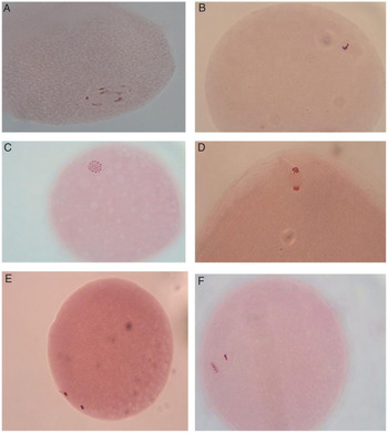
After 22 h of maturation, COCs were removed from the maturation medium and denuded from cumulus cells by 5 min incubation and repeated pipetting in 2.9% sodium citrate. Oocytes (n = 15) in a drop of the appropriate medium were pipetted and mounted onto a glass slide. A coverslip with paraffin wax spots at every corner was put on the centre of a drop of the medium containing the oocytes. Thereafter, oocytes were examined under a stereomicroscope (Nikon SMZ645). The coverslip was pressed down on the oocyte until it was held in place. Oocyte fixation of the oocyte was carried out by placing the slides in a fresh mixture of acetic acid:ethanol (1:3, v/v) for 30 min. Slides were stained with a 1% aceto-orcein stain (1% orcein stain in 40% glacial acetic acid, w/v) for 3 min and washed with aceto-glycerol (3:1, v/v) then examined using phase-contrast microscopy (Leica DM500 with camera Leica ICC50HD) at high magnification (×400). The stage of nuclear maturation (Fig. 1) was defined using the morphology of the nuclear material (Hewitt et al., Reference Hewitt, Watson and England1998).
Stages of nuclear maturation in buffalo oocytes determined by aceto-orcein staining after removal of cumulus cells. (A) Oocyte at germinal vesicle (GV) stage (vesicular nucleus with a distinctive nucleolus surrounded by fine filaments of chromatin). (B) Oocyte at germinal vesicle breakdown (GVBD) stage (chromatin condensation and disappearance of the nuclear vesicle). (C) Oocyte at metaphase of the first meiotic division (MI), formation of bivalents in which chromosomes are arranged equatorially on a metaphase plate. (D) Oocyte at anaphase of the first meiotic division (AI) stage, pulling apart of pairs of chromosomes to opposite ends of the meiotic spindle. (E) Oocyte at telophase of the first meiotic division (TI) stage, the two sets of chromosomes are separated. (F) Oocyte at metaphase of the second meiotic division (MII) stage, characterized by a tightly packed group of chromosomes in the form of the first polar body and another group that are well spread, allowing for the identification of individual chromosomes

Biochemical assay of maturation media
After 22 h of maturation, spent medium from each treatment was collected and preserved at −20°C until analysis. The concentrations of total antioxidants, TAC (Koracevic et al., Reference Koracevic, Koracevic, Djordjevic, Andrejevic and Cosic2001), MDA (Ohkawa et al., Reference Ohkawa, Ohishi and Yagi1979) and H2O2 (Aebi, Reference Aebi1984) were analyzed at wavelengths of 505, 534 and 510 nm, respectively in four runs using commercial kits (Biodiagnostic, Egypt) and spectrophotometry (Spectro UV-VIS Auto, UV-2602, Labomed, USA). All procedures were performed following the manufacturer’s instructions.
RNA isolation and cDNA synthesis
After maturation, control and treated COCs (pool of 25 COCs per replicate, four replicates) were washed twice in phosphate-buffered saline and snap frozen in cryo-tubes containing 20 µl of lysis buffer [0.8% IGEPAL, 40 U/µl RNasin (Promega, Madison, WI, USA), and 5 mM dithiothreitol (DTT) (Promega, Madison, WI, USA)]. Samples were stored in liquid nitrogen at −196°C until RNA isolation. Total RNA was isolated using a PicoPureTM RNA isolation kit (MDS Analytical Technologies GmbH, Ismaning, Germany) following the manufacturer’s instructions. cDNA synthesis used oligo(dT)23 primers, random primers, and SuperScript reverse transcriptase II (Invitrogen, Karlsruhe, Germany). In total, 1 μl of oligo(dT)23 primer and 1 μl random primer were added to 10 μl of RNA sample, and the mixture incubated for 3 min at 70°C, then chilled on ice. Next, 8 μl of master mix containing 4 μl of 5× first strand buffer, 2 μl of 0.1 M DTT, 1 μl of dNTP (10 pmol/μl), 0.3 μl RNase inhibitor and 0.7 μl of SuperScript II™ reverse transcriptase (200 unit/μl) were added to the mixture and incubated for 90 min at 42°C, followed by heat inactivation for 15 min at 70°C. cDNA was stored at −20°C until further use.
Quantitative real-time PCR
Gene-specific primers were designed using PRIMER 3 Software (http://www-genome.wi.mit.edu/cgi-bin/prime/primer3-www.cgi). Primers design was based on cDNA sequences of eight transcripts deposited in GenBank (Table 1). Quantitative analyses of cDNA samples from groups of COCs were expressed relative to GAPDH gene expression (endogenous control) and run in separate wells using the Step One Plus real-time PCR system (Applied Biosystems). Independent qRT-PCR, four replicates per treatment, used a 20-μl reaction volume containing the iTaq SYBR Green master mix with ROX (Bio-Rad Laboratories, Munich, Germany), the diluted cDNA samples and specific forward and reverse primers. Thermal cycling parameters were: 95°C for 10 min, 40 cycles of 95°C for 15 s and 60°C for 1 min. After the end of the last cycle, a dissociation curve was produced by beginning fluorescence acquisition at 60°C and measuring at 7 s intervals until the temperature reached 95°C. Comparative cycle threshold (Ct) was used to quantify expression levels as previously described (Yadav et al., Reference Yadav, Singh, Singh, Saini, Palta, Manik, Singla, Upadhyay and Chauhan2013). Briefly, during data analysis, the Ct value of the housekeeping gene was extracted from the Ct value of the target gene to obtain change in Ct (∆Ct). The ∆Ct value of the target gene sample was subtracted from the calibrator (control) to get ∆∆Ct values. Difference in the transcript abundance for the target genes was calculated using 2−∆∆Ct.
Details of primers used for quantitative real-time PCR

Statistical analysis
Mean percentages of parameters were statistically analyzed using analysis of variance (ANOVA) using the General Linear Model (GLM) procedures in SAS (SAS, 2007). Significant differences among treatments for maturation rate and oxidative biomarkers were tested using Duncan multiple range tests (Duncan, Reference Duncan1955) with significance levels of P < 0.05 and P < 0.0001, respectively. Values are expressed as mean ± standard error (mean ± SE).
Gene expression data were analyzed using the SAS GLM procedure. Differences in mean values were tested among treatment groups using ANOVA followed by Duncan multiple range tests with a significance level of P < 0.05.
Results
In vitro maturation rate (nuclear maturation)
The percentage of oocytes reaching metaphase II stage (MII) was significantly (P < 0.05) higher in maturation medium supplemented with ZnCl2 + Na2SeO3 (92.5 ± 0.37%), ZnCl2 (90.4 ± 1.52%) and Na2SeO3 (89.3 ± 1.89%) compared with the control medium (80.0 ± 1.86%). The percentage of oocytes at the T1 stage was higher (P < 0.05) in the control medium (Table 2).
Effect of zinc chloride and sodium selenite supplementation in maturation medium on in vitro maturation of buffalo oocytes

a,bMeans denoted within the same column with different superscripts are significantly different at P < 0.05.
Total antioxidant capacity and oxidative biomarkers in maturation media
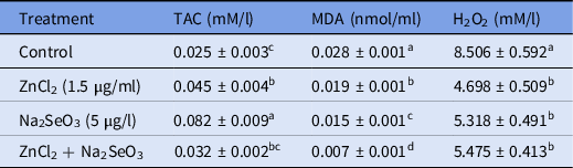
In vitro maturation of buffalo oocytes in medium with ZnCl2 or Na2SeO3 or a mixture significantly (P < 0.0001) increased TAC and decreased both oxidative markers, MDA and H2O2 concentrations, in maturation medium measured after 22 h of maturation compared with the control medium (Table 3).
Effect of zinc chloride and sodium selenite supplementation in maturation medium of buffalo oocytes on total antioxidant capacity and oxidative markers

a–dMeans denoted within the same column with different superscripts are significantly different at P < 0.0001.
H2O2, hydrogen peroxide; MDA, malondialdehyde; TAC, total antioxidant capacity.
Gene expression
Effect of zinc chloride (ZnCl2)
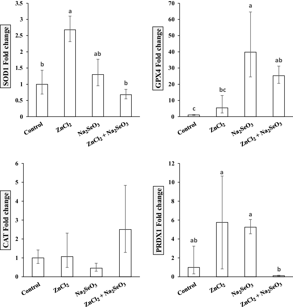
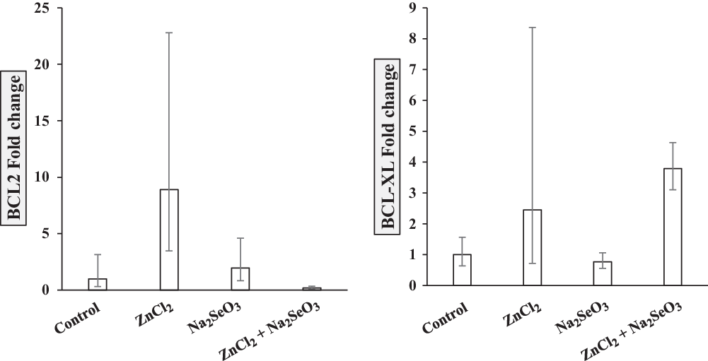
The effect of ZnCl2 supplementation during maturation of buffalo COCs on mRNA expression of selected candidate genes (SOD1, GPX4, CAT, PRDX1, BCL2, BCL-XL, BAX and BID) is depicted in Figures 2–4. A significant increase (P < 0.05) in mRNA expression of SOD1, but insignificant increase in GPX4, BCL2, BCL-XL and PRDX1 genes and insignificant decrease (P > 0.05) in the expression of BAX and BID genes were observed. No change was recorded in CAT gene expression in the treated vs. control COCs.
Differentially expressed (fold change) antioxidant-related genes (SOD1, GPX4, CAT and PRDX1) in control and treated (ZnCl2, 1.5 µg/ml, Na2SeO3, 5 µg/l and ZnCl2 + Na2SeO3) matured buffalo COCs.

Effect of sodium selenite (Na2SeO3):
Supplementation of Na2SeO3 in maturation medium led to a significant increase (P < 0.05) in mRNA expression of GPX4 and insignificant increases in SOD1, BCL2 and PRDX1. CAT, BCL-XL, BAX and BID showed insignificant decreases in supplemented media compared with the control (Figs 2–4).
Effect of zinc chloride and sodium selenite (ZnCl2 + Na2SeO3)
Supplementation of maturation medium with ZnCl2 + Na2SeO3 induced significant upregulation of GPX4, and insignificant increases in BCL-XL and CAT in supplemented media. Dual supplementation insignificantly decreased mRNA expression of SOD1, PRDX1, BCL2, BAX and BID genes (Figs 2–4).
Differentially expressed (fold change) anti-apoptotic genes (BCL2 and BCL-XL) in control and treated (ZnCl2, 1.5 µg/ml, Na2SeO3, 5 µg/l and ZnCl2 + Na2SeO3) matured buffalo COCs.

Differentially expressed (fold change) proapoptotic genes (BAX and BID) in control and treated (ZnCl2, 1.5 µg/ml, Na2SeO3, 5 µg/l and ZnCl2 + Na2SeO3) matured buffalo COCs.

Discussion
Several studies suggest that adding antioxidants to the oocyte maturation medium improves cytoplasmic maturation by reducing oxidative stress through increasing glutathione storage and increasing oocyte protection against oxidative agent damage. The improvement of culture conditions is difficult and depends on the choice of antioxidants, concentration, type and composition of the medium, species, and dynamic variation in the special needs of oocytes as development progresses (Aghaz and Khazaei, Reference Aghaz and Khazaei2017).
Our results revealed that ZnCl2, Na2SeO3 and their combination increased the percentage of matured oocytes at metaphase II compared with the control. Interestingly, enhanced effects were measured following increases in TAC level and decreases in concentrations of oxidative stress markers (MDA and H2O2) in maturation medium. In accordance with the obtained results, GSH-Px activity of oocytes and DNA integrity of cumulus cells increase after Se supplementation during oocyte IVM. Appropriate Se concentrations during IVM are beneficial for yak oocyte maturation and subsequent development (Xiong et al., Reference Xiong, Lan, Li, Lin and Li2018a). Sodium selenite (SS) increases oocyte mitochondrial DNA copy number by decreasing oxidative stress and improving oocyte developmental competence (Ghorbanmehr et al., Reference Ghorbanmehr, Salehnia and Amoushahi2018). The presence of SS in the culture medium of pre-antral follicles in rats increases follicular TAC level and Se-GPx activity and reduces levels of ROS. This activity enhances follicular development in vitro (Abedelahi et al., Reference Abedelahi, Salehnia, Allameh and Davoodi2010).
Zinc is related to oxidative stress. The mineral is essential for various cellular functions and is a cofactor for many enzymes that produce or neutralize cellular ROS (Kloubert and Rink, Reference Kloubert and Rink2015). Insufficient Zn affects intracellular content of GSH and DNA integrity of cumulus cells during IVM of bovine oocytes, with a negative effect on developing preimplantation embryos. Optimal conditions of oocyte IVM depend on the presence of sufficient Zn (Picco et al., Reference Picco, Anchordoquy, De Matos, Anchordoquy, Seoane, Mattioli, Errecalde and Furnus2010). Zn insufficiency leads to increased DNA damage and apoptosis of cumulus cells (CC). In contrast, sufficient Zn in culture medium safeguards DNA integrity and diminishes the ratio of apoptotic to normal cells in CC (Anchordoquy et al., Reference Anchordoquy, Picco, Seoane, Anchordoquy, Ponzinibbio, Mattioli, Peral-García and Furnus2011). Supplementation of Zn during IVM enhances maturation of yak oocytes by increasing the activity of GSH and SOD and reducing ROS (Xiong et al., Reference Xiong, Lan, Li, Lin and Zi2018b). Supplementation with Zn in IVM medium: (i) reduced DNA damage and apoptosis in CC; (ii) increased the activity of SOD; and (iii) did not modify cumulus expansion (Anchordoquy et al., Reference Anchordoquy, Anchordoquy, Sirini, Picco, Peral-García and Furnus2014). Treatment with sufficient zinc during IVM enhanced the potential of porcine embryos by modulating intracellular GSH concentration, ROS levels, and transcription factor expression (Jeon et al., Reference Jeon, Yoon, Cai, Hwang, Kim, Zheng, Lee, Kim and Hyun2014).
Cellular homeostasis is maintained by balancing net growth and death rates of cells (Lockshin and Zakeri, Reference Lockshin and Zakeri2007). Cells respond to adverse conditions by upregulating certain sets of genes and downregulating others. Our results showed an increase in mRNA expression of SOD1 by zinc chloride addition and for GPX4 by sodium selenite addition to maturation medium.
Results may suggest stronger antioxidative and antiapoptotic effects for zinc chloride on buffalo oocyte maturation, consistent with findings of Duprez et al. (Reference Duprez, Roma, Close and Jonas2012) and El-Sayed et al. (Reference El-Sayed, Salem, El-Garhy, Rahman and Kandil2013). Zinc chloride in the present study reduces mitochondrial oxidative stress as reported by Duprez et al. (Reference Duprez, Roma, Close and Jonas2012). Zinc also induces a reduction in the production of ROS in cell cultures (Szuster-Ciesielska et al., Reference Szuster-Ciesielska, Stachura, Słotwińska, Kamińska, Śnieżko, Paduch, Abramczyk, Filar and Kandefer-Szerszeń2000). Furthermore, zinc prevents apoptotic protease caspase-3 expression (Truong-Tran et al., Reference Truong-Tran, Carter, Ruffin, Zalewski and Maret2001), stabilizes the structure of p53 and DNA repair proteins (Chai et al., Reference Chai, Truong-Tran, Ho and Zalewski1999) and subsequently reduces apoptosis. These findings confirm our results regarding downregulation of proapoptotic BAX and BID genes and upregulation of antiapoptotic genes (BCL2 and BCL-XL). Interactions between BCL2 and BAX regulate the release of cytochrome c (cyt c) from mitochondria and establish baseline sensitivity to apoptotic stimuli. A decreased BCL2/BAX ratio is likely to promote apoptosis signalling (Oltval et al., Reference Oltval, Milliman and Korsmeyer1993). Our results indicated that zinc supplementation results in reduced apoptosis process by the BCl2–BID–BAX axis, and the BCL-XL–cytochrome axis, which prevents mitochondrial cells from releasing cyt c and subsequently prevents the release of caspase that triggers programmed cell death. This process is consistent with the upregulation of both BCL2 and BCL-XL genes.
Our results have confirmed that sodium selenite supplementation dramatically increases expression of GPX4 genes nearly 40-fold. Glutathione peroxidases (GPX) have essential biological roles in protecting against oxidative damage and are present in the cytoplasm of nearly all mammalian tissues. The preferred substrate of these enzymes is hydrogen peroxide. Our results are in agreement with Schnabel et al. (Reference Schnabel, Lubos, Messow, Sinning, Zeller, Wild, Peetz, Handy, Munzel and Loscalzo2008), who reported that selenium is a central determinant of antioxidative glutathione peroxidase 1 (GPX1) expression and activity. Furthermore, Bosse et al. (Reference Bosse, Pallauf, Hommel, Sturm, Fischer, Wolf and Mueller2010) indicated that sodium selenite upregulates GPX1 in rat liver cells. Yeo and Kang (Reference Yeo and Kang2007) reported that selenite prohibits H2O2-induced apoptosis of treated cells. This function is associated with H2O2-induced ROS production, the release of cyt c and activation of caspases 3 and 9. The latter authors suggested that selenite activates p44/42 MAPK and prohibits downregulation of BCL2 in selenite-treated cells.
Supplementation of the maturation medium with a combination of both zinc chloride and sodium selenite induced a significant increase in expression level of antioxidative genes GPX4 and an insignificant increase in CAT. This observation is consistent with other studies that suggest that this combination protects cells against oxidative stress (Imed et al., Reference Imed, Fatima and Abdelhamid2009; Tariba et al., Reference Tariba, Živković, Gajski, Gerić, Gluščić, Garaj-Vrhovac, Peraica and Pizent2017; Branca et al., Reference Branca, Morucci, Maresca, Tenci, Cascella, Paternostro, Ghelardini, Gulisano, Mannelli and Pacini2018). Published data on ZnCl2 and Na2SeO3 supplementation for buffalo oocytes is limited, and the present study provides useful information on the beneficial impacts of ZnCl2 and Na2SeO3 individually and in combination during oocyte maturation and helps to elucidate signalling pathways that might be involved.
In conclusion, we provide evidence that supplementation of in vitro maturation medium with zinc chloride and sodium selenite can improve the maturation rate of buffalo oocytes by protecting against oxidative stress and increasing expression levels of antioxidative and antiapoptotic genes.
Ethical approval
This study was carried out under proper research ethics and care and was approved by Mansoura University Research Committee.
Financial support
This research received no specific grant from any funding agency, commercial or not-for-profit sectors.
Conflicts of interest
The authors declare no conflicts of interest.