
Introduction
The extracellular matrix (ECM) provides support for cells and creates an environment where it influences cell growth, shape, and differentiation.[Reference Flemming, Murphy, Abrams, Goodman and Nealey1] Bio-fabrication and the design of engineered fiber scaffolds has been inspired by the natural structure of ECM. The behavior of cell lines, including differentiation, cell adhesion, proliferation, and migration, is influenced by surface topographic properties. Soft lithography, inkjet printing, and photochemistry methods are utilized to construct a surface with specific topographic properties.[Reference Tay, Irvine, Boey, Tan and Venkatraman2] Unidirectional ordered patterns showed great interest in the collagen fibers and the cells found in tendon, ligament, and muscle due to their similarities to native ECM–cell systems.[Reference Kim, Jiao, Hwang, Kim, Kang, Kim and Suh3, Reference Xin, Hussain and Mao4] Based on the fiber rearrangement, random and aligned fibers are used to create structures that contained ECM-mimicking morphologies.[Reference Yoshimoto, Shin, Terai and Vacanti5] Fiber scaffolds can be manufactured with micrometer to nanometer diameter range.
Self-assembly, electrospinning, and phase separation are the major techniques to produce the fiber scaffolds, and out of these techniques, electrospinning is most versatile.[Reference Wade, Bassin, Rodell and Burdick6] However, the electrospinning is limited to a set of organic solvents, needs high voltage, and offers low yield of fibers. Therefore, several methods were reported to fabricate the aligned fiber scaffolds including melt-drawing,[Reference Tan, Tan, Yeong and Tor7] centrifugal spinning,[Reference Wang, Chang, Ahmad, Zheng and Li8] catalyst-free chemical vapor deposition,[Reference Kazantseva, Ivanov, Gasik, Neuman and Hussainova9] unidirectional freeze-drying,[Reference Fereshteh, Fathi, Bagri and Boccaccini10] three-dimensional (3D) extrusion,[Reference Moon, Choi, Koh and Kim11] and bidirectional freezing technique.[Reference Bai, Walsh, Gludovatz, Delattre, Huang, Chen, Tomsia and Ritchie12]
The importance of fibers in cell response and tissue engineering is already proved.[Reference Das, Sharma, Saharia, Sarma, Sarma, Borthakur and Bora13] Specifically, aligned fiber scaffolds showed major advances in cell proliferation and tensile properties. For example, random fibers can be used for the bone or cartilage applications,[Reference Wise, Yarin, Megaridis and Cho14] whereas aligned fibers are used in neurite outgrowth and offer topographic guidance for cellular morphogenesis in structural tissues.[Reference Chaurey, Block, Su, Chiang, Botchwey, Chou and Swami15, Reference Kim, Hwang, Aguilar, Park and Kim16]
Therefore, there is a significant and unmet need for the fabrication of 3D fiber scaffolds to mimic ECM. The 3D fiber scaffolds were fabricated by rolling fiber scaffolds on a metal drum and this method showed obstacles in maintaining homogeneousness in fiber properties.[Reference Wang, Zhang, Fan, Mo, Ruan and Li17] Thus, polycaprolactone-based dual-shaped aligned and random electrospun fibers are developed.[Reference Park, Kim, Lee, Park, Lee, Lee, Jeon and Suh18] Recently, poly(L-lactide)-based aligned fiber scaffolds are prepared by using melt spun and coated with collagen and chitosan.[Reference Feng, Zhang, Zhu and Gao19]
During the past few decades, gelatin-, chitin-, collagen-, alginate-, and zein-based fibers were prepared with or without blending of synthetic polymers by electrospinning.[Reference Fernandez, Torres-Giner and Lagaron20] Gelatin is a natural biopolymer and a hydrolysis product of collagen. Because of its easy availability, biocompatibility, and biodegradability, gelatin has been extensively used for wound healing, nerves, and drug delivery.[Reference Mamidi, Leija, Villela, Isenhart, Barrera and Elías21, Reference Gönen, Erol and Küçükbayrak22] However, poor water resistance and mechanical properties of gelatin fibers lead to rapid degradation.
Zein is a hydrophobic protein of corn and it consists of low polar and high non-polar amino acids. It could be fabricated into films by casting method or hot pressing due to its good solubility in aqueous alcohol solutions and thermoplasticity. Due to its biodegradability, biocompatibility, and nontoxicity, zein was used in the manufacturing of fibers and thin films for biomacromolecules delivery (drug, protein, DNA, gene, or vaccine), antioxidants, anticoagulants, tissue engineering, and biomedical devices.[Reference Corradini, Curti, Meniqueti, Martins, Rubira and Muniz23, Reference Paliwal and Palakurthi24] Therefore, it was postulated that zein could be uniformly dispersed in gelatin to enhance water resistance and tensile properties of gelatin fibers by Forcespinning® (FS).
In this study, first time gelatin:zein aligned 3D fiber scaffolds are fabricated using a versatile, high spinning speed, and readily scalable FS technique.[Reference Sarkar, Gomez, Zambrano, Ramirez, De Hoyos, Vasquez and Lozano25] FS is a sustainable method to produce the fibers with the use of centrifugal force. It can be used for conductive and non-conductive materials. Polymer solution parameters and device configuration can control the surface morphology, the uniformity, and thickness of fibrous scaffolds. It offered high production rate of aligned 3D fibers with enhanced mechanical, prolonged drug release, and biocompatible properties. The effect of gelatin-to-zein ratio was investigated on the chemical and physical properties of fabricated fibers. Berberine drug release was measured and prolonged release rate was observed. Fibroblast cell growth was studied on 3D aligned gelatin:zein fibers and improved cell viability was observed.
Experimental section
Materials
Unless stated otherwise, all materials were obtained from Sigma Aldrich (St. Louis, MO, USA) and used as received. Cell culture consumables and supplements were obtained from Gibco Invitrogen (Carlsbad, CA, USA). CellTiter96®AQueous One-Solution Cell Proliferation Assays were bought from Promega (Madison, WI, USA). Celltiler96® was used as mentioned in the supplied manual.
Methods
Preparation of gelatin:zein (1:1) solution
Gelatin (3.0 g), zein (3.0 g), and berberine chloride (0.5 g) were mixed in acetic acid and stirred for 4 h at 65 °C to produce a clear solution. Similarly, gelatin:zein (1:2, 1:3, and 1:4) solutions were obtained. After that, above solution was injected into a spinneret of forcespinning. The velocity of spinneret and time was optimized as 7000 revolution per minute (rpm) and 20 s, respectively.
Characterizations of fibers
Gelatin:zein fibers were analyzed by Fourier transform infrared (FTIR) (Perkin Elmer Universal ATR Sampling Accessory Frontier). The morphology and diameter of gelatin:zein fiber scaffolds were observed by SEM analysis (ZEISS EVO® MA 25, Ostalbkreis, Baden-Württemberg, Germany). ImageJ analyzer software (ImageJ, NIH, USA) was used to scrutinize the diameter distribution and pore size of the fibers.[Reference Mamidi, Leija, Villela, Isenhart, Barrera and Elías21]
The in vitro degradation of fiber scaffolds was measured by immersing the fibers in Dulbecco's modified Eagle's medium (DMEM) at room temperature. Then, the samples were removed from the solutions and weighed at 10, 15, 20, and 25 days after being dried in a hot oven for 12 h. The weight retention (WR%) of the fibers was measured using the following formula.
 $${\rm WR} \% = \displaystyle{{W_1} \over {W_0}} \times 100,$$
where W 0 is the initial weight of the fibers and W 1 is the weight of the fibers at different days.
The tensile properties of the fibers were measured using a tensile instrument (Instron 3365, Instron, Norwood, MA, USA). The surface wettability of fiber scaffolds was measured using water contact angle (WCA; OCA 15EC).
In vitro drug release
Berberine chloride drug release was measured by mixing 50 mg of gelatin:zein fiber membrane in 20 mL of DMEM. Then, the fibers were placed in the incubator at room temperature and incubated at 50 rpm. At particular period of breaks, the buffer solution (5 mL) was collected and substituted with fresh DMEM solution. Berberine release rate was calculated by the division of total amount of berberine drug with collected drug in the fibers.
Ultraviolet–visible spectroscopy (Agilent Technologies, 89090A) was used to measure the concentration of berberine in the collected samples at a wavelength of 347 nm and triplicate measurements were carried out for the Berberine release.
Cell culture and cell behavior studies
Fibers were cut into small films with the area of approximately 0.1 cm2 and sterilized by UV irradiation for 2 h. Then, human fibroblasts have seeded at a density of 4.8 × 10⁵ cells/cm2 with 100 µg/mL penicillin, 5% (w/v) FBS, and 100 µg/mL streptomycin.
Metabolic activity and proliferation of the fibers were measured using CellTiter96®. Fibroblasts (4.8 × 10⁵ cells/cm2) were seeded on the fibers. On the first, second, and third days, the fibers were transferred into new wells and each well was treated with 20 µL of reagent and incubated under dark conditions for 1 h. A microplate reader (Synergy HT, BioTek, Winooski, Vermont, USA) was used to measure the absorbance of samples and compared with the controller (cells without fibers) to evaluate the percentage of viability. All cell culture experiments were run in triplicate.
Statistical analysis
Mean ± standard deviations (n = 3) was used to express all the quantitative data. One-way analysis of variance and Tukey's post hoc tests were utilized in Minitab17 (Minitab, State College, PA, USA) to measure statistical analysis and P ≥ 0.5 was taken as statistically significant.
Results and discussion
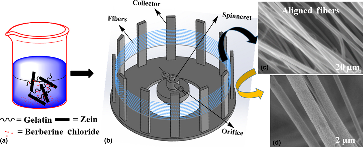
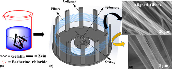
Figure 1(a) shows the preparation of gelatin, zein, and berberine chloride composite solution in acetic acid at 65 °C. Acetic acid was used as a solvent to fabricate gelatin:zein fiber scaffolds because it could dissolve well two proteins. Figure 1(b) shows the diagram of the FS process; it contained a spinneret (internal volume of 2 mL) with two sidewall orifices (orifice diameter of 310 µm) which rotates about its horizontal axis in the center of a fixed collector. The polymer solution fills up into the spinneret and produces fibers that are deposited over the collector (diameter of 300 mm). Initially, gelatin:zein (1:4) fibers were produced to optimize the conditions of FS. As shown in Figs. 1(c) and 1(d), continuous aligned fibers with a diameter of 295 nm were fabricated at 7000 rpm. To control the ability to alter the fiber morphology, gelatin:zein (1:4) fibers were produced at different rotational speeds.
Schematic illustration of the preparation of gelatin:zein composite fiber scaffolds. (a) Preparation of gelatin, zein, and berberine chloride composite solution in 80% acetic acid for 4 h at 65 °C; (b) schematic diagram of FS; and (c, d) SEM images of fiber structures produced from gelatin:zein (1:4) at 7000 rpm in 20 s, respectively.

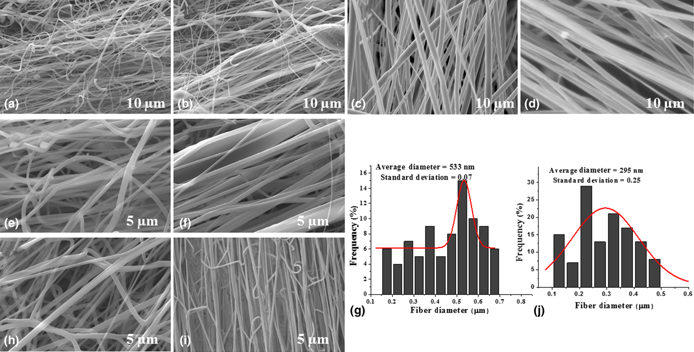
Figure 2 shows SEM images of fiber scaffolds from gelatin:zein composites with various weight ratios of zein protein and 30% (w/v) was the optimized concentration for all the samples. A 1:1 ratio of gelatin:zein produced dense random fibers with few beads at 7000 rpm [Fig. 2(a)]. The bad forcespinnability of this solution could be due to poor chain entanglements in aqueous acetic acid solution. When gelatin:zein (1:2) was used, it provided a mixture of random and aligned fiber scaffolds [Fig. 2(b)]. Whereas, gelatin:zein (1:3) produced aligned fiber scaffolds without beads. In contrast, gelatin:zein (1:4) produced bead-free aligned fiber scaffolds in 20 s.
SEM images of gelatin:zein fibers; (a) (1:1), (b) (1:2), (c) (1:3), and (d) (1:4) at 7000 rpm in 20 s, respectively; scale bar = 10 µm. SEM images of gelatin:zein (1:4) fiber scaffolds at (e) 5000, (f) 6000 rpm in 20 s, and (g) diameter distribution of gelatin:zein (1:4) fibers at rpm 6000. SEM images of gelatin:zein (1:4) fiber scaffolds at 7000 rpm (h) 60 s, (i) 20 s, and (j) diameter distribution of gelatin:zein (1:4) in 20 s; scale bar = 5 µm.
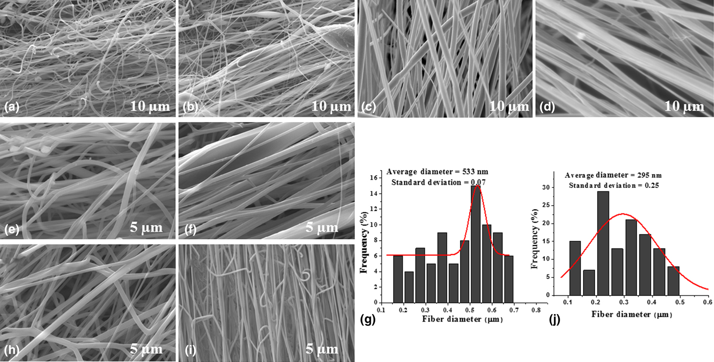
This ratio (1:4) was forcespun successfully into aligned fibers with lower diameter and stable structural form. The reduction in diameter and improvement in the uniformity of fibers were observed with respect to the increment of zein:gelatin concentration (1:1–1:4).
The viscosity of a protein solution was usually proportional to a protein ratio and was important to fabricate high throughput fibrous scaffolds. With low viscosity of protein solution, droplets were observed; it could be because of weak cohesive attractions in it. Whereas, at high viscosity, merged fibers were obtained because the solution was not completely evaporated. Therefore, every protein and solvent has a specific concentration or viscosity to produce desired fibers. In gelatin/zein composite systems, a (1:4) ratio of solution produced the best quality of aligned fiber scaffolds.
Therefore, gelatin:zein (1:4) solution was selected for further optimizations. In addition, rpm of spinneret was also optimized in the fabrication of fibers. For example, gelatin:zein (1:4) solution produced only droplets at 4000 rpm. Whereas, at 5000 rpm, random fiber scaffolds were produced [Fig. 2(e)]. At a higher rpm (6000), gelatin:zein (1:4) solution produced desired fibers with an average diameter of 533 nm [Figs. 2(f) and 2(g)]. At an even higher rpm (7000), lower diameter fibers were obtained. Thus, the high quality fibers were fabricated from gelatin:zein solutions (30 wt.%) at an optimized rpm (7000).
Spinning time of spinneret also played an important role in the production of aligned fiber scaffolds. When 40–60 s wan used, mesh-like fibers were formed and the rate of alignment was reduced [Fig. 2(h)]. This could be due to the simultaneous deposition of multiple fiber scaffolds, as most of the scaffolds could not be stretched and fully not dried. Whereas, when the time was reduced to 20 s, the aligned fiber rate was improved and completely aligned fibers were obtained. Therefore, 30% (w/v) of gelatin:zein, 7000 rpm, and 20 s were the optimized conditions for gelatin:zein solutions.
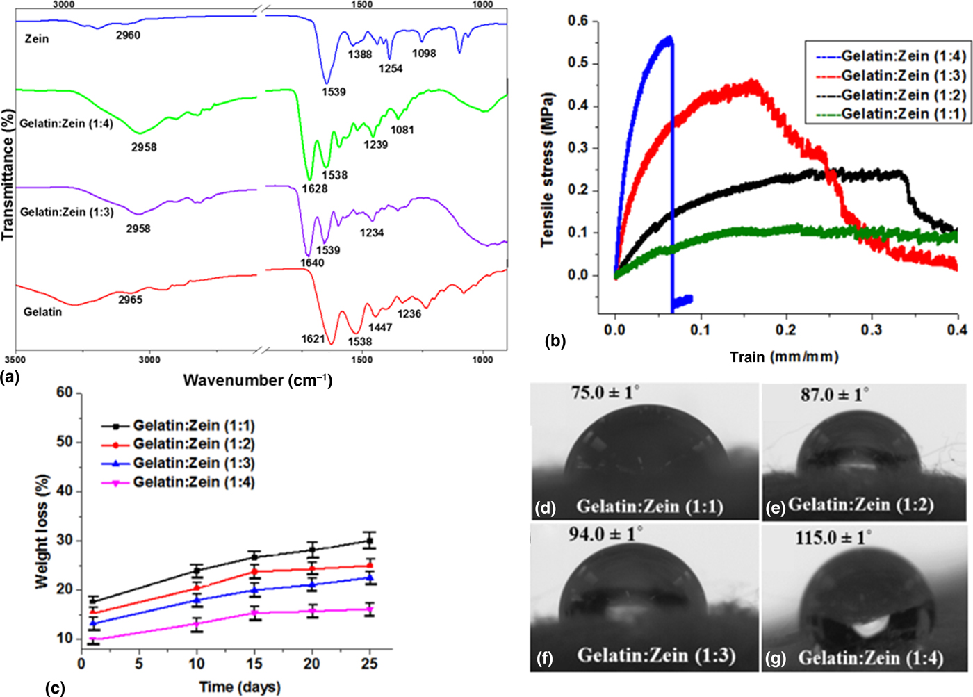
To determine the possible molecular interaction between gelatin and zein protein, FTIR was performed on gelatin, zein protein, and gelatin:zein fiber scaffolds [Fig. 3(a)]. All fiber scaffolds showed common absorption bands of protein at around 1647 cm−1 for amide I and stretching vibration of C=O bond; whereas 1538 cm−1 for amide II and C–N bond, respectively.[Reference Corradini, Curti, Meniqueti, Martins, Rubira and Muniz23] The absorption peaks approximately 2958–2965 cm−1 were designated as C–H stretching vibrations of aliphatic groups. The absorption peaks of N–H bending and C–N stretching of proteins were observed at 1448, 1400–1410, 1239, and 1086 cm−1, respectively. A broad absorption peak in a range of 3500–3100 cm−1 was designated as amide A (N–H stretching vibration). This indicates that there was a hydrogen bonding and good miscibility between gelatin and zein components in the blended fibers.
(a) FTIR spectra of pristine gelatin, zein, and gelatin:zein (1:3 and 1:4) fiber scaffolds. Carbonyl frequency (1539 cm−1) of pristine zein protein was shifted to 1628–1640 cm−1 in the gelatin:zein composites. (b) Tensile curve, (c) in vitro degradation. Data represent mean ± SD (n = 3, Tukey's post hoc test). Statistically significant difference (P < 0.05) was observed in gelatin:zein fibers. (d)–(g) Water contact angles of gelatin:zein (1:1, 1:2, 1:3, and 1:4) fibers, respectively.
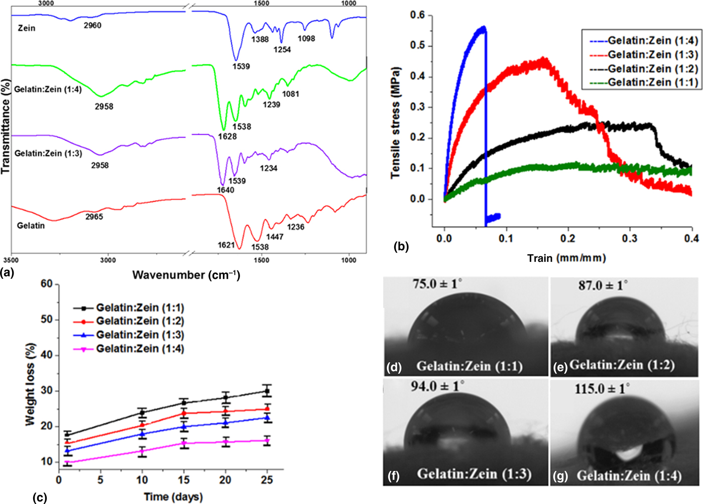
The tensile properties of gelatin:zein fibers were studied using ~1.0 mm thick fiber membrane and Fig. 3(b) represents a tensile curve of gelatin:zein fibers. The results showed improvement in elongation at break and tensile strength of fibers with an increase of zein concentration in gealtin:zein (1:1–1:4) fibers. Specifically, 1.98 MPa of Young's modulus and 0.054 MPa of tensile strength were observed in gelatin:zein (1:1) fibers. In addition, improved Young's modulus (7.1 MPa) and tensile strength (0.21 MPa) were noticed in gelatin:zein (1:2) fibers. Similarly, enhanced tensile strength and Young's modulus were found in both (1:3) and (1:4) of gelatin:zein [Fig. 3(b)]. This was because of electrostatic interactions between zein and gelatin. Thus, gelain:zein fibers were strengthened at the higher concentration of zein.
The degradation results of gelatin:zein (1:1–1:4) fiber scaffolds were measured in DMEM at 37 °C and presented in Fig. 3(c) and the degradation rate of fibers was observed with respect to the time. Gelatin:zein fibers showed discrepancy in the degradation with respect to the concentration of zein protein. Particularly, after 25 days, 29.0% of degradation in (1:1) of gelatin:zein and 25.1% of degradation in (1:2) of gelatin:zein fibers were observed. Further, 22.8% of degradation was noticed after 25 days in (1:3) of gelatin:zein. Whereas, 16.2% of degradation was obtained for (1:4) of gelatin:zein in 25 days. Because of enhanced tensile properties and non-polar functional groups of zein, the rate of degradation was reduced at higher concentration of zein in gelatin:zein (1:4) fibers.
The rate of cell proliferation and adhesion can be directed by the nature of fibers. In order to measure the hydrophobicity of gelatin:zein fibers, WCA was performed and the results were presented in Figs. 3(d)–3(g). WCA of gelatin:zein fibers was proportional to the concentration of zein protein. For example, 75.0°, 87.0°, 94.0°, and 133.5° were recorded in (1:1), (1:2), (1:3), and (1:4) of gelatin:zein, respectively. These results reveal that wettability of gelatin:zein fibers was reduced at a higher concentration of zein protein.
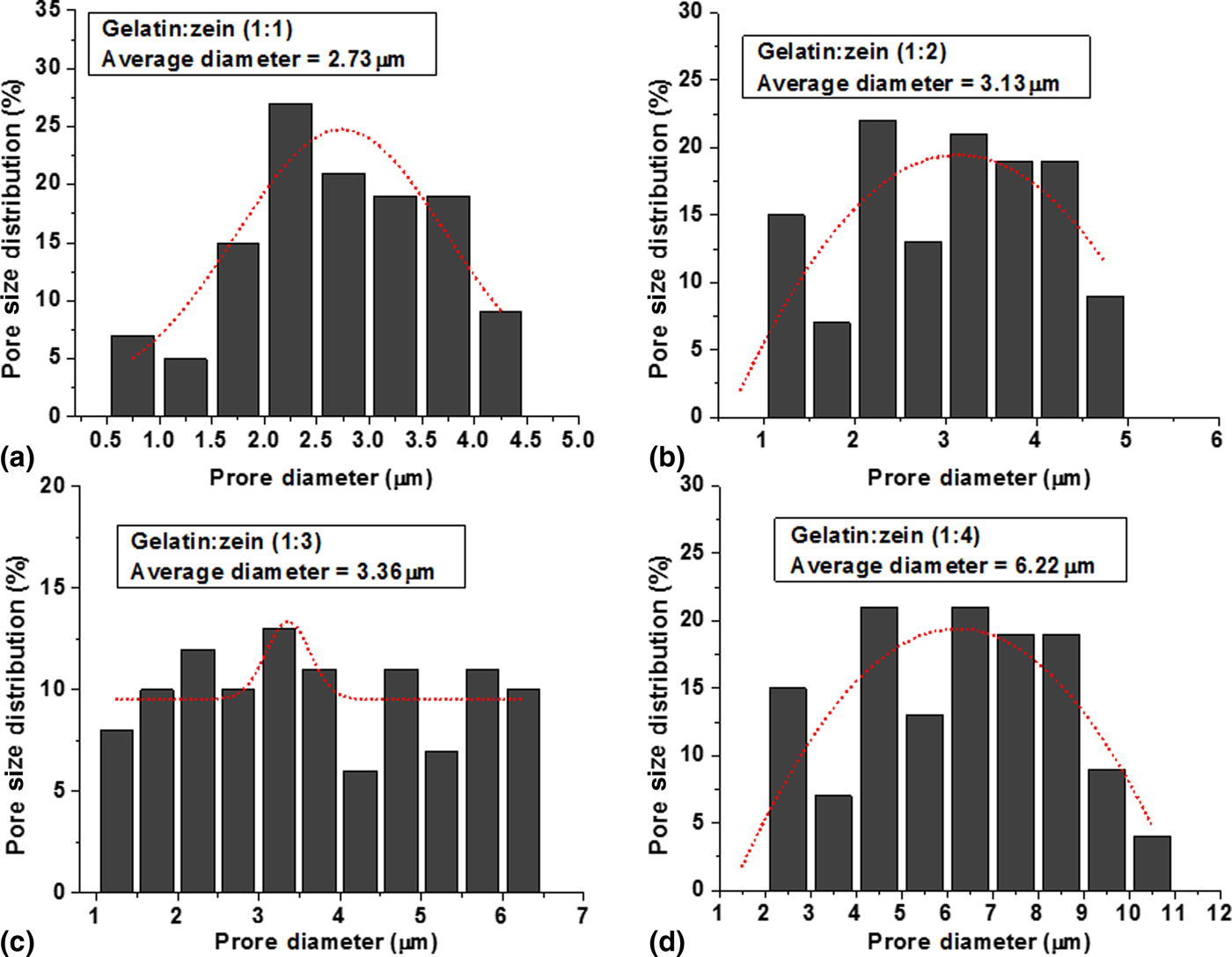
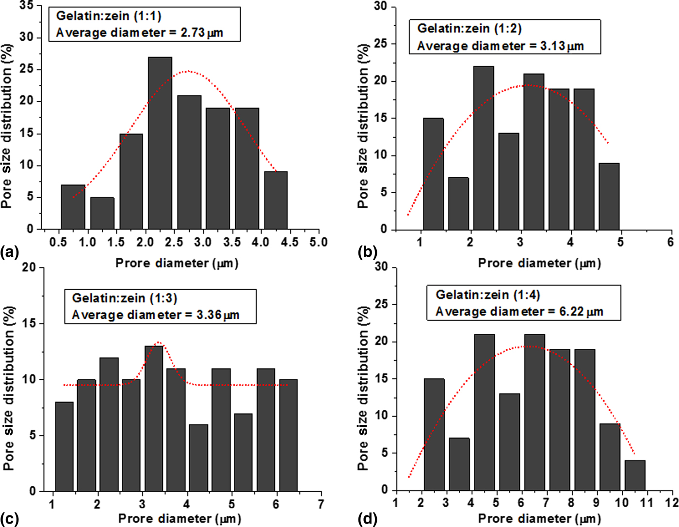
Pore size of zein and gelatin fibers was measured and the results were presented in Fig. 4. As showed in Fig. 4(a), (1:1) ratio of gelatin:zein fibers showed lowest pore size and the average pore diameter was 2.73 µm and the pore distribustion range was found from 0.5 to 4.5 µm. Gelatin:zein (1:2) fibers showed improved pore size (3.13 µm) and pore distribustion was found from 1.2 to 4.98 µm [Fig. 4(b)]. Further, improved pore size was obsreved in (1:3) ratio of gelatin:zein fibers and average pore size was 3.36 µm. The distrubustion of pore size was found in the range of 1.2–6.4 µm [Fig. 4(c)]. Whereas, gelatin:zein (1:4) showed 6.22 µm of average pore diameter and the distribustion of pore size was found in the range of 2.2–10.8 µm [Fig. 4(d)]. Thus, these experimental results showed that the pore diameter of gelatin:zein fibers was gradually increased with the increment of zein concentration.
Pore size distribution of gelatin:zein fiber scaffolds; (a) (1:1), (b) (1:2), (c) (1:3), and (d) (1:4), respectively.
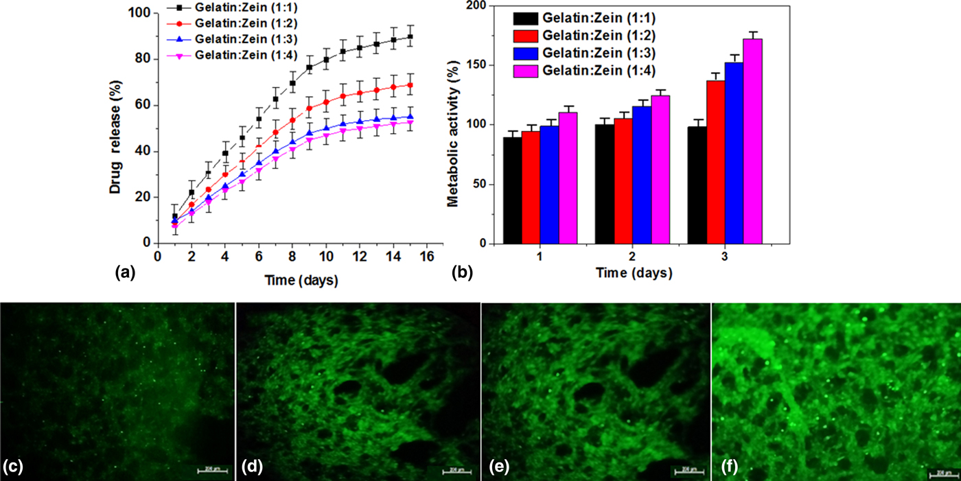
It is reported that nanofibers were vastly useful in the drug delivery including antibacterial or accelerate wound healing process.[Reference Guo, Zhou and Lv26] In this paper, berberine chloride was incorporated into forcespun gelatin:zein fiber scaffolds during the forcespinning. Berberine chloride is an isoquinoline alkaloid and it showed excellent antibacterial effect toward Candida albicans, Bifidobacterium adolescentis, Shigella dysenteriae, Escherichia coli, and Staphylococcus aureus.[Reference Kong, Zhao, Xiao, Jin, Liu and Li27, Reference Yu, Kim, Cha, Kim, Lee, Choi and You28] Fig. 5(a) shows the cumulative drug release of gelatin:zein fibers and the graph was plotted with drug release rate versus time. Gelatin:zein (1:1–1:4) fibers showed slow and sustained drug release within 15 days. After 15 days, approximately 53–89% of the total berberine drug was released from gelatin:zein fibers. Initially, rapid berberine release was observed in gelatin:zein fibers. Gelatin:zein (1:1–1:4) fiber scaffolds showed a sustained berberine release and it demonstrates the electrostatic attractions among berberine, zein, and gelatin. Therefore, gelatin:zein fiber scaffolds can be useful for prolonged berberine drug release.
(a) In vitro berberine drug release measurements from gelatin:zein (1:1–1:4) fiber scaffolds. Data represent mean ± SD (n = 3, Tukey's post hoc test). Statistically significant difference (P < 0.05) was observed in gelatin:zein fibers. (b) Metabolic activity of human fibroblast cells after seeding on gelatin:zein (1:1–1:4) fiber scaffolds for first, second, and third days. Data were normalized against a positive control without the addition of the fiber scaffolds. Confocal microscopy images of human fibroblast cell growth on gelatin:zein fiber scaffolds after 72 h; (c) (1:1), (d) (1:2), (e) (1:3), and (f) (1:4), respectively; scale bar = 200 µm.
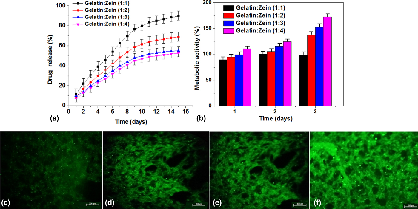
Human fibroblast cells were cultured on gelatin:zein (1:1–1:4) fibers for 1–3 days to measure cell proliferation and the results were presented in Fig. 5(b). The cell metabolic activity was measured using CellTiter96® based cell proliferation assay. On day 1, all the gelatin:zein (1:1–1:4) fibers showed approximately equal metabolic activity [Fig. 5(b)]. After 2 days, the metabolic activity was slightly increased in gelatin:zein (1:3) and gelatin:zein (1:4) fiber scaffolds; the metabolic rate was better than gelatin:zein (1:1 and 1:2) fiber scaffolds. In addition, after 3 days, metabolic activity was significantly increased in gelatin:zein (1:4) fibrous scaffolds than other gelatin:zein (1:1–1:3) fibers. Overall, at higher concentration of zein protein, the metabolic activity was increased in a dose-dependent manner and maximal level reached with gelatin:zein (1:4). This might be because of high wettability and degradation of gelatin:zein (1:1–1:3) fibrous scaffolds.
The cytotoxicity of fibers was measured by incubating the human fibroblasts in culture medium having sterilized mesh of gelatin:zein with different concentrations. Figures 5(c)–5(f) show the confocal images of the incubated fibroblast cells on a 72 h. After 72 h, the cell growth was measured on the surface of all gelatin:zein fiber scaffolds. The morphology observations indicate that all fibroblasts were attached on the surface of fiber scaffolds. The cell growth was gradually increased from 1:1 to 1:4 ratio of gelatin:zein fibers. This observation suggests a positive effect of zein protein on the survival of fibroblasts. A better expansion and elongation was observed on the surface of gealtin:zein (1:4) fibers. This could be explained with the zein presence in the polymer blend. These two materials created 3D aligned fibers that stimulated cell growth. Therefore, the surface morphology, the contact angle with cell media, and the surface chemistry played a key role to improve cell growth. A hypothesis is that good miscibility zein with gelatin produce biocompatible gelatin:zein fiber scaffolds.
Conclusion
The present study shows the production of aligned 3D fiber scaffolds from gelatin:zein using FS. Depending on the gelatin and zein protein blending ratio, the forcespun membrane has amendable fiber morphology, diameter, mechanical, surface wettability, in vitro degradable, and cell viability properties. The combination of zein protein tremendously improved the forcespinnability of gelatin and produced not only aligned fibers but also random fibers at different ratios of zein protein. Gelatin:zein fibers showed improved mechanical and cell viability properties. In addition, sustained berberine drug release rate was observed over 15 days. The mechanical, viability, and sustained drug release results demonstrated that gelatin:zein fiber scaffolds could be useful for drug delivery, hernia repair, tissue regeneration, and wound healing.
Acknowledgment
The authors thank Consejo Nacional de Ciencia y Tecnología de México (CONACyT; Project Number 242269) for financial support. The authors thank Dr. Karina Del Angel for water contact angle.