Complete atrioventricular canal is a congenital malformation that results from the embryonic non-development of endocardial cushions, resulting in a common atrioventricular valve with an absence of cardiac septa. The prevalence of this abnormality is estimated to be between 4–5/10,000 live births according to the United States Center for Disease Control and Prevention.1 The transposition of the great arteries is characterised by atrioventricular concordance with ventriculoarterial discordance as a result of improper rotation of the truncus arteriosus. The prevalence is similar to complete atrioventricular canal, becoming the most frequent cyanotic congenital heart disease. The association between complete atrioventricular canal and transposition of the great arteries is very uncommon with only a handful of reports in the literature. We report the case of an infant with a complete atrioventricular canal with side-by-side transposition of the great arteries and anomalous coronary origins who had undergone early pulmonary banding before total surgical correction at 18 months of age by an arterial switch procedure and a two-patch technique canal repair.
Case report
An 18-month-old male patient presented to our institution with a heart rate of 130, respiratory rate of 30, 74% O2 saturation, and IV/IV systolic murmur on pulmonary foci. Past surgical history revealed a pulmonary artery band implanted in a different hospital at the age of 3 months. Echo showed a side-by-side d-transposition of the great arteries with Type A complete atrioventricular canal with left-to-right shunt, and a properly positioned pulmonary artery band with peak gradient 60 mmHg and mean gradient 40 mmHg with moderate hypertrophy of the pulmonary ventricle. Cardiac catheterisation reported similar results, with good left ventricular function and no anomalous coronary anatomy.
The operation was done through median redo sternotomy with standard cardiopulmonary bypass with bicaval cannulation, under mild hypothermia and cardiac arrest with del Nido cardioplegia. The anatomy was initially observed with side-by-side great vessels and anomalous origin of the coronary arteries (1RL-2Cx).
First, the complete atrioventricular canal was repaired with a two-patch technique, closing the ventricular septal defect with a synthetic Gore-Tex patch (W.L. Gore and Associates, Inc. Newark, Delaware, United States of America), the left-valve component cleft with interrupted mono-filament sutures, and the ostium primum defect with a bovine pericardium patch. A 5-mm atrial septal defect was maintained to vent the right heart in case of right ventricle dysfunction, anticipated due to the long cross-clamp time (198 minutes). This defect would assist left heart drainage in case the patient would require extracorporeal membrane oxygenation. After complete atrioventricular canal repair, a second dose of cardioplegia was given.
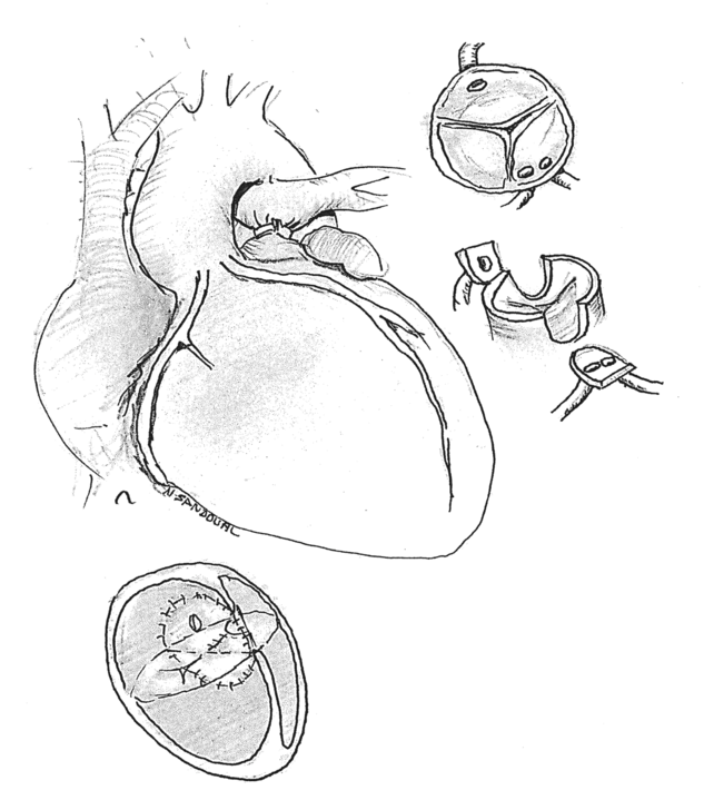
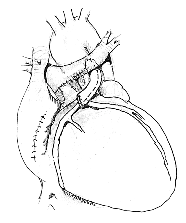
Subsequently, the transposition of the great arteries was corrected with a standard arterial switch technique with Lecompte manoeuvre. Given the anomaly at the origin of the coronary arteries, with the left anterior descending artery and ramus circumflex artery ostia 2 mm apart from each other, making separation with direct anastomosis impossible, an 8-mm Gore-Tex graft was interposed in order to transfer the two-ostium button to the neo-aorta (see Fig 1). The circumflex was transferred in a usual manner (see Fig 2). The narrow segment of the pulmonary artery was resected and re-anastomosed at the neo-pulmonary artery position, moving the anastomosis towards the right pulmonary artery.
Pre-operative cardiac anatomy of our patient with a cAVC and TGA with anomalous coronary anatomy. cAVC=complete atrioventricular canal; TGA=transposition of the great arteries.

Post-operative patient cardiac anatomy, with interposition graft for coronary transfer.

With normal sinus rhythm and electrocardiogram, cardiopulmonary bypass weaning was attempted but severe biventricular dysfunction was noted, confirmed with intra-operative echo showing right-to-left shunt at the atrial level and systemic desaturation. Adequate flow to both coronary arteries was observed. No diagnostic catheterisation was considered at that time given echo results and no electrocardiogram changes. Consequently, extracorporeal membrane oxygenation therapy was started and maintained for 5 days and weaned on post-operative day 6 without complications, with adequate biventricular function, sinus rhythm, and no significant regurgitation of the right or left valve component on echocardiogram. The patient was discharged home on postoperative day 10 with daily anti-aggregation therapy.
The patient has been followed every 3 months with echocardiogram. The last follow-up was 14 months post-operative, showing normal coronary flow and normal biventricular function with mild right atrioventricular-component regurgitation.
Discussion
Atrioventricular canals with conotruncal anomalies are a challenge for surgical repair. The principles of surgery are the construction of an unobstructed anatomical communication between both ventricles and great arteries without significant atrioventricular valve regurgitation.Reference Tchervenkov and Bernier2 The association of transposition of the great arteries with complete atrioventricular canal is rare, normally accompanied by pulmonary stenosis usually managed with a Rastelli operation or following the Fontan pathway.Reference Sakamoto and Galleti3,Reference Kumar, Prabhaker and Wilson4 Bharati et al reported 17 hearts with transposition of the great arteries in an autopsy series of 507 complete atrioventricular canal hearts (3.4%); of 178 transposition heart specimens, Shaher et al found none with complete atrioventricular canal; and Boesen et al noted only 3 complete atrioventricular canal in a review of 132 patients with transposition (2.3%).Reference Bharati and Kirklin5–Reference Boesen7 All hearts in the study by Bharti and colleagues revealed pulmonary stenosis.
In the context of conotruncal anomalies associated with complete atrioventricular canal, there are two approaches: single-stage repair during the neo-natal period or two-stage repair with pulmonary artery band followed by complete repair during infancy.Reference Bautista-Hernandez, Marx and del Nido8 Our patient came from abroad where a two-stage approach was offered as the only surgical option due to socio-economic challenges within the country, performing a pulmonary artery banding thus allowing the child to grow until his referral to our institution for a complete repair. A recent study by Mery et al reports that surgical repair of complex complete atrioventricular canal associated with Tetralogy of Fallot, aortic arch repair, double outlet right ventricle, or total anomalous pulmonary venous return can be performed with similar mortality rates to isolated complete atrioventricular canal, but with high rates of reoperation.Reference Mery and Zea-Vera9 However, transposition of the great arteries associated with complete atrioventricular canal is uncommon and excluded from the outcomes of this study.
There are reports in the literature of transposition of the great arteries associated with complete atrioventricular canal and other anomalies such as persistent left superior caval vein. To our knowledge, this is the first report of this anomaly associated with the aforementioned coronary anatomy and side-by-side great arteries, managed with an arterial switch operation and two-patch technique repair for complete atrioventricular canal, with good surgical results despite the need for short-course extracorporeal membrane oxygenation therapy.Reference Bisoi, Abraham and Venugopal10 The use of a Gore-Tex interposition conduit for the coronary transfer is somewhat controversial; however, we have not found reports in the literature of thrombosis of Gore-Tex conduits in the coronary anatomy, with report of successful use in an arterial switch.Reference Albert, Castilla and Amengual11
Acknowledgements
We would like to acknowledge Andrea Castillo M.D. and Vanessa Romero R.N. for their assistance in gathering the relevant to the patient reported on in this study.
Financial support
The authors received no funding to support the production of this case report.
Conflicts of interest
None.
Ethical standards
Permission was granted by the patient’s family for the publication of this case report.