Introduction
Epithelioid sarcoma is a rare and aggressive malignant neoplasm which was first described by Enzinger in 1970.Reference Enzinger1 Epithelioid sarcoma mainly occurs in young adults and it is rarely found in the head and neck region. A survey of 241 cases treated at the US Armed Forces Institute of Pathology revealed only three cases occurring as primary tumours in the head and neck region.Reference Chase and Enzinger2 The case described in this report is only the second known instance of an epithelioid sarcoma arising in the neck.
Case report
A 56-year-old man was referred to the Kurume University Hospital department of otolaryngology and head and neck surgery after presenting with a three-year history of a sensory disturbance of the right neck and the presence of a neck mass.
A 50 × 40 mm, fixed, nontender mass was palpable in the patient's right neck. The rest of the physical examination findings were normal.
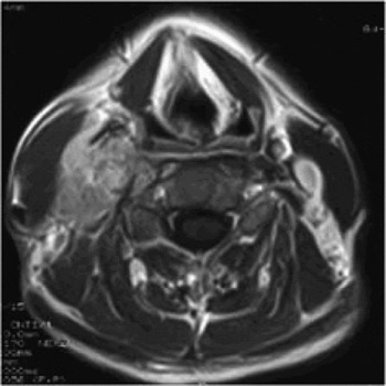
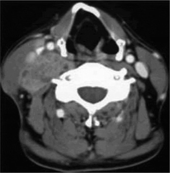
Magnetic resonance imaging showed the mass to have displaced the right common carotid artery and internal jugular vein anterolaterally, and to measure 37 × 51 × 10 mm in size. The tumour was enhanced by gadolinium administration (Figure 1). Computed tomography showed erosion of the C3–C5 vertebral arch (Figure 2).
Axial magnetic resonance imaging scan with T1-weighting and gadolinium administration, showing an enhancing mass which displaces the right carotid artery and internal jugular vein anterolaterally.

Enhanced axial computed tomography scan showing erosion of the C3–C5 vertebral arch.

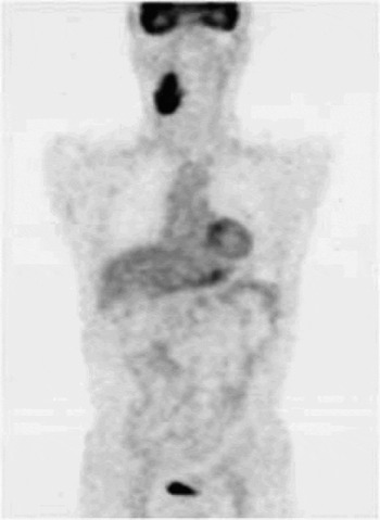
On examination by fluoro-deoxy-glucose positron emission tomography, a high standardised uptake value was noted in the right neck only (Figure 3). An excisional biopsy specimen suggested a diagnosis of either a leiomyosarcoma or myoepithelial carcinoma.
Fluoro-deoxy-glucose positron emission tomography scan showing a high standardised uptake value within tumour in the right neck.

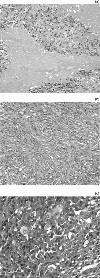
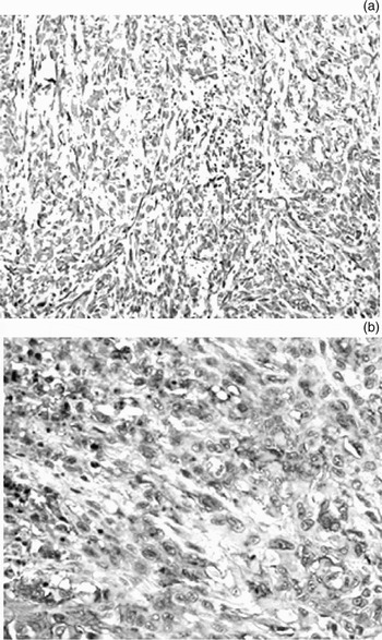
After admission to hospital, the patient underwent a resection of the tumour by means of a conservative neck dissection. Macroscopically, the tumour was yellowish in colour and demonstrated a fibrous consistency. The tumour arose from the scalenous muscle and invaded the soft tissues. The microscopic findings, after haematoxylin and eosin staining, showed a proliferation of epithelioid cells with eosinophilic cytoplasm and atypical spindle cells. Extensive necrosis and fibrous changes were observed. In addition, a few rhabdoid cells were scattered through the specimen (Figure 4).
Photomicrographs of the tumour showing (a) extensive necrosis and the presence of epithelioid cells (H&E; original magnification×100), (b) proliferation of epithelioid cells and atypical spindle cells (H&E; original magnification×200), and (c) a few rhabdoid cells (H&E; original magnification×400).

Immunohistochemical analysis demonstrated positive staining for cytokeratins (CAM5.2) as well as for cytokeratin-multi (AE1/AE3) and epithelial membrane antigen (EMA), vimentin and cluster of differentiation 34 glycoprotein (Figure 5). Staining for S-100 protein was negative. Based on these findings, the tumour was finally diagnosed as an epithelioid sarcoma. Histologically, there was no residual tumour at the surgical margin. However, no safety margin was obtained, so additional radiotherapy (60 Gy) was administered.
Photomicrographs prepared with immunohistochemical staining, showing positive staining for (a) vimentin (original magnification ×100) and (b) cluster of differentiation 34 glycoprotein (original magnification ×400).

Follow-up examinations revealed no local recurrence or distant metastasis for four years.
Discussion
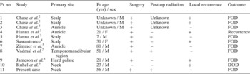
Epithelioid sarcomas are rare, aggressive, malignant neoplasms which were first described by Enzinger in 1970.Reference Enzinger1 Epithelioid sarcoma arises most frequently (58 per cent) in the distal upper extremities and only rarely (1 per cent) in the head and neck.Reference Chase and Enzinger2 Indeed, only 11 epithelioid sarcoma cases, including the current case, have previously been reported in the head and neck. According to previous reports, the primary sites of epithelioid sarcoma in the head and neck are the scalp (n = 4),Reference Chase and Enzinger2–Reference Suwantemee4 the auricle (n = 3),Reference Chase and Enzinger2, Reference Hanna, Kaste, Jenkins, Hewan-Lowe, Spence and Gupta3, Reference Zimmer, Gillman and Barnes5 the neck (n = 2),Reference Kuhel, Monhian, Shanahan and Heier6 the temporo-mandibular region (n = 1)Reference Vadmal, Hajdu and Arlen7 and the hard palate (n = 1).Reference Jameson, Simpson and Towers8
Microscopically, epithelioid sarcoma is characterised by a nodular proliferation of polygonal epithelioid cells with eosinophilic cytoplasm and oval nuclei. Spindle cells are also present and merge with the epithelioid cells. Mitotic figures tend to be common, and intercellular collagen is also prominent in most cases. Calcification, multinucleated giant cells and osseous metaplasia are also observed.Reference Chase and Enzinger2, Reference Batsakis9 Immunohistochemical staining of epithelioid sarcoma demonstrates the expression of vimentin. Most cases demonstrate the presence of cytokeratins, but staining for S-100 protein is usually negative.Reference Batsakis9, Reference Arber, Kandalaft, Mehta and Battifora10
In 1997, Guillou et al. Reference Guillou, Wadden, Coindre, Krausz and Fletcher11 demonstrated a proximal variant of epithelioid sarcoma, which is characterised by the presence of prominent large or rhabdoid cells. Rhabdoid cells are characterised by an eosinophilic cytoplasm containing spheroid perinuclear inclusion bodies. Immunohistochemically, these tumour cells show expression of both cytokeratin and vimentin. The proximal variant of epithelioid sarcoma usually has a more progressive clinical course than classical epithelioid sarcoma.Reference Shiratsuchi, Oshiro, Saito, Itakura, Kinoshita and Tamiya12
It is important to keep in mind that epithelioid sarcoma is a neoplasm with a high rate of local recurrence and metastasis. According to a review of 202 cases by Chase and Enzinger,Reference Chase and Enzinger2 local recurrences were recorded in 77 per cent of cases, and distant metastases to such regions as the lung, lymph nodes or scalp were found in 45 per cent. The overall five- and seven-year survival rates were 59 and 48 per cent, respectively.Reference Schimm and Suit13
Regarding treatment, a wide surgical resection is generally recommended. If the surgical margin is not satisfactory, then post-operative radiation is highly recommended. Schimm and Suit have reported that the rate of local recurrence decreases with the use of radiation therapy.Reference Schimm and Suit13 In a review of previous reports, three patients with epithelioid sarcoma occurring in the head and neck region received radiation and no local recurrence was observed in any of these cases (Table I). These results indicate that the combination of surgery and radiation therapy appears to be the treatment of choice for epithelioid sarcoma of the head and neck.
Review of epithelioid sarcoma originating in the head and neck

Pt = patient; no = number; yrs = years; post-op = post-operative; M = male; F = female; += present; – = absent; FOD = free of disease; DOD = dead of disease