Introduction
The gullet worm, Gongylonema pulchrum Molin, 1857, is a thread-like spirurid nematode found worldwide. A variety of mammals, such as cattle, sheep, goats, camels, pigs, equines, cervids, rodents, bears and primates, serves as the definitive host and dung beetles play a role as the intermediate host (Lichtenfels, Reference Lichtenfels1971; Kirkpatrick et al., Reference Kirkpatrick, Leiby, Abraham and Duffy1986; Anderson, Reference Anderson1992). In Japan, this nematode has been found in cattle (Kudo et al., Reference Kudo, Oyamada and Ito1992; Suzuki et al., Reference Suzuki, Nakamura, Takahashi and Seki1992; Sato, Reference Sato2009), sika deer (Cervus nippon) (Yokohata & Suzuki, Reference Yokohata and Suzuki1993; Kitamura et al., Reference Kitamura, Yokohata, Suzuki and Kamiya1997; Sato, Reference Sato2009), macaques (Macaca fuscata) (Uni et al., Reference Uni, Abe, Harada, Kaneda, Kimata, Abdelmaksoud, Takahashi, Miyashita and Iseki1992, Reference Uni, Kobayashi, Miyashita, Kimura, Kato, Aimi, Kimata, Iseki and Shoho1994), zoo-kept squirrel monkeys (Saimiri boliviensis) (Sato et al., Reference Sato, Une and Takada2005) and humans (Haruki et al., Reference Haruki, Furuya, Saito, Kamiya and Kagei2005; Hara et al., Reference Hara, Nozaki, Makata, Yamada, Yoneda and Takao2010).
The gullet worm occurs in a variety of mammals and the host animals appear to live sympatrically (e.g. deer graze in fields for cattle in some localities). The transmission cycle of the worm may be shared by both domestic and wild animals through accidental ingestion of infected dung beetles or water contaminated with the third-stage larvae (L3) having emerged from dead dung beetles (Kudo et al., Reference Kudo, Oyamada, Okutsu and Kinoshita1996). Furthermore, a high prevalence of the gullet worm in local populations of sika deer in localities where stock farming is prosperous seems to support this view (Yokohata & Suzuki, Reference Yokohata and Suzuki1993).
To elucidate the transmission dynamics of G. pulchrum in domestic and wild ruminants and other wild animals in Japan, we characterized the 18S to 28S ribosomal RNA gene (rDNA) and the cytochrome c oxidase subunit I (COI) of mitochondrial DNA (mtDNA) using specimens from cattle, sika deer, wild boars (Sus scrofa leucomystax), Japanese macaques, a feral Reeves's muntjac (Muntiacus reevesi) and captive squirrel monkeys (Saimiri spp.).
Materials and methods
Collection and examination of cattle and wild animals
Full-length oesophagi of 638 cattle from farms located in 27 prefectures of Japan were collected from a bovine viscera-processing company (Nanko-zouki Co. Ltd, Nanko-minami, Suminoe-ku, Osaka) during February and March 2009. Most of the cattle were Japanese black breed (2.3–2.8 years old) or F1 between Japanese black and Holstein–Friesian breed (1.6–2.5 years old). Full-length oesophagi of two infected cattle of Holstein–Friesian breed from farms in Obihiro, Hokkaido were supplied by Professor H. Furuoka, Obihiro University of Agriculture and Veterinary Medicine. These two cattle were sacrificed on 19 and 26 November 2008. Twenty-seven Honshu sika deer (Cervus nippon centralis) were shot at three localities in Hyogo Prefecture during March 2008 and 12 sika deer (either Kyushu sika deer (C. nippon nippon) or Honshu sika deer) were shot in Kochi Prefecture on Shikoku Island during the hunting season of 2008–2009. In addition, 132 and 24 Honshu sika deer were collected between July 2009 and May 2010 in Hyogo Prefecture and between March 2009 and April 2011 in Wakayama Prefecture, respectively. Furthermore, oesophagi of four Hokkaido sika deer (C. nippon yesoensis) shot in Nakasatsunai Village, Hokkaido in May 2009 and a single dead body of a young Yaku sika deer (C. nippon yakushimae) found on Yakushima Island, Kagoshima Prefecture on 7 August 2010 were examined for the parasite. During the hunting seasons of 2009–2011, 132 wild boars were shot in Hyogo Prefecture. Ten Japanese macaques were collected in Nara Prefecture during November and December 2010 with prefectural permission. Suspensions of lingual scrapings in physiological saline from Bolivian squirrel monkeys (Saimiri boliviensis) and common squirrel monkeys (Saimiri sciureus) kept in two different zoological gardens on Kyushu Island were individually collected according to the method described by Sato et al. (Reference Sato, Une and Takada2005) during the winter of 2009 to 2010. A feral Reeves's muntjac collected on Izu-oshima Island, Tokyo in June 2009 was provided by Dr S. Ozawa, Oshima Island Park, Tokyo Prefectural Office.
The whole oesophagi of cattle, usually ten oesophageal samples per plastic bag, were transported at 4°C to the Laboratory of Parasitology, Yamaguchi University. The oesophagi or carcasses of sika deer, wild boars, Japanese macaques and the feral Reeves's muntjac were sent as frozen materials. In the laboratory, each oesophagus was opened longitudinally and the mucosal layers were peeled from the underlying tissues. The mucosal surface was then carefully checked with the naked eye and, particularly for sika deer samples, worm collections were conducted under a dissection microscope. Individual worms were carefully removed from the oesophageal epithelium using fine forceps and fixed in 70% alcohol or 10% neutral-buffered formalin, except for worms for DNA extraction. Microtubes containing lingual scrapings from the squirrel monkeys were centrifuged and the supernatant was discarded except for 40 μl of physiological saline and precipitate (Sato et al., Reference Sato, Une and Takada2005). The precipitate in the microtube was resuspended and 10–20 μl of the suspension was examined under a light microscope.
Morphological examination of the gullet worm
Specimens fixed in 10% neutral-buffered formalin solution were cleared by the addition of several drops of glycerol. Six female and six male adults from each host category were observed under a light microscope and figures were drawn with the aid of a camera lucida. Measurements were performed on these drawn figures using a digital curvimeter type S (Uchida Yoko, Tokyo, Japan) when necessary. Collected specimens were deposited in the National Museum of Nature and Science, Tokyo, Japan under specimen numbers NSMT-As3501–As3527, As3608–As3639 and As3647–As3671.
DNA extraction, polymerase chain reaction and sequencing
Parasite DNAs were extracted by using single worms each taken from 18 cattle, eight Honshu sika deer, two Kyushu/Honshu sika deer collected in Kochi Prefecture, two Hokkaido sika deer, one Yaku sika deer, four wild boars, two Japanese macaques and one feral Reeves's muntjac, and by using G. pulchrum eggs collected from two colonies of captive squirrel monkeys. In addition, DNA was extracted from two or three worms taken from four Honshu sika deer. DNA extraction was performed using an Illustra™ tissue & cells genomicPrep Mini Spin Kit (GE Healthcare UK, Buckinghamshire, UK) according to the manufacturer's instructions.
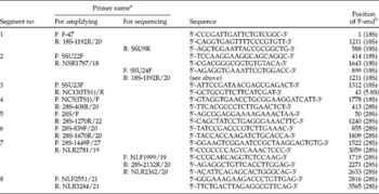
Polymerase chain reaction (PCR) amplification of overlapping rDNA fragments was performed as previously described (Sato et al., Reference Sato, Suzuki, Osanai, Kamiya and Furuoka2006; Halajian et al., Reference Halajian, Eslami, Salehi, Ashraft-Helan and Sato2010) using different primer combinations (table 1). PCR products for sequencing were purified using a High Pure PCR Cleanup Micro Kit (Roche Diagnostics GmbH, Mannheim, Germany). After direct sequencing of PCR amplicons, sequences were assembled manually with the aid of the CLUSTAL W multiple alignment program (Thompson et al., Reference Thompson, Higgins and Gibson1994). For rDNA segments containing internal transcribed spacers (ITS) 1 and 2, the amplicon was cloned into a plasmid vector, pTA2 (Target Clone™; Toyobo, Osaka, Japan), and transformed into Escherichia coli JM109 (Toyobo) according to the manufacturer's instructions. Following propagation, plasmid DNA was extracted using a NucleoSpin® Plasmid kit (Macherey-Nagel GmbH, Düren, Germany) and inserts from multiple independent clones were sequenced using universal M13 forward and reverse primers. Clones were sequenced as follows: six clones for a single G. pulchrum from sika deer, wild boars and Japanese macaques, nine clones for a single G. pulchrum from a steer in Okayama/Tottori Prefecture, 11 clones for a single G. pulchrum from a steer in Oita Prefecture, 11 clones for a single G. pulchrum from a steer in Kagoshima Prefecture and five clones each for G. pulchrum eggs from captive Bolivian and common squirrel monkeys.
Primers used to amplify and sequence eight overlapping segments of rDNA of Gongylonema pulchrum.

a Universal primers used for amplifying and sequencing a certain segment are shown as ‘for amplifying’ and those only for sequencing as ‘for sequencing’. F: forward and R: reverse.
b Relative position of the 5′-end of each primer in G. pulchrum rDNA sequence (DDBJ/EMBL/GenBank accession no. AB495389). The 5′-end of F-47 primer is considered as the beginning of 18S rDNA here.
The COI region of G. pulchrum mtDNA was amplified by a primer pair of BpCoxI-F1 (5′-TTTGGTCATCCTGAGGTTTATATT-3′) and BpCoxI-R1 (5′-ATGAAAATGTCTAACTACATAATAAGTATC-3′). PCRs were conducted in a thermal cycler using the following cycling protocol: 3 min at 94°C, followed by 40 cycles at 94°C for 45 s, 52°C for 1 min and 72°C for 1 min, then a final extension at 72°C for 7 min. The number of worms examined for COI included 18 from cattle in Japan, two from cattle in Iran (Halajian et al., Reference Halajian, Eslami, Salehi, Ashraft-Helan and Sato2010), 14 from Honshu sika deer, two from Kyushu/Honshu sika deer collected in Kochi Prefecture, two from Hokkaido sika deer, five from wild boars, four from Japanese macaques and two from different squirrel monkey colonies. Relationships of different haplotypes based on 369-bp long COI nucleotide sequences were visualized using an automated haplotype network layout and visualization software, HapStar, downloaded from http://fo.am/hapstar (Teacher & Griffiths, Reference Teacher and Griffiths2011).
Nucleotide sequences reported in the present study are available from the DDBJ/EMBL/GenBank databases under the accession numbers AB495389–AB495402, AB513707–AB513730, AB645978–AB646106 and AB646110–AB646129.
Results
Infection levels of G. pulchrum in different hosts
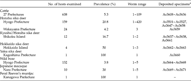
Prevalences of adult G. pulchrum in different hosts are summarized in table 2. Of 638 oesophagi from cattle, 34 had G. pulchrum embedded in the mucosa. These infected cattle occurred in 12 of 27 prefectures examined. A single worm infection was detected in 17 oesophagi and two worms were recovered from five oesophagi. From the remaining 12 oesophagi, more than three worms were found and the maximum recovery was 109 (58 females and 51 males) from a steer of Japanese black breed. Up to 33 of 159 Honshu sika deer shot in Hyogo Prefecture were infected with G. pulchrum. The prevalence of G. pulchrum varied with locality, with all 20 deer shot on Awaji Island being infected, whereas in other localities in Hyogo Prefecture, infections were rare or absent. Five individuals of the 33 infected deer were parasitized with 134–420 gullet worms and the mucosal surface of the oesophagi had grossly visible roughness through the complete length of the organ. The remaining deer were infected with 1–38 gullet worms and the oesophageal mucosa had no gross lesions except for the intramucosal localization of the parasites. Of 24 Honshu sika deer collected in Wakayama Prefecture, one harboured three gullet worms in its oesophagus. From two of 12 Kyushu/Honshu sika deer shot in Kochi Prefecture on Shikoku Island, one and two gullet worms were recovered. From two of four Hokkaido sika deer, one and three gullet worms were recovered. One gullet worm was collected from the Yaku sika deer. Of 132 wild boars shot in Hyogo Prefecture, five individuals harboured 1–5 gullet worms. Of 10 Japanese macaques collected in Nara Prefecture, three animals were infected with one, two or five gullet worms. Microscopic examination of lingual scrapings revealed Gongylonema eggs in 14.2% (16/113) or 12.5% (12/96) of captive Bolivian squirrel monkeys and in 45.7% (21/46) or 46.0% (23/50) of captive common squirrel monkeys in 2009 and 2010, respectively. From the oesophagus of the feral Reeves's muntjac, only a single juvenile worm was found.
Recovery of Gongylonema pulchrum from the oesophagus of a variety of hosts in Japan.
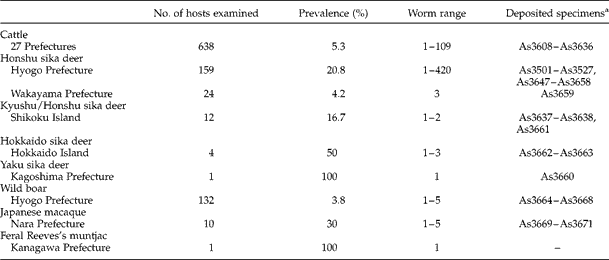
a Specimen ID numbers at the National Museum of Nature and Science, Tokyo.
Measurements of G. pulchrum from sika deer and cattle
Specimens collected from the oesophageal mucosa were characterized by verruciform protrusions on the cuticle near the anterior end. Asymmetrical caudal alae with 4–6 pairs each of pre- and post-anal papillae and distinct lengths of left and right spicules were present in the posterior end of male worms. Consistently, smaller sized worms were collected from sika deer compared to those from cattle. For example, worm length of male worms from sika deer, 21.2–26.9 mm (average 24.0 mm, n= 6) versus those from cattle, 30.7–44.9 mm (average 36.7 mm, n= 6), and worm length of female worms from sika deer, 35.7–51.6 mm (average 43.6 mm, n= 6) versus those from cattle, 67.9–85.1 mm (average 78.5 mm, n= 6). Several other measurements of the worms from sika deer and cattle were clearly different (data not shown) and were in accordance with previous reports for parasites collected from each host species (Kudo et al., Reference Kudo, Oyamada and Ito1992; Yokohata & Suzuki, Reference Yokohata and Suzuki1993).
rDNA of G. pulchrum from different hosts
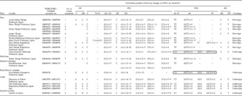
Approximately 6100-bp-long sequences from 18S to 28S rDNA of G. pulchrum from different hosts and localities were compared to identify any unique nucleotide changes related to their origin. The rDNA sequences of G. pulchrum were generally well conserved, with few nucleotide changes detected in 18S, 5.8S and 28S regardless of their origin. Our analysis revealed that there were some unique nucleotide changes in the rDNA of G. pulchrum from cattle and from other origins, which were mainly observed in ITS1 and ITS2 (table 3). The ITS1 and ITS2 regions of G. pulchrum had several repeat units, such as ‘GA’, ‘A’, ‘CA’ and ‘GCT’, in certain regions of their sequences. Sequence analyses of several clones of an amplicon of the ITS region for an individual worm showed that such repeat units were often repeated erratically. However, we did identify a few critical differences in the rDNA nucleotide sequences of gullet worms collected from cattle and sika deer. Specifically, cattle-derived worm genotype (termed ‘cattle-type’) variants were found in the worms from cattle, zoo-kept squirrel monkeys, the feral Reeves's muntjac and Hokkaido sika deer, whereas deer-derived worm genotype (termed ‘deer-type’) variants were found in the worms from the majority of sika deer, wild boars and Japanese macaques (table 3).
Nucleotide changes observed in rDNA of Gongylonema pulchrum of different origins.

* Nucleotide position is expressed relative to the rDNA sequence of G. pulchrum collected from a deer caught on Awaji Island(DDBJ/EMBL/GenBank accession no. AB495394). Gaps are indicated by ‘–’ and blanks indicate no data.
COI of G. pulchrum from different hosts
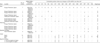
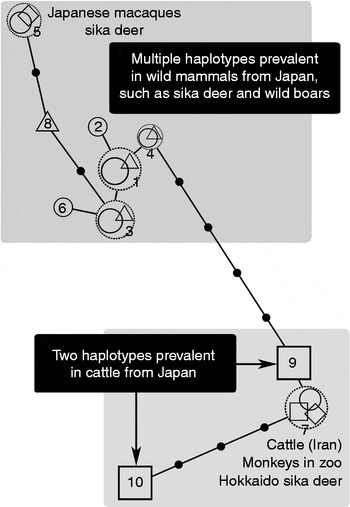
A partial COI region, 369 bp in length, of G. pulchrum mtDNA was successfully sequenced and compared (table 4). The COI nucleotide sequences of G. pulchrum from different origins were divided into ten haplotypes (table 4 and fig. 1). Seven COI haplotypes (#1–#6 and #8) were found in G. pulchrum from a majority of sika deer and wild boars examined in the present study. An identical COI haplotype (#5) was shared by a single sika deer from Wakayama Prefecture and Japanese macaques from Nara Prefecture.
Nucleotide substitutions observed in COI mtDNA of Gongylonema pulchrum of different origins.

* Nucleotide position is expressed relative to the 5′-end of COI sequence of G. pulchrum collected from a deer caught on Awaji Island, Hyogo Prefecture, Japan (DDBJ/EMBL/GenBank accession no. AB513724). Dots denote an identical base to that of G. pulchrum collected from the deer in Hyogo.
Relationships of COI haplotypes (#1–#10, see table 4) of Gongylonema pulchrum found in sika deer (open circle), wild boars (open triangle), Japanese macaques (open diamond), cattle (open square) and captive squirrel monkeys (open diamond). Sizes of symbols are arbitrary, just showing higher frequency in larger symbols. Symbols within a dotted circle denote the same haplotype.

Three COI haplotypes (#7, #9 and #10) were differentiated in G. pulchrum collected from cattle. Haplotypes #9 and #10, differing at five base positions along their 369-bp length, were found in G. pulchrum collected from cattle reared in nine and six prefectures in Japan, respectively. Both COI haplotypes were distributed randomly in Japan, regardless of cattle breed or region. Except for one nucleotide, the 369-bp long COI sequence of G. pulchrum from cattle in Iran was identical to that of haplotype #9 G. pulchrum collected from cattle in Japan. This haplotype (#7) was also found in G. pulchrum collected from Hokkaido sika deer and G. pulchrum eggs from two open-spaced colonies of Bolivian squirrel monkeys and common squirrel monkeys kept in zoological gardens. The amino acid sequences of G. pulchrum COI obtained in this study were identical except in two worms, one from a Kyushu/Honshu sika deer shot in Kochi Prefecture (at the 26th amino acid position, alanine was substituted by threonine) and the other from a wild boar shot in Hyogo Prefecture (at the 57th amino acid position, arginine was substituted by glycine). The COI sequences of the gullet worm from the feral Reeves's muntjac and from the Yaku sika deer were not analysed due to unsuccessful PCR amplification of the gene or exhaustion of the samples.
Discussion
Suzuki et al. (Reference Suzuki, Nakamura, Takahashi and Seki1992) reported different monthly prevalences of G. pulchrum infection in cattle in Hokkaido, Japan, ranging from 4.9 to 13.2%, and Kudo et al. (Reference Kudo, Oyamada and Ito1992) reported different local prevalences of the infection in cattle from the northern part of Japan, ranging from 0 to 24.0%. The age of the examined cattle appears to affect the prevalence of G. pulchrum infection, with Kudo et al. (Reference Kudo, Oyamada and Ito1992) reporting a prevalence of 3.3% (8/242) in cattle aged 2 years or younger, 17.0% (40/235) in 3-year-old cattle and 60.6% (57/94) in cattle aged 4 years or older. Accordingly, the distribution of G. pulchrum in cattle in Japan might not be limited to 12 of the 27 prefectures examined in this study, since only cattle younger than 3 years old were examined and the number of cattle examined from each prefecture was relatively low. However, this study demonstrates that G. pulchrum occurs widely in cattle in Japan, in addition to the northern part of Japan, such as Aomori and Hokkaido (Kudo et al., Reference Kudo, Oyamada and Ito1992; Suzuki et al., Reference Suzuki, Nakamura, Takahashi and Seki1992). Prevalences of G. pulchrum in sika deer were variable in different localities, as suggested previously (Yokohata & Suzuki, Reference Yokohata and Suzuki1993). Similarly, G. pulchrum was found in only five of a total of 132 wild boars, with all five positive wild boars coming from one locality. High prevalences of G. pulchrum infection in pigs and wild boars have been reported in the USA (Zinter & Migaki, Reference Zinter and Migaki1970; Smith et al., Reference Smith, Davidson, Nettles and Gerrish1982) and Iran (Eslami & Farsad-Hamdl, Reference Eslami and Farsad-Hamdl1992). Coombs & Springer (Reference Coombs and Springer1974) reported a higher detection rate of the gullet worm in the tongue (6/9) than in the oesophagus (1/10). As we examined only the oesophagi of wild boars, then it is highly possible that the results presented here are an underestimation of the prevalence of G. pulchrum in wild boar populations in Japan. In contrast, squirrel monkeys in captivity were infected at a high prevalence, as reported previously (Sato et al., Reference Sato, Une and Takada2005), even though the lingual scraping technique detects the minimal number of infected monkeys when considering that parasitism of G. pulchrum in oesophagi could not be detected by this technique.
As mentioned previously, G. pulchrum is found in a variety of mammals, such as cattle, sheep, goats, camels, pigs, equines, cervids, rodents, bears and primates, that serve as the definitive hosts (Lichtenfels, Reference Lichtenfels1971; Kirkpatrick et al., Reference Kirkpatrick, Leiby, Abraham and Duffy1986; Anderson, Reference Anderson1992). Furthermore, successful experimental infections of G. pulchrum L3 of ruminant origin have been reported in rabbits, guinea-pigs and rats (Lucker, Reference Lucker1932; Alicata, Reference Alicata1935; Kudo et al., Reference Kudo, Koreguchi, Ikadai and Oyamada2003, Reference Kudo, Kuratomi, Hatada, Ikadai and Oyamada2005). Based on the apparent low host specificity of G. pulchrum, it is reasonable to speculate that the transmission cycle of the gullet worm may be shared by domestic and wild mammals through accidental ingestion of infected dung beetles serving as an intermediate host. However, the present analyses demonstrate that the rDNA genotypes as well as the COI mtDNA haplotypes of G. pulchrum prevalent in wild animals (including sika deer, wild boars and Japanese macaques) and domestic/captive mammals in Japan are clearly different (tables 3 and 4; fig. 1). This finding indicates that at present G. pulchrum has multiple origins in Japan and its transmission cycle is different between wild and domestic animals. Within the wildlife in Japan, sika deer and Japanese macaques are two known major hosts for the gullet worm (Uni et al., Reference Uni, Abe, Harada, Kaneda, Kimata, Abdelmaksoud, Takahashi, Miyashita and Iseki1992, Reference Uni, Kobayashi, Miyashita, Kimura, Kato, Aimi, Kimata, Iseki and Shoho1994; Yokohata & Suzuki, Reference Yokohata and Suzuki1993). As we examined G. pulchrum from a limited number of Japanese macaques at a single locality, the significance of a special COI haplotype (#5; see fig. 1) of the gullet worms from macaques is unclear, i.e. it is not known whether this haplotype is prevalent in Japanese macaques or multiple haplotypes are shared by macaques and sika deer. Since the rDNA genotype and COI haplotypes of G. pulchrum from wild boars were essentially similar to those found in sika deer and the infection in wild boars was relatively low, these two wild mammals may share the same group of parasites. The rDNA genotype and COI haplotype of G. pulchrum in captive squirrel monkeys appear to be identical to not only those from cattle in Iran, but also those from Hokkaido sika deer. These alien monkeys and/or other captive animals in zoos might introduce the original infection when imported, maintaining its life cycle within the zoo facility. Alternatively, squirrel monkeys and/or other captive animals may newly acquire the infection in the zoo facility.
The sika deer in Japan have been divided into two distinct lineages, the northern and southern groups, by nucleotide sequencing of the D-loop region of mtDNA (Tamate et al., Reference Tamate, Tatsuzawa, Suda, Izawa, Doi, Sunagawa, Miyahira and Tado1998; Nagata et al., Reference Nagata, Masuda, Tamate, Hamasaki, Ochiai, Asada, Tatsuzawa, Suda, Tado and Yoshida1999; Nabata et al., Reference Nabata, Masuda and Takahashi2004). The northern Japan group is distributed from Hokkaido Island to Hyogo Prefecture on Honshu Island, whereas the southern Japan group is in the westernmost part of Honshu Island, Kyushu Island and southward. On the fourth mainland of Japan, Shikoku Island, the distribution of the two sika deer groups is intermingled (Yamada et al., Reference Yamada, Hosoi, Tamate, Nagata, Tatsuzawa, Tado and Ozawa2006). As far as this study's analyses are concerned, we could not find any G. pulchrum genotype or haplotype unique to each sika deer group. Interestingly, however, Hokkaido sika deer were found to be parasitized by G. pulchrum having an rDNA cattle-genotype and #7 COI haplotype. This finding appears to be contradictory to the view mentioned above. Hokkaido sika deer have a history of drastic population decline/near extinction occurring more than 100 years ago as a result of the overexploitation and overharvesting of deer, beginning in 1869, and heavy snowfalls during the winters of 1879 and 1903 (Inukai, Reference Inukai1952; Nabata et al., Reference Nabata, Masuda and Takahashi2004). This is reflected in a reduced genetic variation of current Hokkaido sika deer populations (Nagata et al., Reference Nagata, Masuda, Tamate, Hamasaki, Ochiai, Asada, Tatsuzawa, Suda, Tado and Yoshida1999; Nabata et al., Reference Nabata, Masuda and Takahashi2004). It could be speculated that this severe population decline history of host sika deer induced the extinction of their original G. pulchrum, which was followed by recent re-acquisition of the parasite common in the Holstein–Friesian breed in dairy farms popular in Hokkaido. Indeed, Hokkaido sika deer frequently graze in fields for dairy cattle of the Holstein–Friesian breed. This hypothesis would also account for the scarcity of G. pulchrum parasitism in Hokkaido sika deer reported by Kitamura et al. (Reference Kitamura, Yokohata, Suzuki and Kamiya1997).
When accidental G. pulchrum infection is seen in humans, only a single worm or a fragment of the parasite is available for identification of the causative species (Crusz & Sivalingam, Reference Crusz and Sivalingam1950; Eberhard & Busillo, Reference Eberhard and Busillo1999; Wilson et al., Reference Wilson, Lorente, Allen and Eberhard2001; Haruki et al., Reference Haruki, Furuya, Saito, Kamiya and Kagei2005). Additionally, a stunted development of the parasite in non-bovine hosts (Schwartz & Lucker, Reference Schwartz and Lucker1931; Kirkpatrick et al., Reference Kirkpatrick, Leiby, Abraham and Duffy1986; Yokohata & Suzuki, Reference Yokohata and Suzuki1993; Kudo et al., Reference Kudo, Kuratomi, Hatada, Ikadai and Oyamada2005; Sato et al., Reference Sato, Une and Takada2005) and its wide variation of morphological characters dependent on hosts (Lucker, Reference Lucker1932; Lichtenfels, Reference Lichtenfels1971) makes its identification more difficult. Therefore, a molecular technique-based diagnosis of the species would be advantageous and reliable. This rDNA-based diagnosis of the species may facilitate the identification of G. pulchrum isolated from a variety of animal sources, even under the limitation of sampling for morphological species diagnosis. Furthermore, as shown in this study, such molecular technique-based diagnosis could speculate the possible source of the gullet worm, maintained in either a domestic or sylvatic transmission cycle, at least in Japan.
Acknowledgements
This study was supported in part by research grants from the Morinaga Service Foundation, Tokyo, Japan (2008) and grants-in-aid from the Japan Society for Promotion of Sciences (nos. 19580355, 20570090 and 22580349). We are greatly indebted to Katsutoshi Nishida and Kazumasa Nishida, Nanko-zouki Co. Ltd, for their assistance with the collection of bovine oesophagi.