Introduction
Malignant triton tumour is a subtype of malignant schwannoma comprising mature rhabdomyoblasts scattered throughout sheets of schwannoma cells.Reference Woodruff, Chernik, Smith, Millett and Foote1 The tumour behaves more like a malignant schwannoma than a rhabdomyosarcoma, and the prognosis for long-term survival is grim. Nearly one-third of these tumours develop in the head and neck region. Malignant triton tumour arises sporadically, as well as in association with neurofibromatosis type one,Reference Yakulis, Manack and Murphy2 and usually affects individuals under the age of 35 years.Reference Brooks, Freeman and Enterline3 Rare cases have been reported in the nasal cavities and paranasal sinuses. In this report, we present a unique case of malignant triton tumour of the maxillary and ethmoid sinuses.
Case report
A 47-year-old woman was referred to our hospital in December 2006 complaining of painful swelling of the upper molar area accompanied by progressive facial swelling, of four months' duration, and a three-month history of nasal obstruction, localised facial tenderness and headache, all on the right side. Her past medical history was significant for hypertension over the past five years and type B hepatitis for the past 10 years. The patient reported no vision-related complaints, and the family history was negative for possible congenital lesions.
Physical examination revealed pressure pain on the right cheek. The neck was clinically negative.
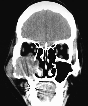
Investigations, including a computed tomography scan of the thorax, disclosed no evidence of metastatic disease. Nasal endoscopy revealed a yellow, mucoid secretion in the right nasal cavity, expansion of the inferior part of the lateral nasal wall, and a yellow, mucopurulent secretion in the middle nasal meatus which was difficult to suction. Computed tomography scans revealed a tumour mass in the right maxillary sinus, with destruction of the anterolateral maxillary sinus bony wall, the orbital floor and extension into the pterygopalatine and pterygomaxillary fossae (Figure 1).
Coronal computed tomography scan showing a tumour mass in the right maxillary sinus with destruction of the anterolateral maxillary sinus bony wall, the orbital floor and extension into the pterygopalatine and pterygomaxillary fossae.

Under general anaesthesia, a lateral rhinotomy was performed. This confirmed that the tumour occupied the entire sinonasal cavity, causing bone destruction of the lateral wall of the right maxillary sinus and the floor of the orbit, with extension into the pterygopalatine and pterygomaxillary fossae. The tumour mass measured 6 × 3 × 4 cm in size and was dumbbell shaped with a smooth surface and clear margins. Complete resection of the lesion and the right lateral nasal wall was performed, from the anterior tip of the turbinates to the eustachian tube and from the cribriform plate and ethmoid roof to the floor of the nose.
On macroscopic examination, the tumour appeared encapsulated and firm and measured 6 cm in diameter. The cut surface showed microcystic, haemorrhagic and necrotic areas.
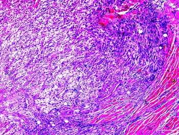
Microscopic examination of the tumour showed a circumscribed, oval lesion composed of interlacing fascicles of compact spindle cells, which was clearly seen to be infiltrating facial striated muscle. Numerous extremely large pleomorphic cells with occasional clear intranuclear cytoplasmic inclusions and abundant eosinophilic cytoplasm were also present (Figure 2).
Photomicrograph showing the tumour as a circumscribed, oval lesion composed of interlacing fascicles of compact spindle cells, which is clearly seen to be infiltrating facial striated muscle. Numerous extremely large pleomorphic cells with occasional clear intranuclear cytoplasmic inclusions and abundant eosinophilic cytoplasm are also present (H&E; original magnification indicated by scale bar corresponding to 100 µm).

Immunostaining was performed on the tumour, including stains for S-100, pan-actin and desmin. The majority of spindle cells exhibited intense expression of S-100. The scattered cells expressed pan muscle actin and desmin. Positivity for α-smooth muscle actin was only observed in association with the walls of small blood vessels.
The morphology and biphenotypic expression of the nerve sheath and skeletal muscle markers confirmed the diagnosis of malignant triton tumour. The final clinical and radiological staging for this tumour was T3 N0 M0 (based on the Internationa Union Against Cancer (UICC) staging system for malignant neoplasms).
Post-operative recovery was uneventful. Post-operative radiation therapy was conducted four weeks after the operation. The patient was reviewed at weekly intervals for the first month and subsequently every two months. Twelve months after the operation, there were no signs of either local tumour recurrence or distant metastasis.
Discussion
Malignant triton tumour was first described in 1932 by Masson and Martin. The term ‘triton tumour’ was first used by Woodruff in 1973. This term was derived from the triton salamander, which is able to regenerate limbs, with both bone and muscle components, if the cut end of the sciatic nerve is inserted into the soft tissues of its back.
Malignant triton tumour is extremely rare, with less than 100 cases documented worldwide. Although approximately 20 per cent of the total cases are reported to occur in the head and neck, there are only two previously reported cases of malignant triton tumour in the sinonasal area.Reference Bhatt, Graeme-Cook, Joseph and Pilch4–Reference Victoria, McCulloch, Callaghan and Bauman7
It is very difficult to make an accurate diagnosis of malignant triton tumour because of a lack of standardised diagnostic criteria. In the current case, nerve sheath differentiation was confirmed by S-100 protein positivity, while rhabdomyoblastic differentiation was confirmed by positivity to desmin. Leiomyosarcoma was also considered in the differential diagnosis, but there was neither morphological support (due to lack of blunt-ended nuclei in the spindle cells) nor immunohistochemical support for this diagnosis. Chromosomal abnormalities have been reported in several patients with malignant triton tumour.Reference Travis, Sandberg, Neff and Bridge8, Reference McComb, McComb, DeBoer, Neff and Bridge9 Cytogenetic testing was not performed in our patient.
Treatment of malignant triton tumour should include complete resection of the tumour, as many times as necessary, or tumour reduction surgery, followed by adjuvant radiotherapy to prevent local recurrence and metastasis. Adjuvant chemotherapy has not been shown to be effective in this tumour.Reference Lang-Lazdunski, Pons and Jancovici10, Reference Bose, Deodhar and Duncan11 Complete resection of malignant triton tumour with or without adjuvant therapy tends to lead to improved survival, compared with incomplete resection with or without adjuvant therapy. Although little is known about the radiosensitivity of malignant triton tumour, and cytotoxic drug therapy has been found to have no value, Reference Dewit, Albus-Lutter, de Jong and Voute12 adjuvant radiation therapy and chemotherapy may play important roles in managing this tumour.
Conclusion
We have presented a case of malignant triton tumour in a rare location, with characteristic radiological, histological and immunohistochemical features. Malignant triton tumours of the paranasal sinuses are rare and difficult to diagnose. Otolaryngologists should be aware of this tumour, which should be included in the differential diagnosis of malignant lesions involving the sinonasal tract.