INTRODUCTION
The family Rhinobatidae contains 45 species distributed mostly in the tropical coastal waters of the Atlantic, Indian and Pacific Oceans (Compagno, Reference Compagno and Hamlett2005), and they are important components of fishing activities in the Atlantic–Mediterranean region (Chaves et al., Reference Chaves, Cova-Grando and Calluf2003; Seck et al., Reference Seck, Diatta, Diop, Guélorget, Reynaud and Capapé2004; Vooren et al., Reference Vooren, Lessa, Klippel, Vooren and Klippel2005; Costa & Chaves, Reference Costa and Chaves2006). In southern Brazil the genus Rhinobatos Linck, 1990 is represented by two species, R. horkelli Müeller & Henle, 1841 and R. percellens (Walbaum, 1752) (Vooren et al., Reference Vooren, Lessa, Klippel, Vooren and Klippel2005). The guitarfish R. percellens is benthonic and inhabits coastal waters from the Caribe to north of Argentina (Menezes et al., Reference Menezes, Buckup, Figueiredo and Moura2003), being very common at Santa Catarina and Paraná (Chaves et al., Reference Chaves, Cova-Grando and Calluf2003; Costa & Chaves, Reference Costa and Chaves2006), where females approach the shore because nursery areas are probably situated there (Lessa, Reference Lessa, Vooren and Lahaye1986; Batista, Reference Batista1987, Reference Batista1991). The objective of the present work was to record and describe an anomalous embryo of R. percellens in Brazilian waters.
MATERIALS AND METHODS
Samples were collected between July 1991 and June 1992 by bottom trawling. Twelve operations of five minutes performed monthly from using bottom trawl nets (20-mm mesh size) in a sand–muddy tidal flat in Paranaguá Bay, southern Brazil (25º30′s 48º13′W). Ten female guitarfish were captured.
A 550 mm total length pregnant female collected on 20 January 1992 showed five developing embryos, one of them with morphological deformities. The five embryos were removed, measured and weighed after blotting. Morphometric measurements were taken to the nearest 0.1 mm according to Compagno (Reference Compagno1984) and McEachran & Carvalho (Reference McEachran, Carvalho, Carpenter and Niem1999). The embryos were deposited in the collection of the Museu de História Natural Capão da Imbuia (MHNCI).
RESULTS
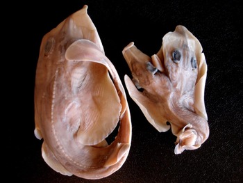
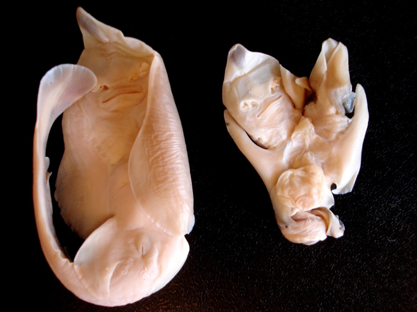
The anomalous embryo presented two well-defined heads, both of them fused in the vertebral column. The lateral margins of the pectoral fins are detached from the heads, therefore the disc is incomplete in both heads (Figure 1). The right head (ventral view) is deformed, and the lateral margins of the rostrum are fused in the ventral surface, consequently the mouth opening is not visible (Figure 2). Eyes seemed to be well-formed, but dorsal, caudal and anal fins were rolled and deformed. Only one vitelinic sac was observed (Figure 2).
Dorsal view of the normal (left) and anomalous (right) embryos of Rhinobatos percellens.

Ventral view of the normal (left) and anomalous (right) embryos of Rhinobatos percellens.

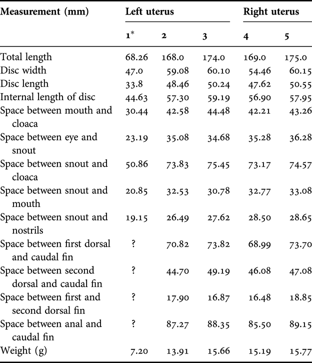
The other embryos were in perfect condition, ranging from 168 to 175 mm total length. The largest embryos displayed newborn features, with vitelinic sac almost totally absorbed, probably reaching the full-term length. Morphometric data of the deformed embryo showed divergences from normal embryos (Table 1). Some measurements were not recorded because of the atypical morphological formation of the anomalous embryo.
Measurements (mm) of four normal embryos and one anomalous embryo (*) of Rhinobatos percellens.

DISCUSSION
Several cases of anomalies in elasmobranch embryos have been recorded, such as pectoral fins not fused to the head (Templeman, Reference Templeman1965; Rosa et al., Reference Rosa, Gomes and Gadig1996), spinal and fin deformities in the axial skeletons (Springer, Reference Springer1960; Du Buit, Reference Du Buit1978; Mancini et al., Reference Mancini, Casas and Amorim2006), skeletal deformities (Heupel et al., Reference Heupel, Simpfendorfer and Bennett1999) and albinism (Saïdi et al., Reference Saïdi, Bradaï, Marouani, Guélorget and Capapé2006). Despite the fact that several causes may be associated with the deformations, cases of anomalies in elasmobranchs generally do not present known causes.
Rosa et al. (Reference Rosa, Gomes and Gadig1996) observed a freshwater stingray Potamotrygon motoro with both pectoral fins anteriorly detached from the head, with its anterior tips barely reaching the level of the rostrum. As the separation of pectoral fins and the head are a normal characteristic in the first stages of a ray's embryonic development (Bigelow & Schroeder, Reference Bigelow and Schoroeder1953), the deformations assigned were probably a congenital abnormality. According to Heupel et al. (Reference Heupel, Simpfendorfer and Bennett1999), besides congenital abnormality, deformations may be associated with several other factors, such as parasitic infection, arthritis, injury, tumours or malnutrition, the last one suggested as a cause of vertebral deformities in teleosts.
Because of the fact that only one anomalous embryo of R. percelles was registered, the cause of the deformities may be associated with malnutrition during the gestation period; however, environmental causes and genetic anomalies cannot be discarded (Mancini et al., Reference Mancini, Casas and Amorim2006).
Despite the fact that teratogeny cases and skeletal deformities have already been reported for elasmobranchs (Gudger, Reference Gudger1933; Babel, Reference Babel1967; Rosa et al., Reference Rosa, Gomes and Gadig1996; Heupel et al., Reference Heupel, Simpfendorfer and Bennett1999; Thornburn & Morgan, Reference Thornburn and Morgan2004; Saïdi et al., Reference Saïdi, Bradaï, Marouani, Guélorget and Capapé2006; Escobar-Sánchez et al., Reference Escobar-Sánchez, Galván-Magaña, Downton-Hoffmann, Carrera-Fernández and Alatorre-Ramírez2008), this malformation is described here as the first occurrence for Rhinobatos percellens. We believe that the morphological abnormalities would prevent this embryo from surviving in the aquatic environment.