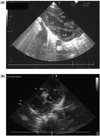
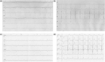
Transcatheter ventricular septal defect closure is successful in >95% of patients and has been widely adopted. However, both early- and late-onset atrioventricular may occur, particularly with perimembranous defects.Reference Carminati, Butera and Chessa1 Here, we document late-onset left bundle branch block after transcatheter closure of a congenital muscular ventricular septal defect. An infant with a body weight of 5.4 kilograms presented with feeding difficulties, sweating and tachypnoea at 6 months of age. Despite treatment with digoxin, furosemide, spironolactone, and propranolol he remained symptomatic. Primary device closure of a 5-millimetre muscular outlet defect (Fig 1a; LV = left ventricle; RV = right ventricle; Ao = aorta; LA = left atrium) was performed using a 6-millimetre muscular ventricular septal defect occluder (Amplatzer, AGA Medical Corporation, United States of America; Fig 1b, arrow) at 9 months of age and 7 kilograms body weight. The procedure was uneventful, and routine follow-up, including electrocardiogram (Fig 2a and b), recorded at 50-millimetre per second, was normal until 3 years later. At that time, the electrocardiogram showed a new complete left bundle branch block (Fig 2c and d). Given the possibility of late conduction defects, as illustrated here, caution should be exercised even when closing muscular ventricular septal defects, particularly if the device will be positioned close to the conduction system. Life-long clinical follow-up is indicated in all patients.
(a) Cross-sectional echocardiogram in the long-axis view showing the muscular outlet defect between the measuring calipers; AO = aorta; LA = left atrium; LV = left ventricle; RV = right ventricle. (b) Four-chamber view showing the Amplatzer muscular ventricular septal defect occluder (arrow) in the correct position; LV = left ventricle; RV = right ventricle.

(a and b) Normal 12-lead electrocardiogram 18 months after closure of the muscular defect; recording speed is 50 millimetres per second. (c and d) Twelve-lead electrocardiogram showing complete left bundle branch block 3 years after closure of the defect; recording speed is 50 millimetres per second.