Implications
The pig model is increasingly used in the field of neuroscience because of the similarities of its brain with human. Indeed, aside from the rodent model there is a critical need for a large animal model ethically acceptable, i.e. excluding non-human primates. In this review we present some experiments dealing with the various procedures achievable in the pig model ranging from image-guided brain surgery to functional brain imaging studies. The recent advances in functional imaging data processing pointed out our partial knowledge of pig brain neuro-anatomy and the requirement for a digitalized atlas matching MRI (magnetic resonance imaging) and histological resources.
Introduction
Swine have been used extensively as a model of human in biomedical researches such as cardiovascular, metabolic and transplantation (Phillips et al., Reference Phillips, Panepinto, Spangler and Westmoreland1982; Larsen and Rolin, Reference Larsen and Rolin2004; Imai et al., Reference Imai, Konno, Nakamura, Shimizu, Kubota, Seki, Honda, Tomizawa, Tanaka, Hata and Saito2006; Groth, Reference Groth2007). In the last decade, an increasing number of studies in the field of neuroscience has been reported (Lind et al., Reference Lind, Moustgaard, Jelsing, Vajta, Cumming and Hansen2007). In addition to the study of the action or metabolism of various pharmacological drugs or neurotransmitters (Bhalla et al., Reference Bhalla, Saxena and Sharma2002; Rosa-Neto et al., Reference Rosa-Neto, Doudet and Cumming2004a; Lind et al., Reference Lind, Olsen, Moustgaard, Jensen, Jakobsen, Hansen, Arnfred, Hemmingsen, Gjedde and Cumming2005b; Minuzzi et al., Reference Minuzzi, Nomikos, Wade, Jensen, Olsen and Cumming2005), pathological models dealing with traumatic brain injury (Grate et al., Reference Grate, Golden, Hoopes, Hunter and Duhaime2003), stroke (Sakoh et al., Reference Sakoh, Ostergaard, Gjedde, Rohl, Vestergaard-Poulsen, Smith, Le Bihan, Sakaki and Gyldensted2001) or neurodegenerative diseases such as Parkinsonism induced by 1-methyl-4-phenyl-1,2,3,6-tetrahydropyridine hydrochloride (MPTP) poisoning (Danielsen et al., Reference Danielsen, Cumming, Andersen, Bender, Brevig, Falborg, Gee, Gillings, Hansen, Hermansen, Johansen, Johansen, Dahl-Jorgensen, Jorgensen, Meyer, Munk, Pedersen, Poulsen, Rodell, Sakoh, Simonsen, Smith, Sorensen, Ostergard, Zimmer and Gjedde2000; Cumming et al., Reference Cumming, Danielsen, Vafaee, Falborg, Steffensen, Sorensen, Gillings, Bender, Marthi, Andersen, Munk, Smith, Moller and Gjedde2001; Dall et al., Reference Dall, Danielsen, Sorensen, Andersen, Moller, Zimmer, Gjedde and Cumming2002) have also been developed.
The major benefit of the swine for neuroscience research remains the size of its brain. It is large enough to allow evoked potential (EP) recordings, neurosurgery and conventional imaging in living animals. Furthermore, the pig has cerebral structures common to other mammalian species and, with relative well-defined cerebral circumvolutions, its brain appears to be comparable to human in terms of anatomy, histology and vascularization (for review: Lind et al. (Reference Lind, Moustgaard, Jelsing, Vajta, Cumming and Hansen2007)). This represents an undeniable advantage compared to the rodent model for multimodality imaging using magnetic resonance imaging (MRI), computed tomography (CT) or positron emission tomography (PET).
In this review, we will present the particularities of the pig brain in anatomical and functional terms with specific reference to primates and rodents. An overview of former works describing the use of functional imaging and neuronavigation in the pig model will present the challenge in using the porcine model compared to other animal models. Finally, we will set in perspective work currently ongoing in our laboratory as examples of the capabilities of the pig model in the field of neurosciences.
Anatomical and functional considerations on the pig brain
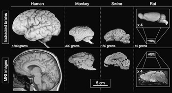
Pig brain is comparable to that of human in gross anatomy (Felix et al., Reference Felix, Leger, Albe-Fessard, Marcilloux, Rampin and Laplace1999), myelination, growth and development (Dickerson and Dobbing, Reference Dickerson and Dobbing1967; Flynn, Reference Flynn1984; Mayhew et al., Reference Mayhew, Mwamengele, Dantzer and Williams1996). Furthermore, a considerable body of evidence indicates that it resembles primate more than rat when considering its cortical convolutions, shape and total number of neocortical neurons (Lind et al., Reference Lind, Moustgaard, Jelsing, Vajta, Cumming and Hansen2007). The rat has a lissencephalic, smooth-surfaced cerebral cortex, whereas the pig has a gyrencephalic, folded brain cortical surface with well-defined circumvolutions (Hofman, Reference Hofman1985) (Figure 1).
Ex vivo anatomical and magnetic resonance imaging (MRI) correlation between brains of four different species: human, rhesus monkey, swine and rat. Brains are displayed at the same scale. Enlarged pictures are provided for the rat. Human, monkey and swine brains are gyrencephalic, contrary to the rat brain, which is lissencephalic. The average brain weight is mentioned for each species. Images of the human brain, monkey brain, monkey MRI are obtained with permission from JC Fournet (University Hospital Sainte-Justine, Montreal, Canada, http://www.humpath.com), CA Pardo-Villamizar (Johns Hopkins University School of Medicine, Baltimore, USA) and C François (Inserm UMR975, Paris, France), respectively. Other images are from our institution.

The global aspect of the pig brain differs somewhat from that of primate since the curvature of the telencephalon is less pronounced and the anterior pole is also less developed (Figure 2). Viewed from above, the pig brain has an elongated oval shape with the hemispheres being widest at the posterior third and the occipital pole being larger than the frontal pole. The olfactory system is also much more developed and occupies a large portion of the anterior part of the brain. Though the cerebral sulci and gyri of the pig have been described in a number of studies (Dellman and McClure, Reference Dellman and McClure1975; Craner and Ray, Reference Craner and Ray1991a and Reference Craner and Ray1991b; Jarvinen et al., Reference Jarvinen, Morrow-Tesch, McGlone and Powley1998; Okada et al., Reference Okada, Lahteenmaki and Xu1999; Jelsing et al., Reference Jelsing, Hay-Schmidt, Dyrby, Hemmingsen, Uylings and Pakkenberg2006; Lind et al., Reference Lind, Moustgaard, Jelsing, Vajta, Cumming and Hansen2007), there has been only a limited number of anatomical and histological studies describing the overall structure and cellular organization of the neocortex in pig. Furthermore, discrepancies between results exist (Campbell, Reference Campbell1905; Stephan, Reference Stephan1951). On the contrary, numerous functional studies dealing with specific regions of the cortex have been performed either in miniature or conventional domestic pig. According to the existing literature, we propose an approximate mapping of the pig’s cortical areas in Figure 2.
Dorsal, lateral and medial (left hemisphere) views of the brain of a Large White × Landrace pig weighing 30 kg, with schematic representations of the cortical areas. The somatosensory cortex (Woolsey and Fairman, Reference Woolsey and Fairman1946; Andrews et al., Reference Andrews, Knight and Kirby1990; Craner and Ray Reference Craner and Ray1991a and Reference Craner and Ray1991b; Okada et al., Reference Okada, Lahteenmaki and Xu1999), auditory cortex (Andrews et al., Reference Andrews, Knight and Kirby1990), visual cortex (Laube et al., Reference Laube, Schanze, Brockmann, Bolle, Stieglitz and Bornfeld2003; Fang et al., Reference Fang, Li, Rudd, Wai, Yew and Yew2006; Gizewski et al., Reference Gizewski, Schanze, Bolle, de Greiff, Forsting and Laube2007), motor cortex (Palmieri et al., Reference Palmieri, Farina, Panu, Asole, Sanna, De Riu and Gabbi1986) and prefrontal cortex (Jelsing et al., Reference Jelsing, Hay-Schmidt, Dyrby, Hemmingsen, Uylings and Pakkenberg2006) have been described with varying accuracy among authors and some results are imprecise or conflicting. To date, no comprehensive functional mapping of the cortical areas has been described in swine.

Functional studies have successfully explored the visual cortex of the pig with EP recordings (Laube et al., Reference Laube, Schanze, Brockmann, Bolle, Stieglitz and Bornfeld2003; Sachs et al., Reference Sachs, Gekeler, Schwahn, Jakob, Kohler, Schulmeyer, Marienhagen, Brunner and Framme2005; Strain et al., Reference Strain, Tedford and Gill2006) or functional MRI (Fang et al., Reference Fang, Li, Rudd, Wai, Yew and Yew2006; Gizewski et al., Reference Gizewski, Schanze, Bolle, de Greiff, Forsting and Laube2007). Exploration of the auditory cortices has also been addressed with EP recordings in miniature pig (Andrews et al., Reference Andrews, Knight and Kirby1990) or cytoarchitectonic techniques in domestic pigs (Plogmann and Kruska, Reference Plogmann and Kruska1990). Using EP recordings, the primary and secondary cortical somatosensory representations (SI and SII cortices) for the snout, the face, the forelimb and the hind limb have been described with varying precision among the studies (Woolsey and Fairman, Reference Woolsey and Fairman1946; Andrews et al., Reference Andrews, Knight and Kirby1990; Craner and Ray, Reference Craner and Ray1991a and Reference Craner and Ray1991b; Okada et al., Reference Okada, Lahteenmaki and Xu1999). With multiunit recordings, it has been shown that the overall organization of the primary somatosensory cortex (SI) was similar to that of other mammals (Craner and Ray, Reference Craner and Ray1991a). Though it is important to specify that the most comprehensive description of the somatosensory cortex in pig currently available (Craner and Ray, Reference Craner and Ray1991a and Reference Craner and Ray1991b) has been made at the neonatal stage (7 days to 2 months) and may not strictly correspond to the adult stage. With anterograde tracing techniques, it has been shown that the somatosensory cortex had marked connectivity with the ventral posterior and the ventral posterior lateral nucleus of the thalamus (Jelsing et al., Reference Jelsing, Hay-Schmidt, Dyrby, Hemmingsen, Uylings and Pakkenberg2006). SI contains a complete and somatotopically organized projection from the body surface progressing from the hind limb to the head with medial to lateral locations in the cortex. The large representation of the rostral snout in the fronto-lateral cortex (Figure 1) is probably related to the important use of the snout by the pig for tactile sensation in exploring its environment. The receptive fields for the rostrum are primarily contralateral (92%), although ipsilateral and bilateral receptive fields have been found for the face (Craner and Ray, Reference Craner and Ray1991a). The motor cortex was found to be immediately caudal to the dorsofrontal prefrontal cortex (PFC) and adjacent to SI without sulcal delineation with the latter (Breazile et al., Reference Breazile, Swafford and Thompson1966; Palmieri et al., Reference Palmieri, Farina, Panu, Asole, Sanna, De Riu and Gabbi1986; Craner and Ray, Reference Craner and Ray1991a; Jelsing et al., Reference Jelsing, Hay-Schmidt, Dyrby, Hemmingsen, Uylings and Pakkenberg2006). Recently, the PFC has been fully described in miniature Göttingen pig in a comprehensive study based on the distribution of reciprocal thalamocortical projections investigated using anterograde and retrograde tracing techniques and cytoarchitectonic criteria (Jelsing et al., Reference Jelsing, Hay-Schmidt, Dyrby, Hemmingsen, Uylings and Pakkenberg2006). On the observation that these areas were lacking a granular layer IV and had topographically organized reciprocal connections with the medio-dorsal nucleus of the thalamus, the PFC was delineated and subdivided into four main regions: the frontopolar, the anterior cingulate, the anterior insular and the dorsofrontal region. The whole PFC in these young minipigs was evaluated to constitute about 24% of the total neocortex volume and 10% of the total brain volume. The hippocampal region (Holm et al., Reference Holm, Geneser and Zimmer1993; Holm and West, Reference Holm and West1994; Saito et al., Reference Saito, Bjarkam, Nakamura and Nemoto1998) and the cerebellum (Larsell, Reference Larsell1954; Lange, Reference Lange1974; Mwamengele et al., Reference Mwamengele, Mayhew and Dantzer1993) have also been investigated in terms of histology, topology or immunohistochemistry.
In addition to global description of the central structures available in stereotaxic atlases of the pig brain (Yoshikawa, Reference Yoshikawa1968; Salinas-Zeballos et al., Reference Salinas-Zeballos, Zeballos and Gootman1986; Felix et al., Reference Felix, Leger, Albe-Fessard, Marcilloux, Rampin and Laplace1999), numerous deep cerebral structures have also been described in details with standard histology and/or immunohistochemistry. Among the comprehensive and recently published list of these studies (for review: Lind et al. (Reference Lind, Moustgaard, Jelsing, Vajta, Cumming and Hansen2007)), we may report some studies dealing with portions of the brainstem (Gillian, Reference Gillian1943; Breazile, Reference Breazile1967), specific mesencephalic nuclei such as the red nucleus (Otabe and Horowitz, Reference Otabe and Horowitz1970), the ventral mesencephalon (Ostergaard et al., Reference Ostergaard, Holm and Zimmer1992) or the raphe nucleus (Niblock et al., Reference Niblock, Kinney, Luce, Belliveau and Filiano2004) as well as diencephalic structures such as the thalamus (Solnitzky, Reference Solnitzky1938), and hypothalamus (Solnitzky, Reference Solnitzky1939; van Eerdenburg et al., Reference van Eerdenburg, Swaab and van Leeuwen1992; Leshin et al., Reference Leshin, Kraeling, Kiser, Barb and Rampacek1995). Description of the basal ganglia in pig has brought some evidence that they are similar to that of primates. It seems that the pig brain has a PFC-basal ganglia-thalamocortical circuit comparable to that described in other animals (Jelsing et al., Reference Jelsing, Hay-Schmidt, Dyrby, Hemmingsen, Uylings and Pakkenberg2006). The pig’s striatum is well developed and, in contrast to rat, individualized into distinct caudate and putamen (Felix et al., Reference Felix, Leger, Albe-Fessard, Marcilloux, Rampin and Laplace1999). The distribution of dopamine in the brain is similar to primate (Rosa-Neto et al., Reference Rosa-Neto, Doudet and Cumming2004a) and dopaminergic neurons of the substantia nigra project in a topographically organized manner to the dorsal and ventral compartments of the striatum as in primate (Agarwal et al., Reference Agarwal, Chandna, Engelking, Lightbown and Kumar1993). In addition, the subthalamic nucleus (STN) in pig has similarities with the STN described in human and other species (rat, cat, non-human primate) when considering its shape, localization and cytoarchitecture (Yelnik and Percheron, Reference Yelnik and Percheron1979; Larsen et al., Reference Larsen, Bjarkam, Ostergaard, West and Sorensen2004). Its orientation and dimensions (length 4.5 mm, width 1.9 mm, height 1.2 mm, volume 6.9 mm3) have been evaluated in minipig (Larsen et al., Reference Larsen, Bjarkam, Ostergaard, West and Sorensen2004). The porcine STN contains medium-sized neurons with uniform Golgi morphology. An immunohistological study revealed glutamate-positive neurons that receive large GABAergic, dopaminergic, and cholinergic inputs and a high number of acetylcholinesterase neurons comparable to findings in human (Larsen et al., Reference Larsen, Bjarkam, Ostergaard, West and Sorensen2004). Due to the importance of this nucleus in surgical treatment for advanced Parkinson’s disease (PD), it is highly probable that in the forthcoming years, pig might become an alternative model to rodent and primate for modelling PD (Mikkelsen et al., Reference Mikkelsen, Moller, Jensen, Pedersen, Harajehi and Pakkenberg1999) and studying deep brain stimulation (Bjarkam et al., Reference Bjarkam, Cancian, Larsen, Rosendahl, Ettrup, Zeidler, Blankholm, Ostergaard, Sunde and Sorensen2004; Dalmose et al., Reference Dalmose, Bjarkam, Sorensen, Djurhuus and Jorgensen2004).
Finally, the cerebrovascular anatomy of the pig shared similarities with human. Indeed, pig and human present a similar circle of Willis though the diameter of the posterior communicating artery is comparable to that of the internal carotid artery in pig. Thus, the connection between anterior and posterior circulation is well developed compared with that of human brain. In contrast to human, the pig presents two middle cerebral arteries originating from the internal carotid artery in each hemisphere, one coursing laterally and another rostrally over the olfactory tract (Imai et al., Reference Imai, Konno, Nakamura, Shimizu, Kubota, Seki, Honda, Tomizawa, Tanaka, Hata and Saito2006).
Brain imaging and neurosurgery in pig
Functional and molecular cerebral imaging
Recently, swine have been used as experimental models for brain imaging studies using PET (Smith et al., Reference Smith, Poulsen, Ishizu, Sakoh, Hansen, Gee, Bender and Gjedde1998; Danielsen et al., Reference Danielsen, Cumming, Andersen, Bender, Brevig, Falborg, Gee, Gillings, Hansen, Hermansen, Johansen, Johansen, Dahl-Jorgensen, Jorgensen, Meyer, Munk, Pedersen, Poulsen, Rodell, Sakoh, Simonsen, Smith, Sorensen, Ostergard, Zimmer and Gjedde2000 and Reference Danielsen, Smith, Andersen, Gee, Bender, Hansen, Hermansen, Ostergaard, Cumming and Gjedde2001b; Ishizu et al., Reference Ishizu, Smith, Bender, Danielsen, Hansen, Wong, Cumming and Gjedde2000; Sakoh et al., Reference Sakoh, Ostergaard, Rohl, Smith, Simonsen, Sorensen, Poulsen, Gyldensted, Sakaki and Gjedde2000b; Cumming et al., Reference Cumming, Danielsen, Vafaee, Falborg, Steffensen, Sorensen, Gillings, Bender, Marthi, Andersen, Munk, Smith, Moller and Gjedde2001; Dall et al., Reference Dall, Danielsen, Sorensen, Andersen, Moller, Zimmer, Gjedde and Cumming2002; Andersen et al., Reference Andersen, Watanabe, Bjarkam, Danielsen and Cumming2005), MRI (Sakoh et al., Reference Sakoh, Rohl, Gyldensted, Gjedde and Ostergaard2000a; Watanabe et al., Reference Watanabe, Andersen, Simonsen, Evans, Gjedde and Cumming2001; Jelsing et al., Reference Jelsing, Rostrup, Markenroth, Paulson, Gundersen, Hemmingsen and Pakkenberg2005; Duhaime et al., Reference Duhaime, Saykin, McDonald, Dodge, Eskey, Darcey, Grate and Tomashosky2006) and functional MRI (Fang et al., Reference Fang, Lorke, Li, Gong, Yew and Yew2005 and Reference Fang, Li, Rudd, Wai, Yew and Yew2006). Functional MRI has been used to evaluate the maturation of the brain in minipig from the early postnatal period to 6 months old. It has thus been possible to determine whether the sequence of temporal development of the visual cortex and its spatial localization are similar to primate (Fang et al., Reference Fang, Li, Rudd, Wai, Yew and Yew2006). In order to facilitate identification of activated cerebral structures in PET studies, a standard stereotaxic coordinate system in miniature Göttingen pig has been developed for voxel-by-voxel analyses across animals, with reference to a common standard MRI template for pig brain (Watanabe et al., Reference Watanabe, Andersen, Simonsen, Evans, Gjedde and Cumming2001; Andersen et al., Reference Andersen, Watanabe, Bjarkam, Danielsen and Cumming2005). To date, functional imaging in pig has rarely been used to evaluate the effects of various physiological stimuli. It has been demonstrated in vivo with [11C]raclopride that striatal dopamine neurotransmission was linked to exploratory behaviour (Lind et al., Reference Lind, Gjedde, Moustgaard, Olsen, Jensen, Jakobsen, Arnfred, Hansen, Hemmingsen and Cumming2005a).
On the contrary to functional imaging, several molecular imaging studies using PET have been performed in the pig (Bhalla et al., Reference Bhalla, Saxena and Sharma2002; Niblock et al., Reference Niblock, Luce, Belliveau, Paterson, Kelly, Sleeper, Filiano and Kinney2005; Minuzzi et al., Reference Minuzzi, Olsen, Bender, Arnfred, Grant, Danielsen and Cumming2006). These investigations have become available, thanks to the development of specific radioligand such as [11C]WAY100635 and [11C]DASB for serotonin transporters (Jensen et al., Reference Jensen, Smith, Bender, Jakobsen, Peters, Nielsen, Olsen, Scheel-Kruger, Wilson and Cumming2003; Cumming et al., Reference Cumming, Møller, Benda, Minuzzi, Jakobsen, Jensen, Pakkenberg, Stark, Gramsbergen, Andreasen and Olsen2007). A PET study with [11C]WAY100635 radioligand has thus evaluated the effects of 3,4-methylenedioxy-methamphetamine (MDMA) (‘ecstasy’) on the serotonin system in Göttingen minipigs (Cumming et al., Reference Cumming, Møller, Benda, Minuzzi, Jakobsen, Jensen, Pakkenberg, Stark, Gramsbergen, Andreasen and Olsen2007). The distribution of dopamine D1 and D2/3 binding sites within the basal ganglia of Göttingen miniature pig has been characterized with PET studies and compared to rhesus monkey (Ishizu et al., Reference Ishizu, Smith, Bender, Danielsen, Hansen, Wong, Cumming and Gjedde2000; Rosa-Neto et al., Reference Rosa-Neto, Doudet and Cumming2004a). Similar dorsal-ventral distribution gradients were found in both species, with predominant D1 binding in the ventral striatum and more D2/3 binding in the dorsal-posterior striatum (Rosa-Neto et al., Reference Rosa-Neto, Doudet and Cumming2004a). Thanks to the development of methods for measuring cerebral uptake and metabolism of [18F]fluoro-l-DOPA (FDOPA) (Danielsen et al., Reference Danielsen, Smith, Gee, Venkatachalam, Hansen, Hermansen, Gjedde and Cumming1999 and Reference Danielsen, Smith, Andersen, Gee, Bender, Hansen, Hermansen, Ostergaard, Cumming and Gjedde2001b), it is possible to analyse the activity of DOPA decarboxylase in living pig and apply this strategy to investigate the effect of neuroleptics (Danielsen et al., Reference Danielsen, Smith, Hermansen, Gjedde and Cumming2001a). Similarly, PET studies with [11C]raclopride, a radioligand binding in competition with the endogenous dopamine, have been conducted to analyse the effect of psychoactive drugs such as amphetamine (Lind et al., Reference Lind, Olsen, Moustgaard, Jensen, Jakobsen, Hansen, Arnfred, Hemmingsen, Gjedde and Cumming2005b), nicotine (Cumming et al., Reference Cumming, Rosa-Neto, Watanabe, Smith, Bender, Clarke and Gjedde2003), MDMA (Rosa-Neto et al., Reference Rosa-Neto, Gjedde, Olsen, Jensen, Munk, Watanabe and Cumming2004b) and lysergic acid diethylamide (Minuzzi et al., Reference Minuzzi, Nomikos, Wade, Jensen, Olsen and Cumming2005). It has been shown that these psychoactive agents reduced the availability of [11C]raclopride binding sites for dopamine D(2,3) receptors in the striatum of Göttingen minipigs. The variation of the regional cerebral blood flow (rCBF) has been used to analyse the effects of MPTP poisoning. As in primates, MPTP-treated pigs develop an experimental syndrome of Parkinsonism (Mikkelsen et al., Reference Mikkelsen, Moller, Jensen, Pedersen, Harajehi and Pakkenberg1999). This model has been associated with a 60% decline in the uptake of FDOPA, a tracer for the activity of DOPA decarboxylase in nigrostriatal fibers of the striatum (Danielsen et al., Reference Danielsen, Cumming, Andersen, Bender, Brevig, Falborg, Gee, Gillings, Hansen, Hermansen, Johansen, Johansen, Dahl-Jorgensen, Jorgensen, Meyer, Munk, Pedersen, Poulsen, Rodell, Sakoh, Simonsen, Smith, Sorensen, Ostergard, Zimmer and Gjedde2000; Cumming et al., Reference Cumming, Danielsen, Vafaee, Falborg, Steffensen, Sorensen, Gillings, Bender, Marthi, Andersen, Munk, Smith, Moller and Gjedde2001; Dall et al., Reference Dall, Danielsen, Sorensen, Andersen, Moller, Zimmer, Gjedde and Cumming2002). It is highly probable that this new model for induced Parkinsonism will challenge the animal models commonly used to date (i.e. MPTP-treated primate and 6-hydroxydopamine lesioned rat).
Stereotaxic and image guided surgery
Several stereotaxic atlases of the pig brain have been described (Yoshikawa, Reference Yoshikawa1968; Szteyn et al., Reference Szteyn, Galert, Dynowski and Hoczyk1980; Salinas-Zeballos et al., Reference Salinas-Zeballos, Zeballos and Gootman1986; Felix et al., Reference Felix, Leger, Albe-Fessard, Marcilloux, Rampin and Laplace1999; Watanabe et al., Reference Watanabe, Andersen, Simonsen, Evans, Gjedde and Cumming2001) as well as specific stereotaxic instruments either for piglets (Salinas-Zeballos et al., Reference Salinas-Zeballos, Zeballos and Gootman1986), pigs weighing between 30 and 40 kg (Poceta et al., Reference Poceta, Hamlin, Haack and Bohr1981; Saito et al., Reference Saito, Bjarkam, Nakamura and Nemoto1998) or adults (Marcilloux et al., Reference Marcilloux, Rampin, Felix, Laplace and Albe-Fessard1989; van Eerdenburg and Dierx, Reference van Eerdenburg and Dierx2002). The stereotaxic frame devoted to the pig developed by Marcilloux et al. (Reference Marcilloux, Rampin, Felix, Laplace and Albe-Fessard1989) was based on the fixation of the skull by the infraorbital ridges, the hard palate and the auditory canals. The ear-bars were carried by a swivelling system allowing adaptation to the orientation of the auditory canals, perpendicular to the sagittal plane in the new-born piglet but oblique in the weaned pig (Poceta et al., Reference Poceta, Hamlin, Haack and Bohr1981; Salinas-Zeballos et al., Reference Salinas-Zeballos, Zeballos and Gootman1986; Marcilloux et al., Reference Marcilloux, Rampin, Felix, Laplace and Albe-Fessard1989). Unfortunately, this system may have some adverse effects on the eardrums and cannot be used with Göttingen mini-pigs. Indeed, in this breed, the auditory canal is almost vertical, and hence the impossibility to fix the head in the Marcilloux stereotaxic frame. This peculiarity plus the need for a system compatible with MRI led Bjarkam et al. (Reference Bjarkam, Cancian, Larsen, Rosendahl, Ettrup, Zeidler, Blankholm, Ostergaard, Sunde and Sorensen2004) to use MRI-compatible localizers fixed on the skull and to develop a head-fixation box made of methyl methacrylate (Plexiglas®) with the head fixated by aluminium screws inserted bilaterally in the zygoma bone.
Stereotaxic surgery requires precise determination of a system of coordinates either based on bony landmarks or intracerebral structures easily identifiable on images. The choice of such a reference system varies between authors. Some of them have chosen to base their procedure on external skull structures as commonly performed in rodents (Paxinos and Watson, Reference Paxinos and Watson2006). This strategy has been used in newborn pigs aged 1 to 12 days (Salinas-Zeballos et al., Reference Salinas-Zeballos, Zeballos and Gootman1986), for description of the PFC in miniature Göttingen pig aged of 3 months (Jelsing et al., Reference Jelsing, Hay-Schmidt, Dyrby, Hemmingsen, Uylings and Pakkenberg2006) and for targeting the hippocampus of Landrace prepubertal pigs weighing less than 30 kg (Saito et al., Reference Saito, Bjarkam, Nakamura and Nemoto1998). Saito et al. (Reference Saito, Bjarkam, Nakamura and Nemoto1998) have reported the reliability of this method and minimal variation in distances between some external bony landmarks. Despite the fact that this strategy might be successful in young pig, there is a too large inter-individual variation in skull morphology in growing animals and these landmarks may vary considerably as the age of the animal increases (Marcilloux et al., Reference Marcilloux, Rampin, Felix, Laplace and Albe-Fessard1989; Bjarkam et al., Reference Bjarkam, Cancian, Larsen, Rosendahl, Ettrup, Zeidler, Blankholm, Ostergaard, Sunde and Sorensen2004). In addition, measures based on bone structures can hardly be applied generally since pig present considerable inter-individual and inter-breed skull variations in size and shape. This becomes even more of a problem in adult miniature Göttingen pig presenting a large pneumatized frontal sinus inducing considerable inter-individual variation on distances between bone and brain structures.
A more precise definition of a stereotaxic coordinates system has been achieved on the basis of stable intracerebral landmarks. Depending on the tools used, two stereotaxic systems have been proposed. Using ventriculography, the horizontal zero plane was defined on the basis of the plane connecting the recessus preopticus to the posterior commissure (CP) and CP as the origin of the system (Marcilloux et al., Reference Marcilloux, Rampin, Felix, Laplace and Albe-Fessard1989; Felix et al., Reference Felix, Leger, Albe-Fessard, Marcilloux, Rampin and Laplace1999). The recessus preopticus was found to correlate closely with the anterior commissure defining a system similar to that commonly used in human and primate (Felix et al., Reference Felix, Leger, Albe-Fessard, Marcilloux, Rampin and Laplace1999). Andersen et al. (Reference Andersen, Watanabe, Bjarkam, Danielsen and Cumming2005) found difficult to identify the two commissures on MRI and defined the referenced plane on the basis of the line connecting the optic chiasma to the pineal gland. The origin of the stereotaxic system was then defined as the centre of the pineal gland. Fortunately, both coordinates systems remain interchangeable by a geometric transformation of rotation and translation. Whatever the procedure and the tools used, stereotaxic surgery has been successfully performed in swine. The hippocampus was targeted in prepubertal pigs weighing 10 kg with a technique based on external skull structures alone such as bregma, sutura coronalis and sagittalis (Saito et al., Reference Saito, Bjarkam, Nakamura and Nemoto1998). Implantation of foetal dopaminergic grafts in the striatum (Danielsen et al., Reference Danielsen, Cumming, Andersen, Bender, Brevig, Falborg, Gee, Gillings, Hansen, Hermansen, Johansen, Johansen, Dahl-Jorgensen, Jorgensen, Meyer, Munk, Pedersen, Poulsen, Rodell, Sakoh, Simonsen, Smith, Sorensen, Ostergard, Zimmer and Gjedde2000; Cumming et al., Reference Cumming, Danielsen, Vafaee, Falborg, Steffensen, Sorensen, Gillings, Bender, Marthi, Andersen, Munk, Smith, Moller and Gjedde2001) or deep brain stimulation of the STN (Bjarkam et al., Reference Bjarkam, Cancian, Larsen, Rosendahl, Ettrup, Zeidler, Blankholm, Ostergaard, Sunde and Sorensen2004; Dalmose et al., Reference Dalmose, Bjarkam, Sorensen, Djurhuus and Jorgensen2004) were performed in miniature pig on the basis of intracerebral landmarks (Figure 3).
Location of the subthalamic nucleus (St) on coronal (a) and sagittal (b) sections. Its coordinates are 5.00 mm ahead the posterior commissure (CP) and 6.00 mm laterally from the midsagital plane respectively. The reference (zero) horizontal plane contains a virtual line connecting the recessus preopticus and the CP. Drawings from Felix et al. (Reference Felix, Leger, Albe-Fessard, Marcilloux, Rampin and Laplace1999) are with permission from Elsevier.

Examples of studies on the porcine central nervous system
To illustrate the intricacies in using the pig model compared to more classical model such as the rodents, we have selected two examples of studies currently ongoing in our laboratory at UMR SENAH, Saint-Gilles, France. These experiments were performed in growing farm pig about 30 kg in weight.
Functional brain imaging during gastric distension
Signals arising from gastric distension have a critical role in the control of food intake (Smith, Reference Smith1998; Ritter, Reference Ritter2004). These signals are likely being transmitted to the brain mainly by the vagal nerve (Phillips and Powley, Reference Phillips and Powley1998; Berthoud et al., Reference Berthoud, Lynn and Blackshaw2001). The contribution of spinal nerves is considered as insignificant (Sengupta and Gebhart, Reference Sengupta and Gebhart1994). However a recent study performed in humans suggests that the situation is not so clear-cut (Vandenbergh et al., Reference Vandenbergh, Dupont, Fischler, Bormans, Persoons, Janssens and Tack2005). The aim of this study was to identify brain areas and nervous pathways involved in the central processing of non-noxious gastric distension signals. Using functional brain imaging, we have investigated brain activity during gastric distension in pigs with intact vagal nerves or after a bilateral vagotomy. Animals were surgically fitted with a permanent access to the stomach through a gastric cannula. This cannula allowed insertion of an inflatable balloon into the stomach so as to achieve a pressure-controlled distension with preset pressure rise time. This was performed by a custom-made inflator using compressed air and controlled by dedicated software acting on high-speed electrovalves through a pressure-controlled loop. The pressures investigated were within the range of that observed after a medium- to large-size meal and were below the nociceptive range (25 mmHg above mean abdominal pressure for a pig).
Brain functional imaging was performed using single photon emission tomography (SPECT). Briefly, this method used the property of 99Tc-HMPAO (d-l hexamethylpropylene amine oxime) to enter into the neuron because it is lipophilic. Though 99Tc-HMPAO is not an ideal tracer of CBF since it underestimates high flow areas, it has the unique capability to allow CBF imaging in conscious animal. Indeed, upon its injection in the bloodstream, it will enter neurons and stay within the cell body because it undergoes a biochemical transformation that changes its lipophilic properties into a hydrophilic one, probably as a result of an action with gluthathione. The kinetic of this transformation is within the order of the minute. Afterwards, it is possible to image the brain radioactivity in anaesthetized animal while the recorded radioactivity distribution reflects the brain perfusion at the time of radiolabel injection. Furthermore, anaesthesia has no adverse affect on the nuclear imaging procedure. As for all functional brain imaging method, the relationship between brain activity and regional vascularization of the brain structures allows from the rCBF to map brain function.
The experimental protocol consisted in two imaging sessions before and after truncal vagotmy. Five Large White × Landrace female pigs aged of 3 months and weighing 30 kg were used in this study. An initial imaging session was performed on animals at different pressures in a randomized order (0, 10 and 20 mmHg). After vagotomy (5 cm above the diaphragm) of the same animals, a second imaging session was made at different pressures as previously described. A 7-day washout was allowed between experiments on the same animal. Gastric distension and SPECT were achieved on halothane-anaesthetized pigs under artificial ventilation after a 20-min resting period without external solicitations. Thirty minutes after its insertion in the stomach through a gastric cannula, the barostat bag was inflated to the test pressure. Concomitant to inflation, an intravenous injection of 99Tc-HMPAO (555 to 740 MBq, Ceretec™, GE Healthcare, Amersham, UK) was performed. Fifteen minutes after radiolabel injection, brain images were acquired in three dimensions by SPECT on an Elscint gamma camera (Elscint Ltd, Haifa, Israel) fitted with a low energy-high resolution collimator. Each image was acquired for 90 s with a step shoot angle of 6° allowing to record about 2 to 5 kcounts/s, which is the formal requirement for three-dimensional (3D) reconstruction from SPECT images.
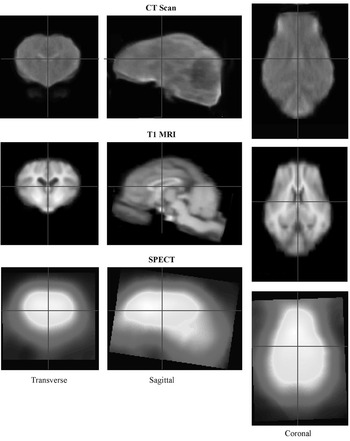
Axial brain slices were reconstructed offline using a filtered back projection with a dedicated Sheep–Logan filter to remove diffusion artefacts. Back projections of brain images were normalized and co-registered using Automated Image Registration algorithm to a SPECT pig template built in our laboratory (Figure 4). This SPECT pig template corresponds to the mean of 16 SPECT obtained in anaesthetized animals of the same breed, age and weight. Briefly, each of the individual SPECT volumes used for the template was initially co-registered to an anatomical segmented image of the brain of the same animal obtained with a CT scanner. This CT volume was, prior its use for SPECT co-registration, co-registered with a T1 MRI pig template calculated by Andersen et al. (Reference Andersen, Watanabe, Bjarkam, Danielsen and Cumming2005). This T1 MRI template was obtained as a mean of 22 MRI’s brain imaging. The intermediate step using CT images is required because the error in co-registering the low resolution SPECT image onto the high resolution MRI image is too large. Using this procedure, it is possible to register the SPECT volume into a referenced volume mimicking the Talairach space. For simplicity and interoperability, we chose to use the stereotaxic space defined by Felix et al. (Reference Felix, Leger, Albe-Fessard, Marcilloux, Rampin and Laplace1999).
Intermodality co-registered templates of the pig brain used to reset each functional volume of individual pigs into the same reference volume. The MRI T1 (magnetic resonance imaging) template was based on 22 pigs (Andersen et al., Reference Andersen, Watanabe, Bjarkam, Danielsen and Cumming2005). The CT (computed tomography) scan and SPECT (single photon emission tomography) templates were based on 16 animals. The fuzzy aspect of the images was due to the intersubject averaging procedure. Co-registration was achieved using the Automated Image Registration algorithm (University of California, Los Angeles, USA) and involved affine and elastic transformations in the three dimensions.

Voxel-based analyses were performed using parametric tests via Statistical Parametric Mapping (SPM2) software adapted to the characteristics of the pig brain, size of which being about 1/3 of that of a human. Regression analyses were performed to identify brain areas of which the activity was associated/correlated with the increase of distending pressure. This brain activity was compared before and after vagotomy. Voxels significantly associated to the increase of the distending pressure were displayed on the MRI template of pig brain.
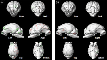
Before vagotomy, the activity of several brain areas was found significantly associated with the increase of gastric distending pressure, only (Figure 5). After vagotomy, only a limited number of brain areas were associated with the increase of gastric pressure. This study demonstrates that spinal afferences encode non-painful gastric distension signals. Furthermore, in intact animal, vagal and non-vagal afferences originating from gastric wall and activated by non-painful gastric distension are involved in the brain representation of post-prandial gastric fullness.
The 3D-rendered images of the front, back, right, left, top and bottom brain views of: (a) intact and (b) vagotomized pigs with activated (red) and deactivated (green) voxels correlated (P < 0.02) with the increase of the fundic distending pressure. Voxels were projected onto the cortical surface of the rendered magnetic resonance imaging template built from 22 animals.

Deep brain stimulation and neuronavigation
Deep brain stimulation (DBS) is widely used as a functional treatment for various movement disorders such as PD, dystonia and essential tremor. New indications are emerging in neuropsychiatry such as obsessive–compulsive disorders or depression (Wichmann and Delong, Reference Wichmann and Delong2006). This therapy is based on continuous high frequency stimulation of a deep structure: STN, globus pallidus internalis or thalamus. Usually, a quadripolar electrode is implanted in the nucleus and high frequency stimulation is provided through a pulse generator implanted subcutaneously in the subclavicular area. This functional therapy has proved its efficacy in severe PD with improvement of motor symptoms, reduction of motor fluctuation and dyskinesia. However, the exact mechanism of action of DBS is still unknown as well as the influence of electrical stimulation on behaviour. Since stimulation of a nucleus affects complex cortico-subcortical loops involved in motor, cognitive and behavioural processes, studies on living organisms remain essential to assess the collateral effects of such a therapy. Among those, excessive weight gain has been observed in patients with PD after stimulation of the STN. Whether this effect is exclusively due to the decrease in energy expenditure consecutive to the improvement of motor symptoms is still unknown. An additional effect on energy metabolism by current diffusion to the hypothalamus or motivational change by activation of the limbic circuit of the basal ganglia may be hypothesized (Perlemoine et al., Reference Perlemoine, Macia, Tison, Coman, Guehl, Burbaud, Cuny, Baillet, Gin and Rigalleau2005; Montaurier et al., Reference Montaurier, Morio, Bannier, Derost, Arnaud, Brandolini-Bunlon, Giraudet, Boirie and Durif2007).
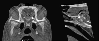
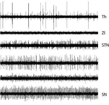
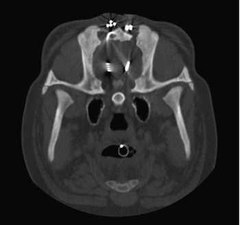
With the aim to study feeding behaviour in implanted animals, we have developed a surgical procedure for electrode implantation in the STN based on intraoperative microrecording and CT images based on neuronavigation. Six Large White × Landrace female pigs aged of 3 months and weighing 30 kg were used in this study. Neuronavigation was based on images acquired with a CT scanner while pigs were maintained in a custom-made stereotaxic frame similar to that described by Marcilloux et al. (Reference Marcilloux, Rampin, Felix, Laplace and Albe-Fessard1989). The CT images were transferred to a DICOM PACS Workstation running OsiriX, an Open Source Dicom viewer (Rosset et al., Reference Rosset, Spadola and Ratib2004) allowing 3D reconstruction, and direct length and angle measurements (Figure 6). The stereotaxic coordinates of the target were calculated on the basis of identification of intracerebral stable landmarks, i.e. the anterior and posterior white commissures, outlined by injection of contrast liquid. In order to define precisely the position of the STN, we further performed microrecordings with three parallel microelectrodes (FHC, Bowdoinham, ME, USA) distant of 2.5 mm from each other (Figure 7). The microelectrode demonstrating the highest neuronal activity was replaced by a chronic electrode (model 3389; Medtronic, Minneapolis, MN, USA) fixed to the skull with dental cement. The electrode’s wire and the extension lead were fixed to the skull (Figure 8). The extension emerged on the midline between the scapulas and was connected to a pulse generator (Kinetra; Medtronic, Minneapolis, MN, USA) fixed on the back of the animal in a harness designed in our laboratory. Pigs recovered quickly from the surgery and continuous high stimulation was maintained for 3 months.
Coronal (left) and sagittal (right) computed tomography scan images of the brain of a Landrace × Large White pig aged of 3 months and weighing 30 kg. The stereotaxic coordinates of the target were defined with ventriculography allowing visualization of stable intracerebral landmarks that were the interhemispheric line and the bicommissural line. This animal underwent implantation of bilateral electrode in the subthalamic nucleus.

Electrophysiological identification of the subthalamic nucleus (STN) with microrecording (Trace duration: 10 s). Thalamic cells were recorded about 5 mm above the STN Thalamus (Th), then after a silent area of about 2 mm that might correspond to the zona incerta (ZI), a noticeable increase in background activity and spontaneous firing discharge were observed. These corresponded to penetration of the electrode tip into the STN. Sustained irregular activity was recorded over 1.4 ± 0.4 mm (mean ± s.d.). The lower border of the nucleus was more difficult to differentiate from activity in the substantia nigra (SN). The latter presented however a more tonic pattern of activity than the STN.

Coronal computed tomography image acquired at the level of the subthalamic nucleus in a pig implanted with bilateral electrodes for 100 days and aged 6 months. The four contacts of the electrode (model 3389 from Medtronic) are clearly viewed on the right side. The radio-opaque artefacts on the skull correspond to the extension lead of the electrodes that are rolled under the skin.

The feeding behaviour of the implanted animals was compared to that of control animals with specific reference to the motivational aspect of feeding. At the end of the experiment, animals were euthanized and the placement of the electrodes was checked using histological examination. It has thus been shown that continuous subthalamic stimulation induced change in the feeding behaviour of the animals with different aspects on the motivation of the animals to feed.
Discussion
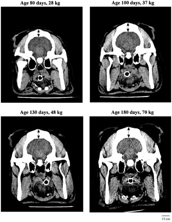
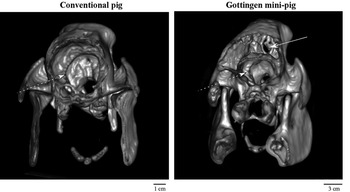
Pigs used in research originate either from common agricultural or research-dedicated breeds, the most common of the latter being the Yucatan and the Göttingen minipigs. Since animals from different breeds present physiological and anatomical variations, they may offer advantages and/or disadvantages depending on the purpose of the study (Bollen et al., Reference Bollen, Hansen and Rasmussen2000). For example, anatomical considerations are crucial for brain surgical intervention. In pig, the brain is protected by a massive skull which changes in size and shape during the growth, in addition to individual variability in skull structure (Poceta et al., Reference Poceta, Hamlin, Haack and Bohr1981; van Eerdenburg and Dierx, Reference van Eerdenburg and Dierx2002). Moreover, there is a dramatic increase in thickness with the animal growing rendering the surgical intervention difficult (Figure 9). For example, the thickness of the skull is about 6 to 7 mm in its middle portion with a maximum of 30 mm at the occipital crest in Landrace pig aged only of 3 months, i.e., before the emergence of the frontal sinus. Since immature pigs grow quickly and mature adults overweight 250 kg, studies duration is limited to 3 or 4 months. Furthermore, they often necessitate restricted caloric intake to limit the weight of the animal. Alternatively, adult minipigs are easily manipulated since they weight less than 60 kg (for Göttingen minipigs). However, these mature minipigs have a large frontal sinus that pneumatizes the skull, which makes the access to the brain tricky (Figure 10). It necessitates oblique trajectories and a wider scalp incision. The development of this frontal sinus starts around puberty and grows rapidly thereafter (van Eerdenburg and Dierx, Reference van Eerdenburg and Dierx2002). Thus, a number of studies have been conducted in agricultural pig but miniature pig might be preferred for chronic or long-term studies for which a significant growth is an issue (Panepinto et al., Reference Panepinto, Phillips, Wheeler and Will1978; Imai et al., Reference Imai, Konno, Nakamura, Shimizu, Kubota, Seki, Honda, Tomizawa, Tanaka, Hata and Saito2006).
Computed tomography scan images of the head of a conventional pig at various age and weight. Irrespective of the age/weight, the size of the brain is unchanged (51, 52, 52 and 53 mm in width). On the contrary, the thickness of the skull (black arrows) is increasing with age/weight (15, 19, 21 and 22 mm). This thickening results in a larger amount of diffused photons for functional imaging studies and an increased difficulty to maintain electrodes in deep brain structures for a large time period. All images shared a similar scale.

Maximal intensity projections of the skull of a conventional pig (left panel) v. a Göttingen minipig of the same weight (26 kg). Note the presence of a large pneumatization of the skull (white arrow) above the brain (doted white arrow) in the Göttingen minipig. This pneumatization makes the access to the brain extremely tricky and requires oblique trajectories whereas vertical ones can be used in the conventional pig.

Recent researches using the pig as a model in neuroscience are mainly imaging studies. This results from the increasing relevance of recent imaging tools to explore brain functions, together with the opportunity to use an animal model with a large developed brain. The pig model appears to be obviously superior to rodent for this purpose especially, because of its compatibility with imaging platform used in human. To date, stereotaxic surgery in pig has undergone a less notable development. As for imaging, pig presents a major advantage that is the size of its brain and the similarities with human brain, but pig is considerably disfavoured by the thickness of the skull bone and the presence of a large median sinus in mature animal. Nevertheless, stereotaxic procedures have been successfully performed in pig, thanks to the development of specific stereotaxic apparatus and atlases.
Paradoxically, though studies with advanced technology have been conducted in pig for years, one has to recognize that fundamental description of the pig brain is still lacking. Precise identification of cerebral structures involved in any specific process requires complete and valid data in conventional and functional neuroanatomy. Though deep structures have been relatively well described in pig (Felix et al., Reference Felix, Leger, Albe-Fessard, Marcilloux, Rampin and Laplace1999), the cortex of the pig has been poorly described and, to date, there is no functional neither structural maps equivalent to Brodmann’s description in primate and human. Nomenclature of cortical sulci and fissures as well the localization of functional cortical areas vary from one author to another (Craner and Ray, Reference Craner and Ray1991a and Reference Craner and Ray1991b; Okada et al., Reference Okada, Lahteenmaki and Xu1999; Lind et al., Reference Lind, Moustgaard, Jelsing, Vajta, Cumming and Hansen2007). Moreover, a number of studies have been restricted to the postnatal ages and only sparse data are available for piglet aged of more than 3 months. When available, these studies have mainly been conducted in miniature Göttingen pig and some discrepancies may occur when one tries to transpose to conventional pig. As a result, precise localization of any activated signal identified by functional imaging remains somewhat imprecise. Another difficulty arises from the wide range of pig breeds that have been used in the different studies inducing considerable anatomical variability and the lack of normative values, e.g. for EP recordings (Strain et al., Reference Strain, Tedford and Gill2006).
In comparison with the other available experimental models (sheep, pig and rat), the non-human primates represent an optimal choice. However, a European directive that was introduced in December 2008 strictly limited the use of non-human primates as animal model (http://europa.eu/rapid/pressReleasesAction.do?reference=IP/08/1632&format=HTML&aged=0&language=FR&guiLanguage=en). In this context, alternative to the non-human primates must be considered. Murine models might be of significant interest for neurological research. However, their brain sizes limit the possibility for functional imaging. The spatial resolution of the best micro PET available today is 2.5 times more than a standard PET scanner able to scan large animals (Suk et al., Reference Suk, Thompson, Labuda and Goertzen2008) whereas the ratio between large animal v. murine brain is ranging between 1 and 3 or less. This limitation is even more important with the wider availability of 3T MRI scanner capable to image large animal brain with a relative spatial resolution comparable to a 7T miniature MRI machine imaging a murine brain. Furthermore, murine brain lacks the development of the cortex in comparison to primates or larger animals. All of these reasons suggest that larger animals such as ruminants (goat and cattle) and pigs might provide an attractive alternative to rodents. For example, the sheep has been successfully used to develop a model for PD using MPTP poisoning (Beale et al., Reference Beale, Higgins, Work, Bailey, Smith, Shinka and Hammock1989; Baskin et al., Reference Baskin, Browning, Widmayer, Zhu and Grossman1994). It has been involved in brain microdialysis studies (Westerink, Reference Westerink1995) or intracranial electroencephalography (Opdam et al., Reference Opdam, Federico, Jackson, Buchanan, Abbott, Fabinyi, Syngeniotis, Vosmansky, Archer, Wellard and Bellomo2002). However, neuroscience studies using mature sheep remain infrequent primarily because of the ruminant-related differences in physiology. For instance, peripheral vagal nutrient sensing has been adapted to forage ingestion and as a consequence the key player is a potassium-sensitive vagal receptor that is absent in non-ruminant species (Cottrell and Iggo, Reference Cottrell and Iggo1984). Furthermore, as a seasonal animal, leptin secretion and sensitivity in the adult sheep is altered during the year, unlike in pigs or humans (Zieba et al., Reference Zieba, Szczesna, Klocek-Gorka, Molik, Misztal, Williams, Romanowicz, Stepien, Keisler and Murawski2008). Finally, there are remaining questions regarding similarities with human in the role of ghrelin in the control of food intake in sheep (Sugino et al., Reference Sugino, Hasegawa, Kurose, Kojima, Kangawa and Terashima2004; Grouselle et al., Reference Grouselle, Chaillou, Caraty, Bluet-Pajot, Zizzari, Tillet and Epelbaum2008). Consequences of differences in digestive physiology overwhelmed the context of digestive behaviour. Indeed, nutrient source and glucose metabolism of the brain, especially the utilization of ketones, differ in sheep in comparison to human and rodent (Lindsay and Setchell, Reference Lindsay and Setchell1976). This may account for the lack of PET studies in sheep with the notable exception of foetal and neonatal lamb (Thorngren-Jerneck et al., Reference Thorngren-Jerneck, Ley, Hellstrom-Westas, Hernandez-Andrade, Lingman, Ohlsson, Oskarsson, Pesonen, Sandell, Strand, Werner and Marsal2001). Additionally, despite studies describing (partially) the functional mapping of the cortex of the sheep (Kirk et al., Reference Kirk, Kitchell and Johnson1987), from our knowledge, this model has never been used for functional neurosurgery or chronic DBS.
Conclusions
Numerous studies with fMRI or PET have proved that the pig is a suitable model for studying various neurotransmitters and radioligands. Stereotaxic surgery is possible despite the thickness of the skull and the presence of a large frontal sinus in some adult animals. Nevertheless, specific points require further improvements to refine the usability of the pig model. A better description of the cortex and its functional areas as well as a better stereotaxic localization of deep structures are absolutely mandatory for brain imaging and stereotaxic surgery. We look forward for high quality atlases for both cortex and deep structures allowing determination of stereotaxic coordinates and localization of activated areas in imaging studies. Especially, there is a need for a digitalized atlas matching MRI and histological resources built using a large number of animals. This atlas should additionally benefit from associated electrophysiological cortical mapping studies.
Acknowledgements
Paul Sauleau was supported by a grant from Medtronic, France and the Scientific Council of the Faculty of Medicine, Rennes, France.