Introduction
The organophosphate pesticide chlorpyrifos (CPF) is an acetylcholinesterase inhibitor that kill pests by blocking the cholinergic neurotransmission (Fukuto, Reference Fukuto1990; Interaction Profile for: Chlorpyrifos, Lead, Mercury, and Methylmercury, 2006). Its sublethal concentration also induces oxidative stress and causes toxic effects to the organisms (Gupta et al., Reference Gupta, Mishra, Sharma, Deepak Balaji, Kumar, Mishra and Chowdhuri2010). CPF pesticides are not only widely used around the world to control agricultural and hygiene pests, but also have posed a serious ecological risk to non-target organisms (Muñiz-González et al., Reference Muñiz-González, Paoli, Martínez-Guitarte and Lencioni2021). These pesticides in the environment can be transported over a medium to high distances due to the drift effect, and become a threat to wildlife in both terrestrial and freshwater ecosystems (Muñiz-González et al., Reference Muñiz-González, Paoli, Martínez-Guitarte and Lencioni2021). Due to its ecological risks and potential health hazards, CPF has been restricted or banned in many countries (Wang et al., Reference Wang, Wang and Li2025). Nevertheless, residues of CPF and other organophosphate pesticides are still frequently detected, as their global production and use persist at high levels (Ikayaja and Arimoro, Reference Ikayaja and Arimoro2024; Karthick Rajan et al., Reference Karthick Rajan, Mohan, Rajarajeswaran, Divya, Thanigaivel and Zhang2024; Tsuchiyama et al., Reference Tsuchiyama, Ito, Taniguchi, Katsuhara, Miyazaki and Kamijima2023).
Evidence in the literature suggests that sublethal CPF can induce the accumulation of reactive oxygen species (ROS) in a wide range of organisms, leading to oxidative damage at the molecular, cellular and tissue levels (Zhang et al., Reference Zhang, Chen, Yang, Kong, Bourguet and Wu2017a). A study in Drosophila (Diptera: Drosophilidae) larvae showed that CPF exposure causes apoptosis and DNA damage through the generation of ROS (Gupta et al., Reference Gupta, Mishra, Sharma, Deepak Balaji, Kumar, Mishra and Chowdhuri2010). These damages are highly involved in the alteration of physiological traits, such as reduced activity levels of the antioxidant defence enzymes, decreased energy budget and delayed metamorphosis, which may further lead to the decrease in larval survival, growth, locomotor activity and some other ecologically relevanttraits collected with the population dynamics (Dinh et al., Reference Dinh, Janssens, Therry, Bervoets, Bonte and Stoks2016; Di Nica et al., Reference Di Nica, González, Lencioni and Villa2020; Verheyen et al., Reference Verheyen, Cuypers and Stoks2023).
In insects, the first line of defence against ROS is provided by superoxide dismutases (SODs), and is inextricably linked with antioxidant enzymes, such as catalase (CAT) and peroxidases (PRXs), at the secondary line (Shi et al., Reference Shi, Yu and Zhang2012). SODs can directly catalyse the reduction of ROS by rapidly dismutate the superoxide to hydrogen peroxide and oxygen (Gretscher et al., Reference Gretscher, Streicher, Strauß, Wielsch, Stock, Wang, Boland and Burse2016). Subsequently, hydrogen peroxide levels in various tissues are maintained by CAT and PRXs (Aidlin Harari et al., Reference Aidlin Harari, Santos-Garcia, Musseri, Moshitzky, Patel, Visendi, Seal, Sertchook, Malka and Morin2020; Corona and Robinson, Reference Corona and Robinson2006; Shi et al., Reference Shi, Zhang, Jia, Zhang, An, Wang, Zhang and Yin2014; Yamamoto et al., Reference Yamamoto, Banno, Fujii, Miake, Kashige and Aso2005). Three typs of SODs are commonly fund in vertebrates and invertebrates: cytoplasmic Cu/ZnSOD (SOD1), mitochondrial MnSOD (SOD2), and secreted Cu/ZnSODs (SOD3) (Zhang et al., Reference Zhang, Guo and Li2017b). Recent studies have further identified SOD domains in a diverse range of proteins. In Bombyx mori (Lepidoptera: Bombycidae), genomic analyisis revealed the presence of a cytoplasmic Cu/ZnSOD (BmSOD1), a mitochondrial MnSOD (BmSOD2), two secreted Cu/ZnSODs (BmSOD3 and BmSOD5), two membrane Cu/ZnSODs (BmSOD4 and BmSOD6) and a cellular copper chaperone for SOD (BmCCS) (Kobayashi et al., Reference Kobayashi, Nojima, Sakamoto, Iwabuchi, Nakazato, Bono, Toyoda, Fujiyama, Kanost and Tabunoki2019). These intracellular and extracellular SODs demonstrated different expression patterns during the larval, pupal, and adult stages, and the extracellular BmSOD3 was up-regulated by the oxidative stress induced by ultraviolet irradiation and bacterial injection. The gene transcripts of CAT and PRX in B. mori were ubiquitously distributed in different tissues (He et al., Reference He, Fang, Zhang, Liu, Wen, Yu, Cheng, Li, Huang, Ai and Wu2024; Yamamoto et al., Reference Yamamoto, Banno, Fujii, Miake, Kashige and Aso2005). CAT and PRX activities can be up-regulated by different adverse situations and play an important role in protecting against the induced oxidative stress (Huang et al., Reference Huang, Zou, Zhan, Hao, Lu, Jing, Li, Xu and Wang2023; Yuan et al., Reference Yuan, Xu and Sima2015).
Protohermes (Megaloptera: Corydalidae) is a species-rich genus of dobsonflies whose larvae are exclusively predacious and aquatic (Liu et al., Reference Liu, Hayashi and Yang2013). Protohermes species are widely distributed in East and Southeast Asia. In the freshwater environments, Protohermes larvae are of great ecological importance due to their euryphagous and predatory habits, which make this group contribute to the equilibrium of aquatic communities (Rivera-Gasperín et al., Reference Rivera-Gasperín, Ardila-Camacho and Contreras-Ramos2019). Furthermore, Protohermes insects are also prey for other invertebrates, fish and some terrestrial vertebrates, thus playing an important role in food web dynamics (Ishikawa et al., Reference Ishikawa, Hayashi, Sasaki, Chikaraishi and Ohkouchi2017). In addition, Protohermes species are vulnerable to water contamination from agricultural and industrial pollutants and are used as bioindicators in environmental impact assessments (Ishii et al., Reference Ishii, Miura, Jo, Tsuji, Saito, Koarai, Hagiwara, Urushidate, Nishikiori, Wada, Hayashi and Takahashi2022). Molecular markers have been identified in Protohermes species and used to analyse the toxic effects of environmental pollutants on these ecologically important aquatic predators (Wen et al., Reference Wen, Yang, Huang and Huang2022; Yang et al., Reference Yang, Huang, Wen, Huang, Liu and Zhang2023).
Molecular biomarkers are widely used as tools for accessing the toxic effects of CPF exposure on non-target organisms in freshwater environment (Muñiz-González et al., Reference Muñiz-González, Paoli, Martínez-Guitarte and Lencioni2021). In Protohermes xanthodes, gene expression of intracellular SODs (PxSOD1 and PxSOD2) were altered by CPF exposure and played an important role in response to CPF-induced oxidative stress (Yang et al., Reference Yang, Wen, Zou, Huang, Wu and Huang2024). However, the effects of pesticides on the gene expression of secreted antioxidant enzymes and antioxidant enzyme activity in the extracellular space of Protohermes insects still remain unclear. In this study, we identified two secreted SODs in the aquatic insect P. xanthodes. The molecular and biochemical effects of sublethal CPF on these SODs were analysed by real-time quantitative polymerase chain reaction (RT-qPCR) and catalytic assay, respectively.
Materials and methods
Insects
Protohermes xanthodes larvae were collected from Donghe National Wetland Park (28°17ʹN, 109°28ʹE). The larvae were kept in tanks under a 14:10 h L:D photoperiod for 1 month. The water in the tanks was 5 cm in depth and kept at a constant temperature of 20 ± 1°C. The larvae were fed with live chironomid larvae which were purchased from a fishing tackle shop.
To determine the subulethal effects of CPF, P. xanthodes larvae with a head capsule width of 4 mm were exposed to 0.42, 4.2, and 8.4 μg/L CPF for 96 h, and 0.168, 0.42, and 1.68 μg/L CPF for 14 d, respectively. The CPF concentrations used in the pesticide treatment are a series of graded dilutions of 8.4 μg/L CPF aqueous solution according to the 96 h LC20 and near the rang of CPF concentration (not detected to 0.414 μg/L) in the field (Tang et al., Reference Tang, Yang, Tam, Tao and Dai2019; Yang et al., Reference Yang, Wen, Zou, Huang, Wu and Huang2024). Subulethal CPF exposure was performed out in a simulated freshwater environment: glass tank (30 cm in length, 20 cm in width and 15 cm in height) containing 1000 mL of CPF aqueous solution and 300 cm3 of sediment obtained from Donghe National Wetland Park. Larvae treated with deionised water were used as a no Cd control. Each treatment was biologically repeated three times. The samples for RNA isolation were immediately frozen in liquid nitrogen. The samples for extracellular antioxidant enzyme activity assays were dissected on the ice.
RNA isolation and first-strand cDNA synthesis
The samples for gene clone and expression analysis were ground into a powder and the total RNA was isolated by using a Total RNA Extractor (Sangon Biotech, China) according to the manufacturer’s instructions. First-strand cDNA was synthesized from the total RNA using Easy Script® One-Step gDNA Removal and cDNA Synthesis SuperMix (Transgen biotech, China).
Identification of SOD genes
Putative SOD genes were identified from P. xanthodes larval transcriptomes (BioProject accession: PRJNA1013730) (Yang et al., Reference Yang, Wen, Zou, Huang, Wu and Huang2024). PCR was performed using a thermocycle instrument (Bioer, China) in a final volume of 25 μL reaction mixture containing 12.5 μL 2 × PCR Master Mix (Cwbiotech, China), 0.5 μL of each primer (10 mM) (Table S1), 2 μL sample cDNA, and 9.5 μL H2O.
Conserved domains and putative protein family were predicted in HMMER (https://www.ebi.ac.uk/Tools/hmmer/). The signal peptide (SP) was predicted in SignalP-6.0 (https://services.healthtech.dtu.dk/services/SignalP-6.0/). Cell-PLoc 2.0 (http://www.csbio.sjtu.edu.cn/bioinf/Cell-PLoc-2/) was used to predict the subcellular localisation of the identified antioxidant enzymes. Molecular weight (Mw) and the theoretical isoelectric point (pI) were predicted in the ExPASy Proteomics Server (https://www.expasy.org/tools/). Transmembrane helices in proteins was predicted in TMHMM – 2.0 (https://services.healthtech.dtu.dk/services/TMHMM-2.0/). Insect SODs used for phylogenetic analysis are shown in Table S2. Maximum-likelihood tree of SODs were constructed in MEGA 11 software by using the bootstrap method with 1,000 bootstrap replicates. Three-dimensional structures were predicted by HelixFold3 (Du et al., Reference Du, Su, Wang, Ye, Wei, Peng, Anishchenko, Baker and Yang2021). The quality of the protein structures was measured using the Ramachandran plot in UCLA-DOE LAB-SAVES v6.0 (https://saves.mbi.ucla.edu/) (Laskowski et al., Reference Laskowski, Rullmannn, MacArthur, Kaptein and Thornton1996). Putative reaction cavity was predicted by using ProteinPlus (https://proteins.plus/).
Real-time quantitative polymerase chain reaction
RT-qPCR was performed using a QuantStudio 1 (Thermo Fisher Scientific, USA) in a final volume of 20 μL reaction mixture containing 10 μL 2 × SYBR Green PCR Master Mix (Cwbiotech, China), 0.5 μL of each primer (10 mM), 2 μL sample cDNA, and 7 μL DEPC H2O. Tubulin alpha chain (α-tubulin) and tubulin beta chain (β-tubulin) were used as reference genes (Yang et al., Reference Yang, Wen, Zou, Huang, Wu and Huang2024). The primers used for RT-qPCR are listed in Table S1. The 2-ΔΔCt method was used for the relative quantification analysis of gene expression. All data were normalized to α-tubulin and β-tubulin expression levels. The gene expression level in the no CPF control was used to calibrate the relative fold changes.
Determination of MDA content and antioxidant enzymes
Whole bodies of P. xanthodes larvae were used to measure the MDA content, SOD activity and CAT activity, in order to determine the oxidative stress induced by sublethal CPF. The larvae were ground in an ice bath. Malondialdehyde Aontent Assay Kit (Sangon Biotech, China), SOD Activity Assay Kit (Sangon Biotech, China) and CAT kit (Nanjing jiancheng Bioengineering Institute, China) were used for the measurements according to the manufacturer’s instructions. To determine the SOD activity in the extracellular space, P. xanthodes larvae were dissected on the ice and the haemolymph was collected for the catalytic assay. The haemolymph was centrifuged for 8 min, 8,000 rpm at 4°C to remove the haemocytes (Cao et al., Reference Cao, Wang, Rogers, Hartson, Kanost and Jiang2020). SOD Activity Assay Kit (Sangon Biotech, China) was used to determine the haemolymph SOD activity. Each treatment was biologically repeat three times.
Statistical analysis
All data were analysed using one-way analysis of variance (ANOVA) with Tukey’s honest significant difference tests in SPSSAU online service (https://spssau.com/en/indexs.html) (Tukey, Reference Tukey1949; Zou et al., Reference Zou, Zhang, Wen, Zhang, Li, Yuan, Tang, Xiao, Shen, Tian, Huang and Yang2025). All data are presented as the mean of triplicates ± standard error (SE).
Results
Identification of extracellular SOD genes in P. Xanthodes
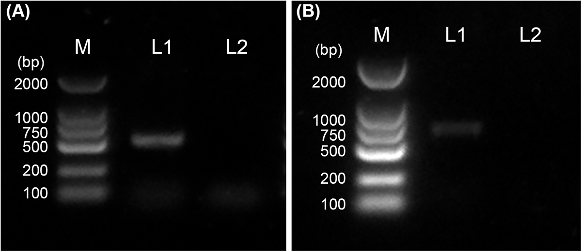
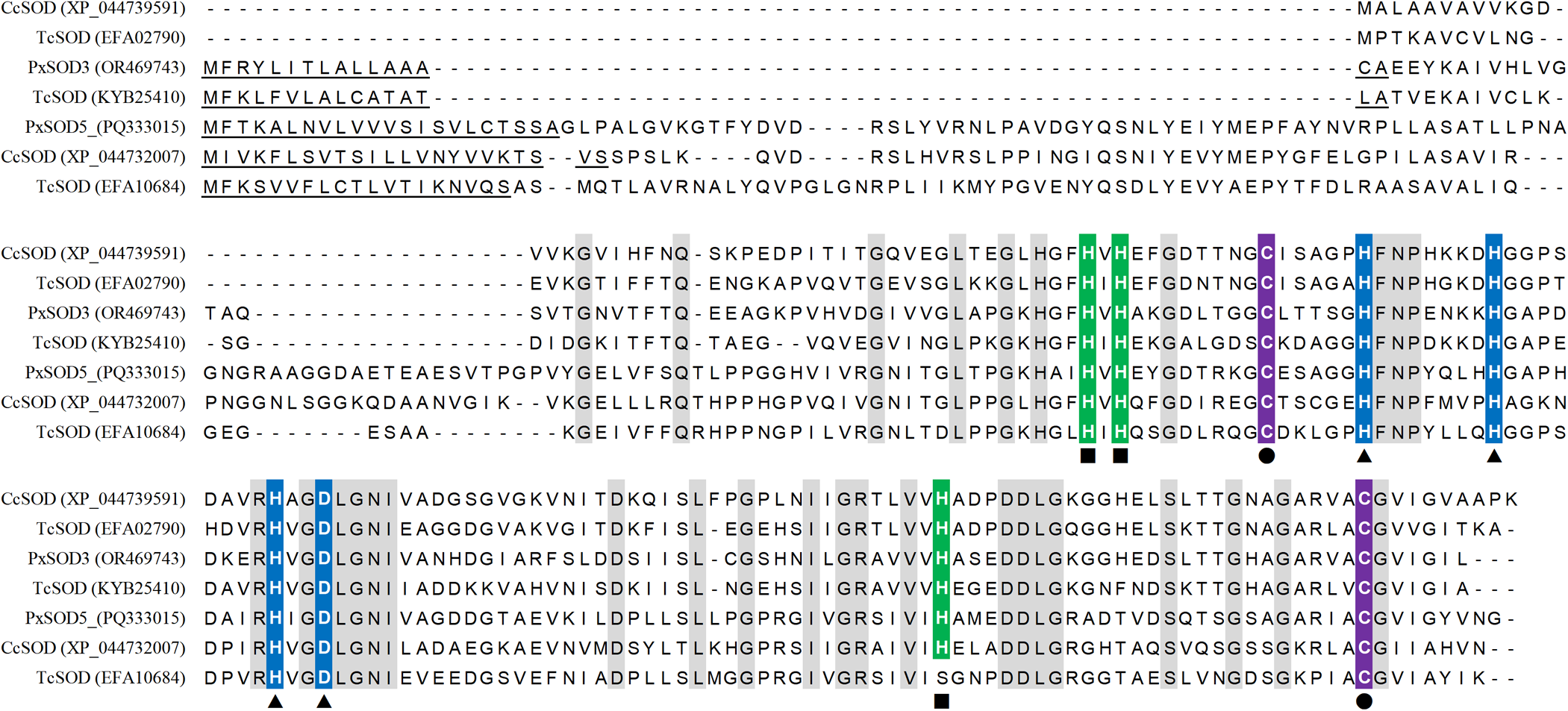
Putative extracellular SOD genes (PxSOD3 and PxSOD5) were identified in P. xanthodes. Their ORF were varied by gene clone and sequencing (fig. 1), and then deposited in GenBank under accession numbers PQ333015 and OR469743, respectively (Table S3). PxSOD3 consists of an ORF of 513 bp encoding a protein of 170 amino acids. The PxSOD5 ORF is 735 bp and encodes a protein of 244 amino acids. The predicted molecular weights of PxSOD3 and PxSOD5 are 17.61 and 25.44 kDa, respectively, and the theoretical isoelectric points are 6.07 and 5.61, respectively. PxSOD3 and PxSOD5 belong to Cu/ZnSODs and share high protein sequence identity (51.03%). Each of them contains an N-terminal signal peptide sequence, suggesting that PxSOD3 and PxSOD5 are secreted proteins (fig. 1). Histidine and aspartic acid residues at the conserved sites are predicted to be involved in the binding of catalytic copper and zinc ions. Two conserved cysteines are involved in the formation of a disulfide bond (fig. 2).
Gene clone of secreted PxSODS. (A) Gene clone of SOD3. (B) Gene clone of SOD5. M, DNA marker; L1, PCR products; L2, negative control without template.

Alignment of PxSODS with Cu/ZnSODS from Chrysoperia carnea and Tribolium castaneum. N-terminal signal peptides are underlined. Zinc and copper binding sites are indicate by triangles and squares, respectively. Conserved cys involved in disulfide bond formation are marked with circles.

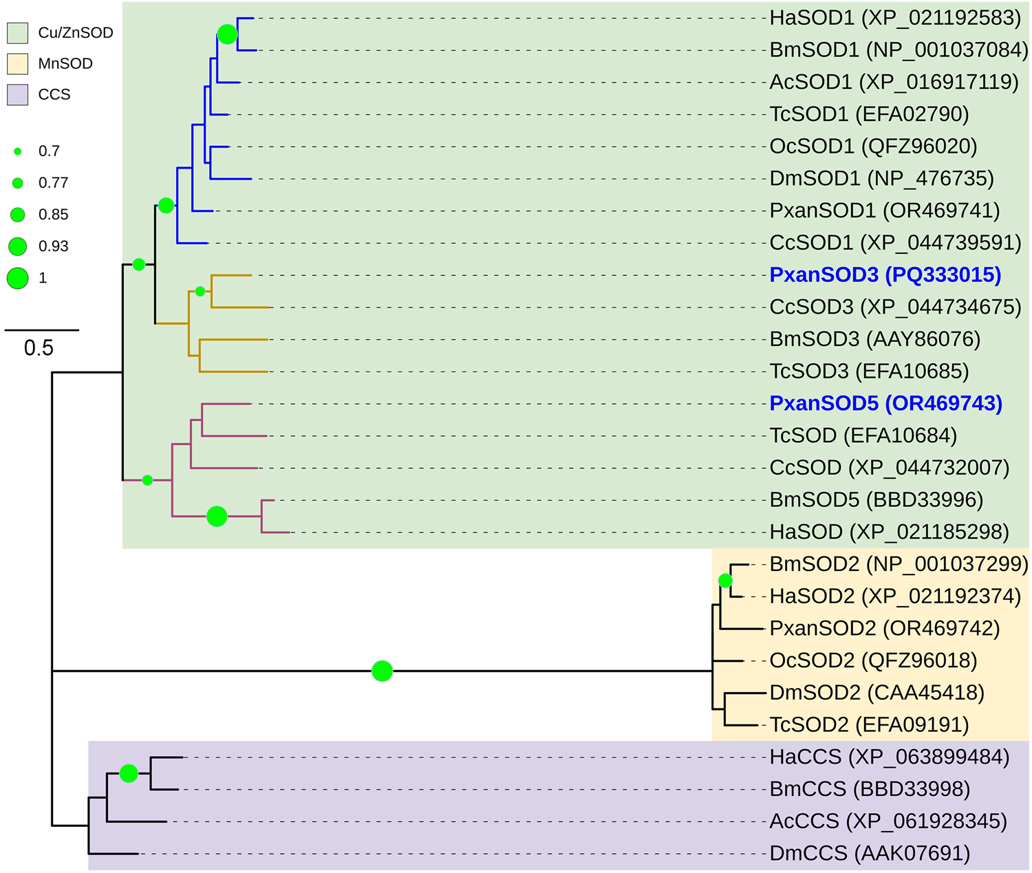
Physiological analysis of insect SODs and chaperone for SOD showed that insect SODs can be divided into two groups consisting of Cu/ZnSODs and MnSODs, respectively (fig. 3). The Cu/ZnSODs are further classified into three groups: cytoplasmic Cu/ZnSODs and two groups of extracellular Cu/ZnSODs. Both PxSOD3 and PxSOD5 are independent of the clade of P. xanthodes cytoplasmic Cu/ZnSOD PxSOD1. PxSOD3 is in a branch of extracellular Cu/ZnSODs, which contains Cu/ZnSODs from Chrysoperia carnea (Neuroptera Chrysopidae) (XP_044734675) and Tribolium castaneum (Coleoptera: Tenebrionidae) (EFA10684). PxSOD5 belongs to another extracellular Cu/ZnSODs and shares close relationship with Cu/ZnSODs in C. carnea (XP_044732007), T. castaneum (Coleoptera: Tenebrionidae) (EFA10684) and B. mori (BBD33996).
Neighbour-joining physiologic tree of insect SODS and copper chaperone for SOD. Insect sods are assigned to three subfamilies: Cu/ZnSODS, MnSODS, and copper chaperone for SOD (CCS). Genbank accession numbers are given in the brackets after the SODS names. Bootstrap values below 70% are not shown.

Protein structure of PxSOD3 and PxSOD5
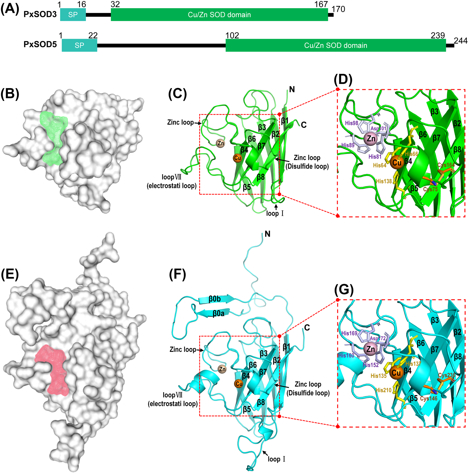
PxSOD3 and PxSOD5 contain signal peptide sequence at the N-terminus. Each of them contains a conserved Cu/ZnSOD domain (pfam00080) (fig. 4A). In addition, PxSOD5 contains an extended N-terminal domain.
Protein structure of PxSOD3 and PxSOD5. (A) Putative domain structure of PxSOD3 and PxSOD5. (B) Surface of PxSOD3. Putative reaction centre is marked by greed shadow. (C) Ribbon diagram of the structure of PxSOD3. (D) Ion-binding sites and disulfide bond in PxSOD3. (E) Surface of PxSOD5. Putative reaction centre is marked by red shadow. (F) Ribbon diagram of the structure of PxSOD5. (G) Ion-binding sites, disulfide bond, and transmembrane domain in PxSOD5. SP, n-terminal signal peptides. N, N-terminus. C, C-terminus.

Three-dimensional structures of PxSOD3 and PxSOD5 were predicted by HelixFold3. Putative reaction channel near the ion binding sites were found in these proteins (fig. 4B,E). The overall structures of PxSOD3 and PxSOD5 are flattened barrels mainly composed of 8 β-sheets. In the structure of PxSOD3, the zinc loop and the electrostatic loop (loop VII) are protruding from the barrel (fig. 4C). Cys75 at the zinc loop and Cys146 at β8 form a disulfide bond that fixes the zinc loop on the barrel (fig. 4D). Three histidines (His81, His89 and His98) and an aspartic acid (Asp101) at the zinc loop are predicted to bind a structural zinc ion. Histidine residues at β4 (His64), the zinc loop (His66) and β7 (His138) are predicted to bind a catalytic copper ion (fig. 4D). PxSOD5 contains three loops protruding from the barrel, including the loop I between β1 and β2, the zinc loop between β4 and β5, and the electrostatic loop (loop VII) between β7 and β8 (fig. 4F). The zinc loop is disrupted into two substructures by a disulfide bond formed by two conserved cysteines at the zinc loop (Cys146) and β8 (Cys236). One substructure is called disulfide loop. The other substructure contains four conserved residues (His152, His160, His19 and Asp172) that are predicted to form a zinc binding site (fig. 4G). In addition, His residues at β4 (His135), zinc loop (H137) and β7 (His210) are predicted to bind a catalytic copper ion (fig. 4G).
CPF-induced oxidative stress
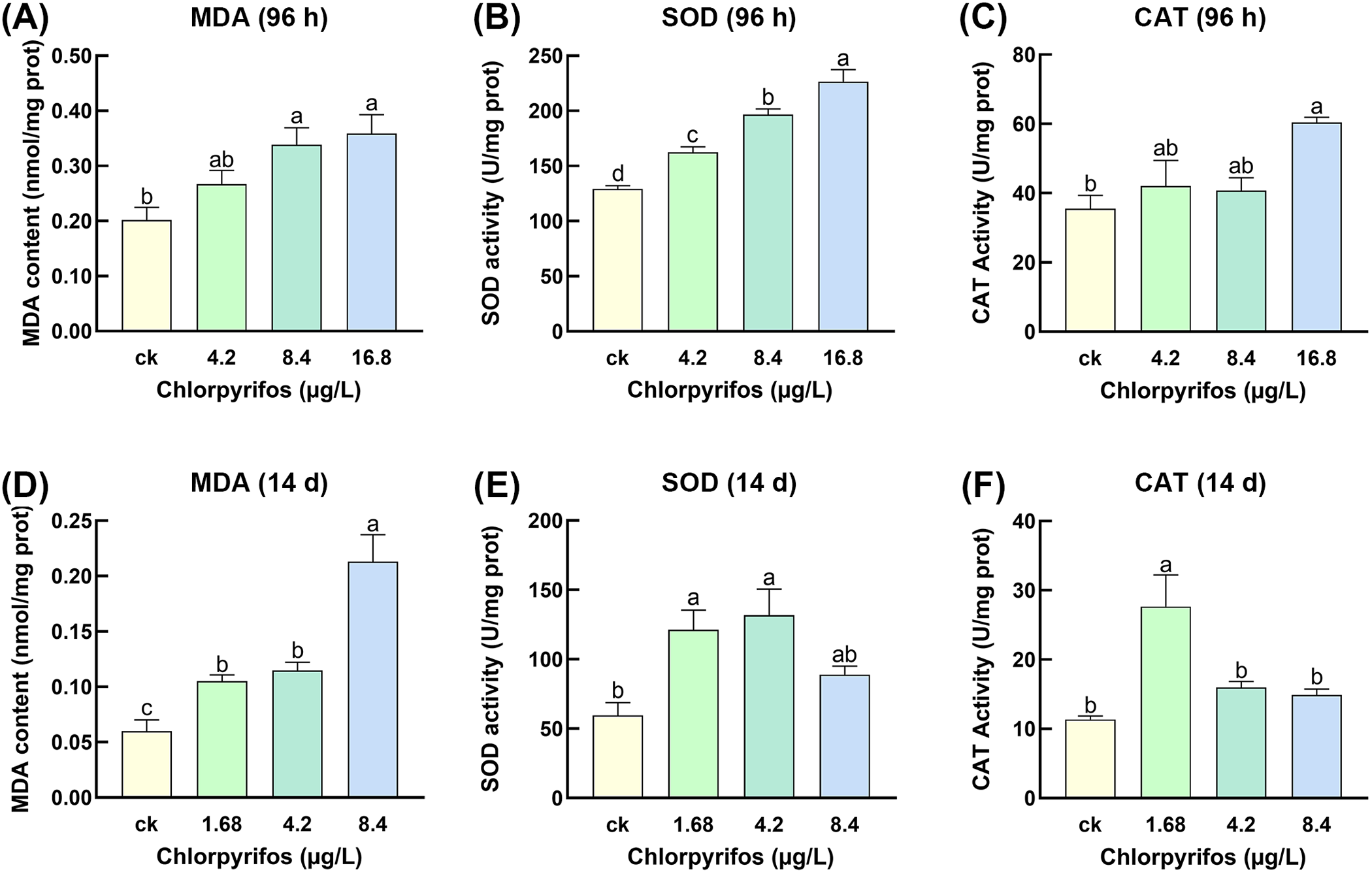
After the exposure to sublethal CPF in a simulated freshwater environment, MDA content, CAT activity, and SOD activity were determined to reveal the oxidative stress induced by CPF in the P. xanthodes larvae (fig. 5). In the 96 h CPF treatment, both MDA content and SOD activity were both increased in the sublethal CPF groups in a dose-dependent manner (fig. 5A,B). CAT activity was slightly up-regulated in 4.2 and 8.4 μg/L sublethal CPF, and was significantly higher in the 16.8 μg/L CPF group than that in no CPF control (fig. 5C). After longer term (14 d) pesticide treatment, MDA levels were also increased in the sublethal CPF groups (fig. 5D). SOD activity was significantly upregulated in the 0.168 and 4.2 μg/L CPF groups (fig. 5E). CAT activity was significantly higher in the 0.168 μg/L CPF treatment and slightly increased in the 4.2 and 8.4 μg/L CPF groups (fig. 5F).
Effects of sublethal CPF on the MDA content and antioxidative enzyme activities. (A) MDA content in response to CPF exposure (96 h). (B) SOD activity in response to CPF exposure (96 h). (C) CAT activity in response to CPF exposure (96 h). (D) MDA content in response to CPF exposure (14 d). (E) SOD activity in response to CPF exposure (14 d). (F) CAT activity in response to CPF exposure (14 d). Significant differences are marked with letters (P < 0.05, one-way ANOVA). All values are mean ± SE.

Extracellular PxSODS expression and haemolymph SOD activity in response to CPF
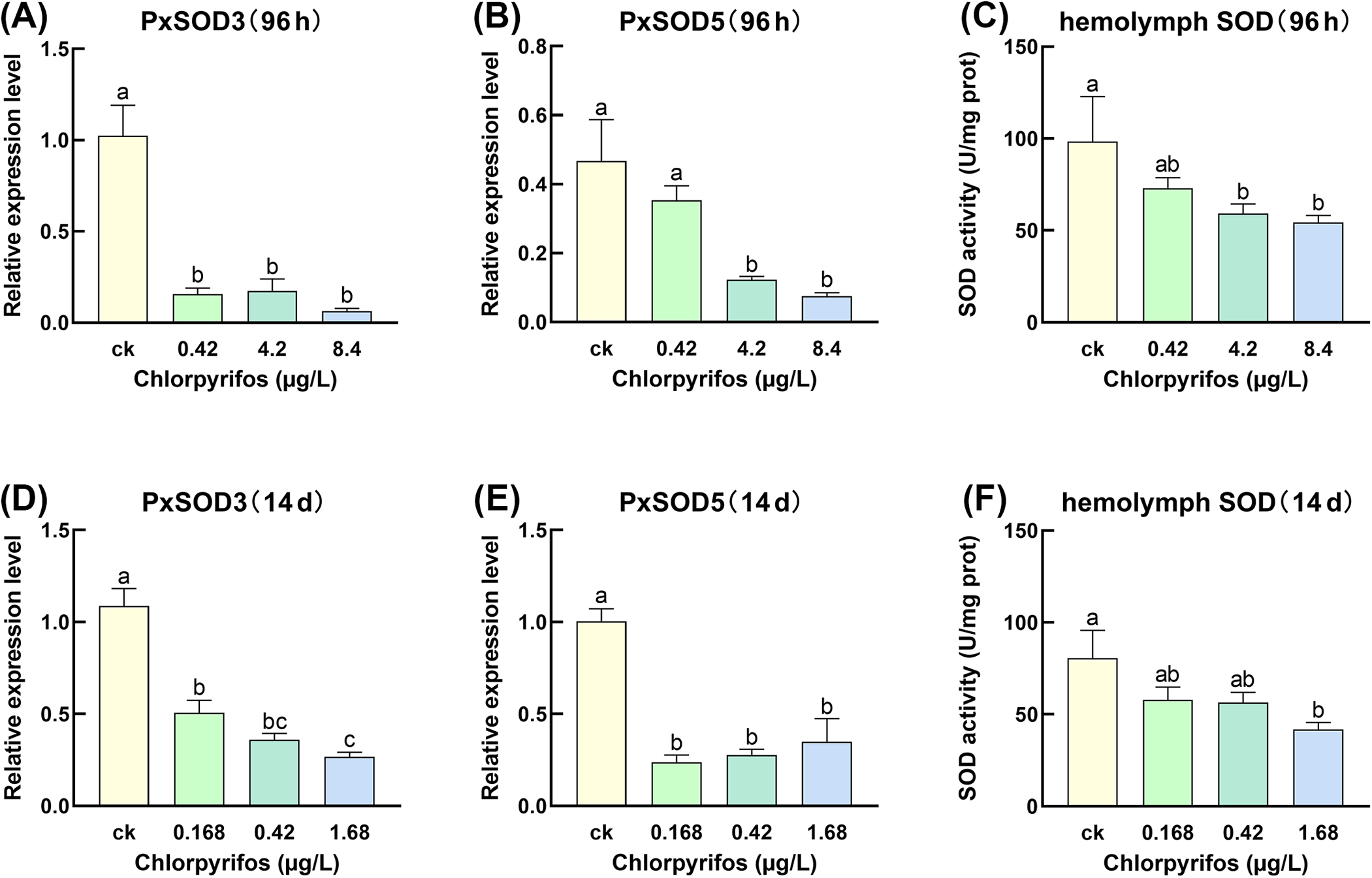
The effects of sublethal CPF on the gene expression of putative extracellular SODs and haemolymph SOD activity in P. xanthodes larvae were determined by RT-qPCR and biochemical assay, respectively (fig. 6). PxSOD3 and PxSO5 were generally down-regulated by CPF exposure. After exposure to CPF for 96 h, PxSOD3 expression levels in the 4.2 and 8.4 μg/L CPF groups are significantly lower than that in the no CPF control (fig. 6A). PxSOD5 expression was significantly downregulated in the 4.2, 42, and 84 μg/L CPF groups (fig. 6B). In the 14 d CPF treatments, both PxSOD3 and PxSOD5 were significantly downregulated in the 0.168, 4.2 and 1.68 μg/L CPF groups (fig. 6D,E). Furthermore, the haemolymph SOD activity were also downregulated by sublethal CPF in both 96 h and 14 d exposure treatments (fig. 6C,F).
Effects of sublethal CPF on the extracellular SOD gene expression and haemolymph MDA content and SOD activity. (A) PxSOD3 expression in response to CPF exposure (96 h). (B) PxSOD5 expression in response to CPF exposure (96 h). (C) Haemolymph SOD activity in response to CPF exposure (96 h). (D) PxSOD3 expression in response to CPF exposure (14 d). (E) PxSOD5 expression in response to CPF exposure (14 d). (F) Haemolymph SOD activity in response to CPF exposure (14 d). Significant differences are marked with letters (P < 0.05, one-way ANOVA). All values are mean ± SE.

Discussion
Pesticides and other harmful compounds enter the tissues of insects via dietary (gut) and waterborne (gill and cuticle) pathways and spread throughout the body mainly via extracellular fluids (Wen et al., Reference Wen, Yang, Huang and Huang2022). Therefore, the extracellular space is easily to be exposed to harmful compounds and suffers from induced oxidative stress, which is represented by a variety of ROS (Mamboungou et al., Reference Mamboungou, Fernandes, Vieira and Rocha2024). In the present study, we identified two secreted SODs in the aquatic predate insect P. xanthodes, and predicted their structural base for ROS degradation. After exposure to different concentrations of sublethal CPF for 96 h and 14 d, the MDA content, CAT activity and SOD activity were generally up-regulated in P. xanthodes larvae, indicating the CPF-induced oxidative stress. But the SOD activity in haemolymph and the expression levels of PxSOD3 and PxSOD5 were down-regulated by sublethal CPF. These results suggest that PxSOD3 and PxSOD5 are candidate extracellular antioxidant enzymes that may reduce ROS in the extracellular space. Sublethal CPF reduces secreted SOD gene expression and extracellular SOD activity, which may lead to the oxidative stress in the extracellular space.
SODs catalyse the dismutation of O2− to H2O2, and the peroxides are further inactivated by CAT and some other antioxidant enzymes (Shi et al., Reference Shi, Yu and Zhang2012). Extracellular SODs are essential for protection against extracellular oxidative damage (Cardoso et al., Reference Cardoso, Silva, Ulian de Araújo, Tanaka, Tanaka and Garratt2004; Zhang et al., Reference Zhang, Guo and Li2017b). In P. xanthodes, PxSOD3 and PxSOD5 contain the signal peptide sequence at the N-terminus and are predicted to be extracellular antioxidant enzymes. Both of them contain a highly conserved Cu/ZnSOD domain, characterized by a flattened barrel formed by antiparallel β-sheets and several loops protruding from the barrel, which is the core structure of both extracellular and intracellular Cu/ZnSODs (Moustafa et al., Reference Moustafa, Perbandt, Liebau, Betzel and Falke2022). This domain contains the reaction centre of Cu/ZnSODs, which have a Cu2+ ion relevant for catalysis and one structural Zn2+ ion; seven His and one Asp residues at the conserved sites are involved in the formation of ion coordination spheres. PxSOD5 contain the extended N-terminal domain which may be formed by coils and two antiparallel β-sheets. In the vertebrates, extracellular Cu/ZnSODs with an extended N-terminal region have been characterized as multimeric enzymes stabilized by hydrophobic interactions within the N-terminal region (Carlsson et al., Reference Carlsson, Marklund and Edlund1996; Due et al., Reference Due, Petersen, Valnickova, Østergaard, Oury, Crapo and Enghild2006). We therefore propose that the N-terminal domain in PxSOD5 may contribute to the inter-protein interaction in the extracellular space. PxSOD3 contains the Cu/ZnSOD domain close to the N-terminal signal peptide seqence. This sggests that the mature PxSOD3, with signal peptides removed, is primarily composed of the Cu/ZnSOD domain. This structural feature has also been found in the cytoplasmic Cu/ZnSOD (SOD1) and the extracellular Cu/ZnSOD (SOD3) of other insects. In B. mori, BmSOD1 and the mature form of BmSOD3 share similar structural features with mature PxSOD3. BmSOD1 and BmSOD3 were mainly expressed during the last-instar larvae and adults, and may play a role in scavenging the excessive ROS during adverse conditions (Kobayashi et al., Reference Kobayashi, Nojima, Sakamoto, Iwabuchi, Nakazato, Bono, Toyoda, Fujiyama, Kanost and Tabunoki2019). Therefore, we suggest that PxSOD3 and PxSOD5 have the structural basis for ROS scavenging and may confer extracellular antioxidant activity in this aquatic predator.
SODs are the first line of defence against ROS and play a crutial role in maintaining the oxidative homeostasis, working in concert with CAT and PRXs. Secreted members have been identified in SODs, TPXs, GPXs, and GSTs, suggesting a comprehensive extracellular antioxidant system that protects the tissues and biological materials from the damage caused by extracellular oxidative stress (Durand et al., Reference Durand, Pottier, Siaussat, Bozzolan, Maïbèche and Chertemps2018; Shi et al., Reference Shi, Zhang, Jia, Zhang, An, Wang, Zhang and Yin2014; Zhang et al., Reference Zhang, Guo and Li2017b). Extracellular SODs were widely distributed in insects from a wide variety of families (Parker et al., Reference Parker, Parker and Keller2004; Zhang et al., Reference Zhang, Guo and Li2017b). Their catalytic properties towards superoxide are critical for balancing the ROS level in the extracellular matrix. Hydrogen peroxide produced in the distribute reaction can be scavenged by extracellular PRXs. In B. mori, BmPrx4 has been characterized as an extracellular TPX and plays a role in maintaining the redox equilibrium state to reduce oxidative damage caused by pesticide (Shi et al., Reference Shi, Zhang, Jia, Zhang, An, Wang, Zhang and Yin2014). Rhodnius prolixus (Hemiptera, Reduviidae) extracellular GPX, RpSeGPx, was widely expressed in various organs and functioned in the regulation of haemolymph redox homeostasis. In Apolygus lucorum (Hemiptera: Miridae), GPXs increase the host plant susceptibility by reducing plant ROS accumulation (Dong et al., Reference Dong, Jing, Shen, Wang, Zhang, Liang, Nyawira, Xia, Zuo, Wu, Wu, Dou and Xia2020). In addition, a conserved group of secreted GSTs have been identified in the olfactory tissues of several insect orders (Zou et al., Reference Zou, Zhang, Wen, Zhang, Li, Yuan, Tang, Xiao, Shen, Tian, Huang and Yang2025). These GSTs are primarily expressed in the antennae of male and female adults and play a role in the olfactory system by degrading odorant compounds, which may also contribute to protecting the olfactory tissues from harmful compounds (Durand et al., Reference Durand, Pottier, Siaussat, Bozzolan, Maïbèche and Chertemps2018; Huang et al., Reference Huang, Fan, Liu and Feng2017). In this study, sublethal CPF induced oxidative stress in P. xanthodes larvae; extracellular SOD expression and SOD activity in the haemolymph were also downregulated. These results suggest that low concentrations of CPF may cause toxic effects by inducing oxidative stress in this aquatic insect. The extracellular space may be an important site for CPF to induce oxidative stress because of the increase in extracellular SOD gene expression and catalytic activity.
Pesticides released into the environment can cause oxidative stress in the non-target insects and may also disrupt the gene expression patterns in a variety of biological processes (Macedo et al., Reference Macedo, Batista, Souza, Dafre, Farina, Kuca, Posser, Pinto, Boldo and Franco2024; Parra Morales et al., Reference Parra Morales, Alzogaray, Cichón, Garrido, Soleño and Montagna2017). A previous study in P. xanthodes showed that CPF can inhibit acetylcholinesterase activity in the larvae and downregulate the expression levels of cholinergic neuronal genes. In Philosamia ricini (Lepidoptera: Saturniidae), sublethal CPF showed adverse effects on the overall nutritional physiology and genotoxicity in the larvae, which could reduce the larvae survivability (Kalita et al., Reference Kalita, Haloi and Devi2016). In this study, the expression of extracellular SODs and haemolymph SOD activity in P. xanthodes larvae are downregulated by exposure to sublethal CPF, suggesting the effects of low concentrations of CPF on the extracellular antioxidant system. Extracellular Cu/ZnSODs play a major role in maintaining the ROS balance in the extracellular space and are essential for antioxidant pathways that protect the cells and tissues from the oxidative damage (Blackney et al., Reference Blackney, Cox, Shepherd and Parker2014; Xikeranmu et al., Reference Xikeranmu, Abdunasir, Ma, Tusong and Liu2019). Their gene expression and catalytic activity are highly correlated with a variety of essential physiological processes. In whiteflies, extracellular SOD was highly expressed in invasive species and functionally involved in host adaptation (Gao et al., Reference Gao, Li, Xu, Yan, Jiu, Liu and Wang2015). The extracellular SOD in Microdera punctipennis (Coleoptera: Tenebrionidae) was up-regulated by cold stress and may contribute to cold tolerance (Xikeranmu et al., Reference Xikeranmu, Abdunasir, Ma, Tusong and Liu2019). In B. mori, BmSOD3 was enriched in the midgut and Malpighian tubules and plays a key role in the response to bacterial, fungal, and viral invasion (Shi et al., Reference Shi, Shen, Ren and Yang2021). The extracellular SOD in Dastarcus helophoroides (Coleoptera: Bothrideridae) showed a fluctuated expression during development, and helps to eliminate oxidative damage products accumulated during ageing and may retard aging (Zhang et al., Reference Zhang, Guo and Li2017b). These facts suggest that exposure to CPF may has extensive effects on various biological processes in insects by disrupting the gene expression and catalytic activity of extracellular SODs and may further affect the population dynamics. Therefore, CPF contamination has been recognized as an important driving factor for global insect diversity decline (de Beeck et al., Reference de Beeck, Verheyen and Stoks2018; Yang et al., Reference Yang, Ma, Yan, Liu, Diao and Dai2019; Zafar et al., Reference Zafar, Van Wijngaarden, Roessink and Van den Brink2011).
Supplementary material
The supplementary material for this article can be found at https://doi.org/10.1017/S0007485325100151.
Acknowledgements
We thank to the funds from Natural Science Foundation of Hunan Province (grant number 2024JJ7416, 2020JJ5454).
Author contributions
X.W. conceived the study. X.L.H. and Z.W. designed the experiments. X.W., Y.L., J.Y., Y.Z., X.R.H., J.Z., and M.Z. performed the experiments. Y.L., X.H.W., and W.Z. analysed the data. X.W. wrote the manuscript. X.L.H. and Z.W. revised the manuscript. All authors read and approved the final manuscript.
Competing interests
None.