INTRODUCTION
Spionidae is one of the most diverse polychaete families, and their members can be easily recognized by the presence of a pair of long and grooved ciliated palps, although these structures are easily lost when collected. Other characteristic traits of the family are the presence of hooded hooks in a variable number of chaetigers (usually combined with bilimbated capillaries in the same or different chaetigers), the postchaetal lamellae associated with both noto- and neuropodia in anterior segments, and the branchiae lying dorsally to notopodia. Spionids use their palps either to collect food from the water column or to selectively collect particles from sediment surface (Blake, Reference Blake, Blake, Hilbig and Scott1996). Members of this family may be dominant in soft bottoms from intertidal to abyssal depths, usually inhabiting tubes constructed of sediment and mucus, although many species bore calcareous substrata instead.
During the Antarctic summers of 2002–2003 and of 2005–2006, the Spanish cruises BENTART to the Bellingshausen Sea were conducted on board RV ‘Hesperides’ and surveyed an area extending from the Antarctic Peninsula (70°W) to the Thurston Island (100°W), with a northern limit defined by the bathymetric line of 2000 m and a southern one by the border of permanent ice cover (Ramos & Moya, Reference Ramos and Moya2003). In the study, numerous individuals of a great number of polychaete species were identified, several of which pertained to Spionidae. Most of the specimens were identified as belonging to well known Antarctic species, but some others were assigned to Aonidella cirrobranchiata (Day, Reference Day1961), a species never recorded from the Austral Ocean. In the present paper a taxonomic account of this taxon is given.
MATERIALS AND METHODS
The objective of the project was to get a complete picture of the benthic community at each station and consequently a number of sampling devices were employed at every site: an Agassiz trawl for epibenthic fauna, a box-corer grab for endofauna and a sledge equipped with three superimposed nets (0.5 mm gauge) for suprabenthic fauna. Further details on the sampling procedures can be found in Ramos & Moya (Reference Ramos and Moya2003), Saiz et al. (Reference Saiz, García, Manjón, Parapar, Peña-Cantero, Saucède, Troncoso and Ramos2008) and San Vicente et al. (Reference San Vicente, Munilla, Corbera, Sorbe and Ramos2009). All the Aonidella specimens herein reported were collected by means of the suprabenthic sledge.
The material retained in the nets of the suprabenthic sledge was sorted using stereomicroscopes in the ship; polychaetes were identified to family level and fixed in 4% formalin in seawater. Specimens were examined using a stereomicroscope and a compound microscope with differential contrast optics (Nomarski) provided with a drawing tube for the figures. Body width measurements exclude parapodia, branchiae or cirri. One specimen was dehydrated and gold coated for SEM study following standard procedure and the remainder of the specimens were preserved in 70% ethanol and deposited in Museo Nacional de Ciencias Naturales de Madrid (MNCNM). In order to accurately identify the Antarctic specimens, type material was borrowed from the British Museum of Natural History. The holotype is apparently lost (Macioleck, 2000), but two paratypes (BMNH ZK1961.9.480/1) and some material collected in the same area and time and determined by J.H. Day are still available for examination.
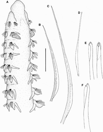
Aonidella cirrobranchiata (Day, Reference Day1961) MNCN 16.01/11621. (A) Anterior end, dorsolateral view; (B) chaetiger 4, anterior notochaeta; (C) chaetiger 4, posterior notochaeta; (D) chaetiger 20, capillary notochaeta; (E) chaetiger 20, notopodial hooded hooks; (F) chaetiger 30, neuropodial hooded hook. Scale bar. A: 0.76 mm; B–F: 48 µm.

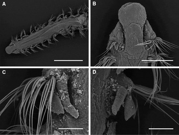
Additional material of Aonidella cirrobranchiata (Day, Reference Day1961). (A) Anterior end, dorsal view; (B) detail of prostomium and first chaetiger, dorsal view, same specimen; (C) chaetiger 9, dorsal view, showing branchia and notopodial postchaetal lobe, same specimen; (D) chaetiger 10, dorsal view, showing branchia and noto- and neuropodial postchaetal lobes, same specimen. Scale bars. A: 1 mm; B: 0.3 mm; C, D: 0.1 mm.

MATERIAL EXAMINED
MNCN 16.01/11621, 70°14′31.2″S–95°1′58.8″W, lowest net of suprabenthic sledge, fine sand, 1958 m, 20 January 2006, 17 anterior fragments plus two median fragments of nine and 23 chaetigers respectively; an additional specimen dehydrated for SEM, from the same sample. BMNH 1961.19.572-595, off South Africa, Stations 228R, 232P and 233 R, 27 specimens, det. J.H. Day
DESCRIPTION
Largest specimen incomplete, an anterior fragment 8.75 mm long, 0.4 mm wide for 23 chaetigers; posterior end lacking in all the specimens. Body squared in section, with intersegmental furrows ill-defined on dorsal and ventral surfaces; colour in alcohol white with notopodial postchaetal lamellae of chaetiger 2 pigmented in red. Prostomium (Figures 1A & 2A, B) broadly rounded anteriorly, flattened; not fused with peristomium; eyes, occipital tentacle, and caruncle absent; bifid nuchal organ extending to posterior end of chaetiger 1 (Figures 1A & 2B). Peristomial lateral wings absent; peristomium partially fused to chaetiger 1 (Figures 1A & 2B). Palps not observed. Notopodial postchaetal lamellae triangular and placed laterally (Figures 1A & 2C, D); smallest on chaetiger 1 (Figures 1A & 2B); much larger and distinctly pigmented in red on chaetiger 2; not pigmented and slightly shorter along the rest of body (Figures 1A & 2A). Neuropodial postchaetal lamellae similar but smaller than notopodial ones; small on chaetiger 1; triangular and larger along the rest of body, not being distinctly larger on chaetiger 2 (Figures 1A & 2D). Chaetae of anterior segments narrowly limbate capillaries, striated, with fine granules imbedded in shaft; arranged in two rows in each bundle, with chaetae of anterior row (Figure 1B) slightly curved and shorter than chaetae of posterior one (Figure 1C); parapodia of first chaetiger with about 6 capillaries in notopodium and 10 in neuropodium; following chaetigers with fascicles of about 20 capillary notochaetae and 25 capillary neurochaetae; number of capillary chaetae decreasing after branchial region. Noto- and neuropodial hooks bidentate, with main fang surmounted by a smaller, straight distal tooth and with a narrow hood (Figure 1E, F); at first accompanied by 3–4 fine capillaries (Figure 1D), then only hooks. Neuropodial hooks (Figure 1F) from chaetigers 18–19, up to 8 per fascicle; notopodial hooks (Figure 1E) from about chaetiger 30, up to 6–7 per fascicle. Branchiae (Figure 1A) from chaetiger 2, numbering 10 pairs; cirriform, 1 ½–2 times as long as notopodial postchaetal lamella, not reaching mid-dorsal line when abated (Figure 2C, D); attached to base of corresponding notopodia and free from postchaetal lamella. Pygidium absent on all specimens studied.
REMARKS
The collected specimens are accurately referred to genus Aonidella Maciolek in López-Jamar (Reference López-Jamar1989) (composed of only two species) based on their flattened, rounded prostomium lacking occipital tentacle and posterior caruncle. They differ clearly from A. dayi Maciolek in López-Jamar (Reference López-Jamar1989), from both sides of the North Atlantic, the Mediterranean Sea and Japan (López-Jamar, Reference López-Jamar1989; Maciolek, Reference Maciolek2000), in that the hooded hooks of this species bear two distal teeth instead of one. On the contrary, the specimens herein reported fit quite well the diagnosis of A. cirrobranchiata (Day, Reference Day1961), a species described from South Africa (Day, Reference Day1961), and subsequently reported from the Bay of Biscay (Maciolek, Reference Maciolek2000). Some slight differences between the collected specimens and both the original description and previous reports were noted, but they are a matter of interpretation. Thus, Maciolek described the capillary chaetae of anterior chaetigers as broadly limbate, but they have been herein considered to be narrowly limbate; the comparison of Antarctic and South African specimens showed them to be identical. Likewise, the presence of a pair of slightly enlarged, reddish pigmented notopodial postchaetal lamellae on chaetiger 2 was observed in some of the specimens loaned by the British Museum, although it was not mentioned in any previous description. Finally, the higher number of hooks per parapodial lobe in mid-body chaetigers in Antarctic specimens might be caused by their larger size and seems not important enough to erect a new species.
Aonidella cirrobranchiata has a very irregular distribution. The original description was based on a number of specimens collected west of Cape of Good Hope (South Africa) from moderate depths (144). Shortly after, several specimens were collected in the area, from depths not extending beyond 400 m (Day, Reference Day1961, Reference Day1967). Out of this area, just a single specimen was collected prior the present work (Maciolek, Reference Maciolek2000). This record was from Bay of Biscay (46°17.5′N–04°45.2′W) and the sample was collected from much deeper bottoms (1336 m). The present record enlarges the geographical range of the genus and the species to the Southern Ocean, and it is also from a distinctly deeper sample than those of the type locality. At first glance, the collection of typically benthic spionids by means of a suprabenthic sledge seems surprising. However, since small amounts of sand were also retained within the net, their presence might be explained by resuspension of the bottom sediment during the towing procedure.
ACKNOWLEDGEMENTS
The present study was financially supported by the Spanish Ministerio de Ciencia y Tecnología through the research projects ‘Estudio integrado de la biodiversidad bentónica del Mar de Bellinghausen y península Antártica (Antártida del Oeste). Primera campaña de muestreo a bordo del RV “Hespérides” REN2001-1074/ANT’ and ‘Estudio integrado de la biodiversidad bentónica del mar de Bellingshausen y península Antártica. Segunda campaña de muestreo a bordo del RV “Hespérides” CGL2004/01856’. The author wishes to express his gratitude to the crew of RV ‘Hesperides’ as well as to the research staff of the project, especially to Dr Ana Ramos and Dr Julio Parapar, for their collaboration both aboard the ship and on land. Emma Sherlock, from the Natural History Museum in London, and Javier Sánchez, Francisco Yagüe and Patricia Álvarez, from Museo Nacional de Ciencias Naturales in Madrid are gratefully acknowledged for the loan and management of the studied material. The useful comments of two anonymous referees are sincerely acknowledged, too.