Introduction
Sinonasal malignant melanoma is uncommon and carries a poor prognosis. The onset of symptoms is often slow and insidious, so patients often present late with advanced disease.
Because this tumour is uncommon, it is difficult to establish an evidence-based management approach from the literature, particularly as regards endoscopic resection versus other techniques.Reference Lund, Howard and Wei1 Most cases are presented in the form of isolated case reports, with only a few small series examining data collected over many years.Reference Thompson, Wieneke and Miettinen2
At present, surgery with or without adjuvant radiotherapy remains the treatment of choice. The role of chemotherapy in treating metastasis remains unclear.
The current study reports 24 patients with sinonasal malignant melanoma treated over a 25-year period, and reviews the current literature. A literature review was conducted because, whilst many of our patients had recurrent disease and did poorly, others did well; however, this did not appear to relate to their treatment.
Materials and method
We studied the medical records of all patients with sinonasal malignant melanoma treated at Queen's Medical Centre Nottingham over 25 years. Cases were identified from the hospital pathology archives and relevant medical records subsequently reviewed. Data obtained included patient demographics and the anatomical site and extent of disease, including evidence of local and distant metastasis. For each individual case, we recorded the type of treatment, timing of disease recurrence and overall survival.
We also reviewed the Medline and PubMed databases for English language articles concerning mucosal malignant melanoma of the nasal cavity and paranasal sinuses. If a single centre had published more than one case series, only the most recent series was reviewed.
Results
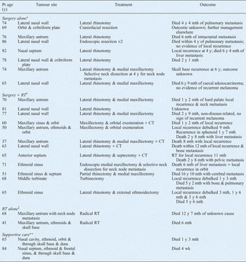
Our results are summarised in Table I. Twenty-six patients were identified, but only 24 complete medical records were available for analysis. These 24 patients presented between October 1982 and October 2007 and comprised 11 men and 13 women. The mean age at presentation was 67.5 years (range, 37–86 years). All patients presented with nasal symptoms, with a mean duration of 18 weeks (range, one to 104 weeks).
Patient data and outcome, by treatment modality

*n=8; †n = 12; ‡n = 2; **n = 2. For surgery alone: mean 5-y survival = 37.5 per cent; mean overall survival = 4 y; median survival = 4 y 4 mth. For surgery plus radiotherapy: mean 5-y survival = 25 %; mean overall survival = 3 y 6 mth; median survival = 2 y 8 mth. Pt = patient; y = years; mth = months; RT = radiotherapy; CT = adjuvant chemotherapy; wk = weeks
Regarding primary anatomical site, seven cases arose from the maxillary sinus, eight from the lateral nasal wall, three from the ethmoid sinuses and two from the nasal septum. In four cases, disease was so extensive at presentation that it was not possible to discern from where it had arisen. Three patients had cervical lymph node metastasis at presentation, and one patient had widespread metastasis to the liver, spleen, bone and brain. No consistent or accurate staging system was recorded for any patient.
Treatment modalities included surgery with or without post-operative radiotherapy, radiotherapy alone, and supportive care. Adjuvant chemotherapy was used in four cases with very advanced disease. The overall five-year survival for all patients was 30 per cent. The mean survival was three years and eight months (median, two years and eight months).
Looking at the different treatment groups, 12 patients had surgery with post-operative radiotherapy; these patients' overall five-year survival was 25 per cent, with a mean survival of three years and six months (median, two years and eight months).
Eight patients were treated with surgery alone; of these, three lived beyond five years (37.5 per cent), with a mean survival of four years (median, four years and four months).
Two patients were treated with radiotherapy alone. One of these patients had a tumour of the maxillary antrum with cervical lymph node metastasis. This patient developed petit mal seizures following treatment, thought to be due to radiation injury. Nonetheless, she survived for 12 years and seven months, and died with no evidence of local recurrence or distant metastasis. The other patient had very extensive disease involving the maxillary and ethmoid sinuses, with extension to the skull base. This patient died six months after radiotherapy having had no response to treatment.
Two patients were managed palliatively after presentation. One patient had systemic metastases involving the liver, spleen, bone and brain, and survived only four weeks. The other patient had a tumour that had breached the dura, and elected to forego treatment. This patient survived for one year and three months.
Discussion and literature review
Incidence
Primary sinonasal tract mucosal malignant melanomas are rare, accounting for 0.3 to 2 per cent of all malignant melanomas and approximately 4 per cent of head and neck melanomas.Reference Freedman, De Santo, Devine and Weiland3–Reference Thompson, Morgan and Bradley9 The incidence of mucosal malignant melanoma is lower in areas where the incidence of cutaneous malignant melanoma is high; furthermore, the incidence of the mucosal form does not appear to be increasing, while the incidence of the cutaneous form has significantly increased in recent decades.Reference Pandey, Mathew, Iype, Sebastian, Abraham and Nair10 Sinonasal malignant melanoma has an equal sex distribution, and a higher proportion (10.4 per cent) identified in Afro-Caribbean compared with Caucasian populations.Reference Thompson, Morgan and Bradley9
The National Cancer Data Base report by the American College of Surgeons Commission on Cancer and the American Cancer Society reported that, of 84 836 melanomas seen between 1985 and 1994, only 1.3 per cent arose from mucosal surfaces.Reference Chang, Karnell and Menck7 Of these, 55 per cent were in the head and neck. Sinonasal malignant melanoma appears to present later in life than cutaneous malignant melanoma, with a mean patient presentation age of 64.3 years; patients over the age of 60 years do less well.Reference Thompson, Morgan and Bradley9
Malignant melanomas arising from the nasal cavity are more common than those arising from the sinuses. Approximately 80 per cent of sinonasal malignant melanomas are found within the nasal vault and 20 per cent in the paranasal sinuses. The tumour is nearly always a primary lesion which arises de novo rather than from a pre-existing naevus. The most common sites are the lateral nasal wall, particularly the inferior and middle turbinates, followed by the nasal septum. Sometimes the lesion is so extensive at presentation that it is difficult to clearly establish from where it has arisen.Reference Iversen and Robins4, Reference Chiu and Weinstock6, Reference Lentsch and Myers11 Tumours tend to grow slowly and present late.Reference Manolidis and Donald5, Reference Lund12 Up to a third of patients present with neck node metastases or distant metastases, which are usually rapidly fatal.Reference Dauer, Lewis, Rohlinger, Weaver and Olsen13
Aetiology and pathogenesis
Melanocytes are derived from neural crest tissue and are distributed throughout the upper aerodigestive tract in all races. They are found in the mucosa, superficial and deep stroma of the turbinates and septum, and the secretory glands, and are particularly abundant around the olfactory cleft neuroepithelium.
The precise aetiology of sinonasal malignant melanoma is unclear, although the presence of long-term melanosis (i.e. pigment in the mucosa) is the most clearly established association in the case of oral cavity tumours.Reference Mangararo, Hammond, Dalton and Williams14 Occupational exposure to formaldehyde has been reported as a possible risk factor in several series.Reference Thompson, Wieneke and Miettinen2, Reference Brandwein, Rothstein, Lawson, Bodian and Urken15, Reference Holmstrom and Lund16 This chemical is used in various manufacturing processes to make products including adhesives, foam insulation, paints, cosmetics and textiles; it is also found in tobacco smoke.
Studies of oral mucosal malignant melanoma have demonstrated a mutation of the KIT proto-oncogene, which encodes the protein C-KIT, and this may play a part in the early stages of tumour development.Reference Rivera, Nagatsuka, Gunduz, Cengiz, Gunduz and Siar17
Clinical presentation can include nasal obstruction (usually unilateral) and epistaxis. Symptoms are often ignored by the patient for many months. Later, a mass (usually, but not always, pigmented) may be visible within the vestibule, sometimes large enough to cause external swelling of the nose. Late signs include proptosis, ophthalmoplegia causing double vision, and epiphora.
Mucosal melanomas of the sinonasal tract tend to be more aggressive and have a poorer prognosis than their cutaneous counterparts, with five-year survival rates of 22 to 46 per cent. This may relate to diagnostic delay due to the tumour's hidden site and non-specific presenting symptoms.Reference Thompson, Wieneke and Miettinen2, Reference Huang, Liao, Kan and Chen18, Reference Bridger, Smee, Baldwin, Kwok and Bridger19 A review from the Mayo clinic found a statistically significant survival benefit if the primary lesion was located on the nasal septum as opposed to the sinuses or lateral nasal wall.Reference Dauer, Lewis, Rohlinger, Weaver and Olsen13 The overall poor prognosis is attributable to both local recurrence and distant metastasis.Reference Rinaldo, Shaha, Patel and Ferlito20
Investigation
Computed tomography of sinonasal malignant melanoma typically shows soft tissue opacification with or without erosion of surrounding bone. Magnetic resonance imaging is useful in discerning a soft tissue mass due to retained secretions within the paranasal sinuses, and in assessing skull base involvement. Imaging the neck, chest and abdomen is mandatory to establish the presence of regional and distant metastasis; positron emission tomography scans are possibly more sensitive in detecting such metastases.Reference Gorres, Stoeckli, von Schulthess and Steinert21
Histology
Many previously reported case diagnoses must be viewed with caution, as a lack of histochemical, immunochemical and ultrastructural support brings many such diagnoses into question.Reference Thompson, Wieneke and Miettinen2 Making an accurate diagnosis of sinonasal malignant melanoma can be difficult, in the light of a differential diagnosis which includes sinonasal undifferentiated carcinoma, poorly differentiated carcinoma, adenocarcinoma, lymphoma, rhabdomyosarcoma, angiosarcoma, neuroendocrine carcinoma, olfactory neuroblastoma and plasmacytoma.
Histologically, most sinonasal malignant melanoma tumours comprise large, epithelioid cells with abundant eosinophilic cytoplasm and round nuclei showing eosinophilic nucleoli, or spindle cells. About a third of tumours have undifferentiated, small, round, blue cells that may be mistaken for lymphoma. There may be a variety of morphological tumour cell types, but the majority of cells have cytoplasmic pigment. It may be more difficult to make a diagnosis when the tumour is amelanotic and/or shows surface ulceration.
The immunohistochemical profile of sinonasal malignant mucosal melanoma is identical to that of cutaneous lesions. Markers include S-100 protein, HMB-45 (antibody), melan-A, microphthalmia transcriptase factor, tyrosinase, vimentin and cytokeratin (Fontana–Masson stain). Positivity for S-100 is not always present, so positivity for a series of markers is needed to make the diagnosis. PNL-2 (antibody) shows promise as a more specific marker.Reference Morris, Wen, Nonaka, DeLacure, Kutler and Huan22, Reference Billings, Wang, Sercarz and Fu23
Certain histopathological features of sinonasal malignant melanoma are predictors for poor survival, and include vascular invasion, necrosis and a polymorphous tumour cell population. However, because these three parameters are associated with each other, none are independently predictive of outcome by multivariate analysis. In the diagnosis of sinonasal malignant melanoma, little prognostic significance has been found for tumour thickness, level of invasion, ulceration, mitotic index or nerve involvement. This demonstrates that the histological prognostic features relevant to cutaneous malignant melanoma differ significantly from those for mucosal malignant melanoma.Reference Prasad, Patel, Hoshaw-Woodard, Escrig, Shah and Huvos24, Reference Patel, Prasad, Escrig, Singh, Shaha and Kraus25
However, the Ki67 antigen has been found to be a useful marker for proliferative activity, and has been used with some success as a prognostic marker in cutaneous melanoma.Reference Cuevas, Jones and Wright26 In one study of 27 patients with mucosal melanoma, Kim et al. demonstrated a survival benefit in patients who had less than 35 per cent Ki67 staining, compared with patients with more than 35 per cent staining.Reference Kim, Kim, Kim, Kim, Lee and Rhee27
Staging
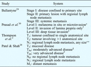
Accurate staging of sinonasal malignant melanoma can be difficult, but it is important for documentation purposes as it enables risk stratification for any given stage of the disease; it also enables comparison of treatment modalities. However, various staging systems for sinonasal malignant melanoma have been used to date (Table II), which makes it difficult to extract meaningful prognostic information from the available literature.
Staging systems for sinonasal malignant melanoma

*No invasion, 75% 5-year survival; †52% 5-year survival; ‡23% 5-year survival. **Ipsilateral, bilateral or contralateral. §Involving deep soft tissue, cartilage, bone or overlying skin. #Involving brain, dura, skull base, lower cranial nerves, masticator space, carotid artery, prevertebral space or mediastinal structures. T = tumour; N = node; M = metastasis
In the seventh edition of the The American Joint Committee on Cancer (AJCC) cancer staging manual, mucosal malignant melanoma of the upper aerodigestive tract was given its own tumour–node–metastasis (TNM) staging system, which has now been in use since January 2010. To reflect this tumour's aggressive behaviour and poor outcome, even disease limited to the mucosa is staged as T3, and as such T1 and T2 lesions do not exist, while T4a and T4b stages represent advanced disease.Reference Patel, Shah, Edge, Byrd, Carducci and Compton28
Previously used staging systems appear frequently in the literature and are worth discussing.
In 1970, Ballantyne described a system which has been used by many clinicians over the years because of its simplicity.Reference Ballantyne29 The disadvantage of this staging system was that most patients presented with early stage I disease, so the system offered limited prognostic information.Reference Prasad, Busam, Patel, Hoshaw-Woodward, Shah and Huvos31
Prasad et al. recognised this limitation, and advocated a staging system based on the degree of invasion of the mucosa.Reference Prasad, Patel, Huvos, Shah and Busam30 They found that this was a significant independent predictor of survival which gave more reliable prognostic information, particularly in the patient group with Ballantyne's stage I disease.
A system proposed by Thompson et al. for the US Armed Forces Institute of Pathology used a TNM-like classification which utilised regional lymph node status and distant metastasis as predictors of biological behaviour.Reference Thompson, Wieneke and Miettinen2 However, use of this system has not been widespread, with many favouring the AJCC TNM classification for sinonasal malignancy. This latter system was not tumour type specific but gave more emphasis to tumour size and extent, and therefore probably provided more accurate prognostic information.
Treatment and results
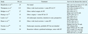
There is no consensus on how to manage mucosal malignant melanoma.Reference Prasad, Patel, Huvos, Shah and Busam30 As a consequence, patients continue to be treated differently in different centres (Table III).
Published data on sinonasal malignant melanoma: summary

Pt = patients; mth = months; y = years; RT = radiotherapy; CT = chemotherapy
The primary treatment modality is surgical resection with a wide margin, as incomplete resection has been shown to be a predictor of poor survival.Reference Lee, Shimizu, Tran, Juillard and Calcaterra32, Reference Penel, Mallet, Mirabel, Van and Lefebvre33 However, it is nearly always impossible to perform a truly radical resection, because of surrounding vital structures and also the surgeon's desire to achieve local control without loss of function or cosmetic disfigurement. Prophylactic neck dissection in an N0 neck is not performed, as the incidence of occult nodal metastases is relatively low.Reference Patel, Prasad, Escrig, Singh, Shaha and Kraus25
Historically, malignant melanoma has been regarded as radioresistant. However, some studies have suggested that radiotherapy for sinonasal malignant melanoma may improve local control. Despite this, no formal randomised trials have been published, and there is no good evidence that such treatment prolongs survival. Owens et al. used adjuvant radiotherapy in a small series of 11 patients, and found that whilst this treatment did not significantly improve overall survival, it did decrease the local recurrence rate.Reference Owens, Roberts and Myers34 A large, retrospective review by Lund et al. assessed whether surgery combined with radiotherapy conferred any survival benefit over radical local excision alone. The authors concluded that the overall patient survival was poor and did not appear to be improved by the addition of radiotherapy.Reference Lund, Howard, Harding and Wei35
Thompson et al. treated patients with surgery and adjuvant radiotherapy, in a series of 115 cases, and found that the specific type of treatment did not seem to influence the overall outcome. There was no significant difference in outcome between patients treated by surgery, radiotherapy or chemotherapy, in any combination.Reference Thompson, Wieneke and Miettinen2
Wada et al. retrospectively studied 31 patients with head and neck mucosal melanoma who had received radiotherapy. Ten had macroscopic residual disease following surgery and 21 received radiotherapy alone. These authors reported a 29 per cent complete response rate and a 58 per cent partial response rate, but overall survival was no better than in other published series.Reference Wada, Nemoto, Ogawa, Hareyama, Yoshida and Takamura36
Teman et al. treated 46 patients with sinonasal malignant melanoma and 23 with malignant melanoma of the oral cavity and pharynx. They concluded that patients with small tumours who received post-operative radiotherapy had better local disease-free survival than patients with larger tumours who did not receive radiotherapy.Reference Teman, Mamelle, Marandas, Wibault, Avril and Janot37 This benefit is however questionable, given that the patients who received radiotherapy had less advanced disease.
Both Patel et al. and Nandapalan et al. found that radiotherapy did not provide any local control in their series.Reference Patel, Prasad, Escrig, Singh, Shaha and Kraus25, Reference Nandapalan, Roland, Helliwell, Williams, Hamilton and Jones38 Another study of mucosal malignant melanoma of the head and neck suggested that radiotherapy increases the local and regional disease-free period but does not improve overall survival.Reference Krengli, Masini, Kaanders, Maingon, Oei and Zouhair39
Local recurrence of sinonasal malignant melanoma is a major problem, and reported rates vary. Possible explanations include incomplete resection, multifocal tumour, submucosal lymphatic spread, implantation during surgery, and transformation of cells at the periphery of the resection. Huang et al. found a mean time from surgery to local recurrence of five months, and a mean surgery to regional recurrence time of 7.45 months.Reference Huang, Liao, Kan and Chen18
The times at which regional and distant metastasis occur following treatment, and the five-year survival rates, vary between series. In a series of 61 patients reported by Dauer et al. from the Mayo clinic, where radical surgery with and without adjuvant radiotherapy was advocated, the median survival time was 19 months and the overall five-year survival 22.1 per cent.Reference Dauer, Lewis, Rohlinger, Weaver and Olsen13 The most common treatment was surgical excision alone; these authors concluded that wide local excision is the treatment of choice, and that some patients ‘may’ benefit from post-operative radiotherapy.
Huang et al. also utilised radical surgery with and without adjuvant radiotherapy, in 15 patients; they found that distant metastasis occurred an average of 10.3 months after treatment, and that the overall five-year survival was 33 per cent.Reference Huang, Liao, Kan and Chen18 Thompson et al. advocated similar management; in their series of 115 patients, 45 per cent were alive at a mean of 2.3 years following treatment, and the overall five-year survival was 22 per cent.Reference Thompson, Wieneke and Miettinen2 Bridger et al. used initial radical surgery with post-operative radiotherapy, and reported a mean survival time of 4.3 years and an overall five-year survival of 46 per cent, in their series of 27 patients.Reference Bridger, Smee, Baldwin, Kwok and Bridger19
Nicolai et al. treated 14 patients with endoscopic resection and three with craniofacial endoscopic resection. Tumour recurred in nine of the endoscopic group (64 per cent) and two of the craniofacial endoscopic group (67 per cent), after a mean follow up of 34.1 months. Survival rates could not be calculated from this information, which was taken from a large, mixed group of 184 patients with skull base tumours.Reference Nicolai, Battaglia, Bignami, Villaret, Delù and Kharais40
In a more recent, prospective study by Lund et al., 11 malignant melanoma tumours were resected endoscopically.Reference Lund, Howard and Wei1 Three patients had previously undergone lateral rhinotomy. Following endoscopic resection, one patient underwent further endoscopic resection and another underwent lateral rhinotomy. Two patients had neck dissections. The projected five-year survival for these 11 patients was 80 per cent overall, but as the mean follow up was only 36 months, this needs to be supported by longer term follow up. Only 36 per cent of patients were disease-free. This is similar to other published series, and would suggest that the five-year survival is unlikely to be as good as 80 per cent, given the natural history of the disease. Lund et al. did suggest that endoscopic removal of malignant melanoma is as effective as removal by other means, but emphasised that craniofacial resection remains the ‘gold standard’ for tumours that reach or traverse the skull base.
Ganly et al. reported that craniofacial resection in patients with malignant melanoma of the skull base had a mortality rate of 6 per cent and a complication rate of 26 per cent, which is comparable to other malignant tumours of the skull base. However, malignant melanoma is associated with a much poorer overall survival, disease-specific survival and recurrence-free survival.Reference Ganly, Patel, Singh, Kraus, Bridger and Cantu41
Whatever the treatment, the outcome of sinonasal malignant melanoma remains poor. Brandwein et al., from Mount Sinai Medical University, performed a retrospective study and a meta-analysis of the English language literature from 1977 to 1995.Reference Brandwein, Rothstein, Lawson, Bodian and Urken15 Of the 25 patients in their own series, aged between 23 and 83 years (mean age, 65 years), 44 per cent were disease-free at five years. These authors also identified a total of 163 cases from the literature, and found a five-year median survival for all patients of 36 months. They reported that the mean survival for patients with sinonasal malignant melanoma remains poor despite advances in imaging, surgical techniques and adjuvant therapies.
• Sinonasal malignant melanoma is still associated with a poor outcome
• Patients often present late
• The initial resection should be wide, with the intention to cure
• Local recurrence is common
• Radiotherapy may help local control but does not appear to affect survival
• Endoscopic removal may be as effective as other removal methods, but craniofacial resection remains the ‘gold standard’ where tumours contact or traverse the skull base
• Novel biological techniques are emerging and hold promise
As our understanding of the molecular biology of sinonasal malignant melanoma improves, new targets for novel therapeutic techniques are emerging. Immunological treatment has included administration of interleukin-2 and interferon α as adjuvant treatments, but no improvement in life expectancy has been shown. The use of vaccination, T cell receptor gene transfer, adoptive cell transfer, tumour-infiltrating lymphocytes and monoclonal antibodies is currently being tested.Reference Burns, Frankel, Hinrichs, Zheng, Xu and Feldman42, Reference Halama, Zoernig and Jaeger43 Several biological modulators (e.g. multi-tyrosine kinase inhibitors and poly-adenosine diphosphate ribose polymerases) are currently being evaluated for use in patients with mucosal melanoma; however, the results of such treatments have not yet translated into clinical benefit for patients.Reference Eustace, Mahgoub, Tryfonopoulos, O'Donovan and Crown44