Cardiac catheterisation continues to play a major role in the management of patients with common arterial trunk and transposition of the great arteries. Although non-invasive imaging modalities have largely obviated the need for diagnostic catheterisation in these patients in the neonatal period, it can still provide valuable information in some cases, and balloon atrial septostomy remains an important intervention in the pre-operative management of patients with transposition of the great arteries. Cardiac catheterisation with possible transcatheter intervention during long-term post-operative management can be used to diagnose and treat a number of potential problems including stenosis of right ventricle to pulmonary artery conduits, stenoses in the branch pulmonary arteries, narrowing of systemic and pulmonary venous baffles, and coronary artery lesions. We review the current role of diagnostic and interventional catheterisation procedures in patients with common arterial trunk and transposition of the great arteries.
Transposition of the great arteries in the neonate
Foetal and postnatal transthoracic echocardiography can accurately diagnose transposition of the great arteries and other associated anomalies such as ventricular septal defects, aortic arch abnormalities, and aberrations in coronary artery branching patterns. Currently, diagnostic catheterisation is rarely needed to evaluate these patients before undergoing an arterial switch operation. Balloon atrial septostomy was first described in 1966 and was the first non-surgical interventional procedure in congential heart disease.Reference Rashkind and Miller 1 This creates a larger atrial septal communication and allows mixing of systemic and pulmonary venous blood as palliation in newborns.
The procedure can be performed through either a femoral vein or umbilical vein, although the femoral venous approach typically results in better tearing of the septum primum and improved atrial mixing.Reference Mullins 2 Balloon atrial septostomy was initially performed in the cardiac catheterisation laboratory under fluoroscopic guidance; however, it is now more commonly performed at the bedside with transthoracic echocardiography. This has been shown to be safe and cost-effective, eliminates radiation exposure, and allows the procedure to be performed quickly in a critically ill patient without the need for transport to the catheterisation laboratory. Complications are rare, but can include transient bradycardia and hypotension, perforation of the atrial appendage, mitral valve injury, balloon rupture, and femoral vein occlusion. More recently, concern was raised about the potential risk of stroke associated with balloon atrial septostomy. McQuillen et alReference McQuillen, Hamrick and Perez 3 performed pre-operative brain magnetic resonance imaging in 29 patients with transposition of the great arteries. They found evidence of brain injury including focal infarct, intraventricular haemorrhage, and focal white matter injury in 12 patients, all of whom had undergone balloon atrial septostomy. None of the 10 patients who did not have a septostomy had abnormalities on magnetic resonance imaging, and they concluded that balloon atrial septostomy was associated with an increased risk of stroke in these patients. In a subsequent study from the Children's Hospital of Philadelphia, pre-operative brain magnetic resonance imaging showed no evidence of stroke and 38% with periventricular leukomalacia.Reference Petit, Rome and Wernovsky 4 Furthermore, they did not find pre-operative balloon atrial septostomy to be a risk factor for brain injury in their series. They found lower arterial oxygenation and longer time to surgical intervention to be significant risk factors for pre-operative brain injury, and concluded that balloon atrial septostomy may actually be beneficial in preventing brain injury in these patients by improving arterial oxygen content.
Evaluation of common arterial trunk in the neonate
Diagnostic cardiac catheterisation and angiography are rarely needed in these patients because of advanced non-invasive imaging techniques that can accurately diagnose truncus arteriosus, as well as evaluate the pulmonary artery anatomy, common truncal valve, and aortic arch. Furthermore, surgical repair of the common arterial trunk is now commonly performed earlier in the neonatal period.Reference Ebert, Turley and Stranger 5 In the occasional patient who presents later in infancy, diagnostic catheterisation can be performed to evaluate the pulmonary vascular resistance. An indexed pulmonary vascular resistance greater than eight is associated with a much higher operative risk of morbidity and mortality.Reference Marcelletti, McGoon and Danielson 6
Post-operative cardiac catheterisation of patients with common arterial trunk
Catheter-based re-intervention is common in patients with a common arterial trunk following surgical repair, most commonly to treat stenoses in the branch pulmonary arteries or right ventricle to pulmonary artery conduit. In a series of 143 patients who underwent surgical repair of truncus arteriosus, the freedom from re-intervention was 68% and 48% at 1 and 2 years after surgery.Reference Lund, Vogel and Marshall 7 In this series, 109 patients required re-intervention, 73 of which were catheter based. The most common interventions performed were balloon angioplasty with or without stent placement in the right ventricle to pulmonary artery conduit, branch pulmonary arteries, or both. Smaller patient size at the time of surgery and smaller right ventricle to pulmonary artery conduit diameter are associated with a shorter freedom from re-intervention in these patients.
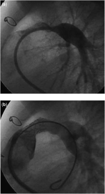
Stent placement in the right ventricle to pulmonary artery conduit can be performed safely, significantly decreases both the right ventricular systolic pressure and the pressure gradient from the right ventricle to the pulmonary artery distal to the stenosis, and can delay the need for further surgical intervention (Fig 1). The Boston Children's paediatric cardiology group reported their experience with stent placement in the right ventricle to pulmonary artery conduit, including 58 patients with common arterial trunk in their series of 221 patients.Reference Peng, McElhinney and Nugent 8 The median freedom from surgical conduit replacement in their series was 2.7 years, with an increase to 3.9 years in patients who were at least 5 years of age at the time of stent implantation. Stent fractures were common in this series, but did not cause any reported haemodynamic sequelae. There were no procedure-related deaths. More recently, the Melody valve has gained Food and Drug Administration approval in the United States for implantation in right ventricle to pulmonary artery conduits and offers a viable alternative to surgical right ventricle to pulmonary artery conduit in patients with a conduit diameter of at least 18 mm. In the initial United States Melody Valve trial, 14 of 124 patients had a diagnosis of common arterial trunk.Reference McElhinney, Hellenbrand and Zahn 9
( a ) A 9-month-old patient after repair of common arterial trunk with an 8-mm-diameter right ventricle to pulmonary artery conduit. There is severe stenosis of the conduit and proximal left pulmonary artery. ( b ) Repeat angiogram after balloon angioplasty of the left pulmonary artery and implantation of a 9-mm-diameter Genesis stent.

Post-operative cardiac catheterisation of patients with transposition of the great arteries
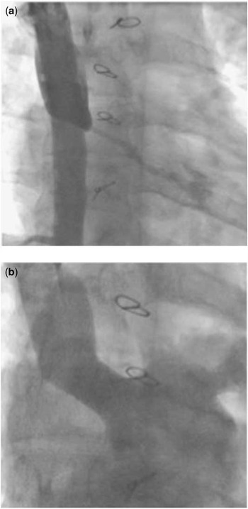
Although the need for early catheter-based intervention is not as common in patients with transposition of the great arteries compared with common arterial trunk, cardiac catheterisation for both diagnosis and intervention still plays an important role in the long-term management of these patients. In older patients who underwent an atrial switch operation, commonly encountered problems include obstruction in the systemic or pulmonary venous baffle, residual intracardiac shunts, and interatrial baffle leaks. Balloon angioplasty with stent implantation can be performed to successfully alleviate stenosis in systemic and venous baffles (Fig 2). Covered stents can also be implanted to simultaneously treat stenosis and residual shunts within the baffle. Various septal occluders can be used to treat residual interatrial baffle leaks. Transcatheter closure of residual ventricular septal defects with the Amplatzer muscular ventrical septal defect occluder can also be performed in patients with a haemodynamically significant residual ventricular level shunt.
( a ) A 31-year-old patient with transposition of the great arteries who previously underwent a Senning procedure. Angiogram in the superior caval vein demonstrates severe stenosis at the superior aspect of the systemic venous baffle (arrow). Note significant contrast flow into the azygous vein. ( b ) Repeat angiogram after implantation of a MaxLD stent (EV3) expanded to 20 mm. The superior limb of the systemic venous baffle is now widely patent with contrast filling in the left atrium. No flow is seen in the azygous vein.

In patients who have undergone the arterial switch operation problems that can be encountered during long-term follow-up include stenosis of the branch pulmonary arteries, supravalvar pulmonary stenosis, residual intracardiac shunts, and coronary artery stenosis. Stenosis of the proximal branch pulmonary arteries can be difficult to treat in the cardiac catheterisation laboratory. Balloon angioplasty has a high incidence of treatment failure or recurrence. Stent implantation has been shown to achieve improved diameter of the stenotic area and improved reduction of pressure gradients versus balloon angioplasty alone in a small series of patients.Reference Formigari, Santoro and Guccione 10 However, not all areas of stenosis are amenable to stent implantation because of the anatomical relationship to adjacent structures, which can lead to potentially serious problems such as coronary artery compression or stent erosion into the ascending aorta. Balloon angioplasty of supravalvar pulmonary stenosis after an arterial switch operation has been described, although lack of success has been reported presumably due the fact that the size of the pulmonary annulus may limit the maximum diameter of the angioplasty balloon which can be used, thus limiting the potential for therapeutic relief of obstruction.Reference Saxena, Fong and Ogilvie 11 Coronary artery lesions including ostial stenosis, and proximal main vessel stenosis, have a reported incidence of up to 5–8% and are more common in patients with Yacoub class B through E coronary artery branching patterns.Reference Ou, Mousseaux and Azarine 12 , Reference Angeli, Formigari and Pace Napoleone 13 Although these abnormalities may be clinically silent, they can cause significant long-term morbidity and mortality in patients following the arterial switch. Cardiac catheterisation with coronary artery angiography can be used to evaluate coronary artery lesions, although multi-slice computed tomography is less invasive and has been shown to accurately detect lesions in patients with coronary artery pathology.Reference Ou, Mousseaux and Azarine 12 Balloon angioplasty has been performed in patients with coronary stenosis following arterial switch operation.Reference Angeli, Formigari and Pace Napoleone 13 This can be performed safely with no adverse events reported, and with vessel patency as documented angiographically. Myocardial perfusion studies and exercise stress testing can be performed for follow-up in these patients following intervention.
Three-dimensional rotational angiography has recently been described for use in the cardiac catheterisation laboratory in patients with congenital heart disease.Reference Pedra, Fleishman and Pedra 14 , Reference Glatz, Zhu and Gillespie 15 This allows accurate delineation of the three-dimensional anatomy of vascular structures outside the heart and is quickly becoming a valuable imaging modality for patients with congenital heart disease as it allows rapid diagnosis of lesions involving extracardiac arteries and can help determine the feasibility of potential interventions in these patients depending on the relationship of vascular structures to each other and with other non-vascular structures such as a mainstem bronchus. As the technique for obtaining images for three-dimensional reconstruction has been refined, it now allows for decreased contrast administration and lower radiation exposure to obtain images of complex vascular structures in patients with congenital heart disease.
Summary
Cardiac catheterisation remains an important component in the long-term management of patients with transposition of the great arteries, and common arterial trunk for diagnosis of potential anatomical problems, and to perform interventions when feasible.