INTRODUCTION
Human schistosomiasis is a parasitic disease currently afflicting over 200 million people in 76 tropical and subtropical countries worldwide (World Health Organization, 2011). Drug treatment like praziquantel is central to the control of schistosomiasis and prognosis is good and full recovery can be achieved if patients receive early treatment. However, drug therapy cannot stop re-infection and repeated and widespread drug use may potentially lead to the emergence of drug-resistant parasite strains; concerns over this issue have already been raised (Melman et al. Reference Melman, Steinauer, Cunningham, Kubatko, Mwangi, Wynn, Mutuku, Karanja, Colley, Black, Secor, Mkoji and Loker2009; Caffrey and Secor, Reference Caffrey and Secor2011). On the other hand, prevention of the disease by interrupting the pathogen transmission also plays an essential part in control of schistosomiasis. Human schistosomal infection results from skin penetration by cercariae, the larval form of schistosome, during contact with contaminated water. The primary risk of infection comes from exposure to contaminated water due to agricultural, domestic and recreational activities. Protecting water sources against parasite contamination and reducing infective water contact are of vital importance in blocking transmission of schistosomiasis. To this purpose, water decontamination which includes elimination of aquatic schistosome cercariae and their intermediate snail hosts poses a key strategy. Although a few agents like niclosamide are currently available that can provide cercarial decontamination and prevention of disease transmission, they are either of considerable toxicity to humans or are unstable in an aqueous environment, which limits their application (Liang et al. Reference Liang, Seto, Remais, Zhong and Yang2007; Park et al. Reference Park, Shin, Kang, Hwang and Cho2011).
Silver is a noble metal with prominent disinfectant properties that have been recognized since early days in history. The use of silver and silver compounds to curb microbial infection and treat infectious diseases is time honoured (Russell and Hugo, Reference Russell and Hugo1994; Stephen-Haynes and Toner, Reference Stephen-Haynes and Toner2007). However, the advent of antibiotics and their success against infectious diseases has largely thrown silver into oblivion. With the emergence and rapid development of nanotechnology, silver has lately been engineered into nano-scale particles that possess various unique properties, among which are superb activities against various microbes, like bacteria, viruses, parasites etc. (Elechiguerra et al. Reference Elechiguerra, Burt, Morones, Camacho-Bragado, Gao, Lara and Yacaman2005; Pollini et al. Reference Pollini, Paladini, Catalano, Taurino, Licciulli, Maffezzoli and Sannino2011; Seil and Webster, Reference Seil and Webster2012; Xiu et al. Reference Xiu, Zhang, Puppala, Colvin and Alvarez2012). Exploitation of silver nanoparticles (AgNPs) has attracted great attention and efforts in various fields. However, it is still the antibacterial activities of the new form of silver that largely take the centre stage in the healthcare arena. Commercialization of AgNPs is already a reality and a wide variety of products taking advantage of the disinfectant and antibacterial properties of AgNPs have already come into clinical practice (Cheng et al. Reference Cheng, Yang and Zhao2004; Lansdown et al. Reference Lansdown2006; Cohen et al. Reference Cohen, Stern, Vanni, Kelley, Baumgart, Field, Libertino and Summerhayes2007). By contrast, the interactions of AgNPs with multicellular parasitic organisms largely remain a field yet to be tapped into. Only a few studies provided scanty information on silver ions (Ag+), which can be released from AgNPs. A previous study has demonstrated that Ag+ at concentrations above 0·09 mM was toxic to Schistosoma mansoni cercariae and Ag+ below 0·09 mM was non-toxic but inhibited their penetration into linolenic acid-impregnated agar (King and Highashi, Reference King and Highashi1992).
In light of the above, it is of great interest to us to investigate the effects of AgNPs on schistosome cercariae and explore their application potential in the control of schistosomiasis, particularly with respect to the blocking of pathogen transmission. The present study was hence carried out to examine the responses of schistosome cercariae to exposure to AgNPs in terms of cercarial viability, behavioural and morphological change, infectivity and fecundity. Cercariae of Schistosoma japonicum were used and Ag+ treatment was also tested as a control of AgNPs.
MATERIALS AND METHODS
Ethics statement
All animals were maintained in the university animal facilities and handled in compliance with the guidelines of the Jianghan University Ethics Committee for the use of laboratory animals and are in accordance with the medical research regulations of Ministry of Health, China. Animal handling and experimental procedures were approved by Jianghan University Ethics Committee for the use of laboratory animals.
Schistosome cercariae
A Chinese strain of Schistosoma japonicum (strain from Hubei Province, China) was used throughout this study. Cercariae were shed from infected Oncomelania hupensis snails in spring water at 28 °C and used within 2 h of emergence.
Silver nanoparticles and characterization
AgNPs were provided by Plasma Chem GmbH, Germany. An AgNP suspension at a concentration of 1 mg mL−1 in deionized water was prepared by probe sonication for 30 min on ice and serial 1:2 dilutions were then made in deionized water and subjected to characterization under experimental conditions. Particle size and zeta potential were measured by Dynamic Light Scattering (DLS) using a Zeta Sizer (NicompTM 380 ZLS, PSS. Nicomp, USA). Supernatant samples were taken from AgNP suspensions 2 h after sonication for analysis of silver and silver ion contents. Element contents of silver in the supernatants were measured by an atomic absorption spectrometer (AAS, AAnalyst800, Perkin Elmer, USA). Ag+ concentrations were determined using a silver ion-selective electrode (ISE, ELIT8211, NICO2000, UK). A calibration curve was constructed from diluted AgNO3 solutions with Ag+ concentrations ranging from 0 to 1·25 μM. AgNP dispersions were visually inspected using a transmission electron microscope (TEM, CM30, FEI, Japan). The microscope was operated in the bright field mode at an acceleration voltage of 300 kV and images were recorded with a CCD camera attached.
Exposure of schistosome cercariae to AgNPs
AgNPs at 1 mg mL−1 and serial 1:2 dilutions until 15·6 μg mL−1 were prepared as mentioned above and placed in a 96-well plate, 300 μl per well. Then 40 ± 1 S. japonicum cercariae freshly shed from the snails were transferred into each well and incubated for the required periods of time. As a comparison, fresh cercariae were incubated with concentrations of AgNO3 (serial 1:2 dilutions were made of an initial concentration of 10 μM).
Observation of cercarial behaviour and assessment of cercarial viability
Over the course of AgNPs incubation, cercariae were directly observed under a dissecting microscope (SZX10, Olympus, Japan) and changes in behavioural pattern (swimming, contraction) and morphology (tail shedding) were recorded with a CCD recorder. In addition, cercariae exposed to AgNPs and non-exposed samples that served as controls were fixed in a 10% glutaraldehyde and processed for tegumental examination by scanning electron microscopy (SEM, CX21, Olympus, Japan) (Shaw and Erasmus, Reference Shaw and Erasmus1987). To determine the viability of cercariae, staining with fluorescein diacetate (FDA) was used. FDA is a non-fluorescent derivative of fluorescein, which can diffuse into cells of viable cercariae and be hydrolysed by cytoplasmic non-specific esterase into fluorescein that stains the cercariae fluorescently green. Non-viable cercariae will lose cellular esterase activity and thus will not be stained. FDA staining for assaying viability of schistosomes of S. mansoni has recently been validated (Peak et al. Reference Peak, Chalmers and Hoffmann2010). In our study, 1 μL of FDA (Sigma-Aldrich, USA, 1 μg mL−1 in acetone) was added to each well of cercariae at the end of the incubation with AgNP. After incubation in the dark for 3 min, the cercariae were observed under a fluorescence microscope (DM5000, Leica, Germany).
Evaluation of infectivity and fecundity of cercariae
The ability of AgNP-treated cercariae to invade into, and develop and breed in the host was evaluated by animal experiments. Six-week-old female BALB/c mice (supplied by Hubei Provincial Laboratory Animal Centre, China) were randomly grouped. After anaesthesia of the mice, 40 ± 1 AgNP-treated cercariae, with or without tails, were transferred onto the shaved abdominal skin of each animal. Cercariae suspended in pure water were used as the control. The animals were euthanized at day 42 post-infection. Worms were collected by portal perfusion (Ruppel et al. Reference Ruppel, Shi and Moloney1990) and counted under a dissecting microscope. Livers were removed following recovery of the worms and the large left lobe of each liver was digested at 37 °C for 3 h in 20 mL of 5% potassium hydroxide. Three 0·1 mL samples from each digested liver were counted for egg number and a mean value was determined. The total number of eggs per liver and the liver eggs per worm pair were calculated (Moloney et al. Reference Moloney, Doenhoff, Webbe and Hinchcliffe1982).
Determination of cercarial secretion
Linoleic acid was used to stimulate secretion of cercarial acetabular glands. Specifically, approximately 300 freshly shed cercariae were transferred into each well of a 24-well plate containing 350 μL AgNPs or AgNO3 solution; the bottom surface of each well had previously been painted with linoleic acid (0·9 g mL−1, Sigma, USA) and air-dried. The plates containing the cercariae were maintained at 37 °C for 1 h. The water containing all cercarial materials was then collected in 1·5 mL Eppendorf tubes and the larvae were allowed to sediment on ice. The supernatant containing cercarial secretions were spun for 2 min at 3000 g to eliminate cercarial debris. Protein concentrations in the supernatants were measured using the BCA test (Pierce, USA) (Bahgat et al. Reference Bahgat, Francklow, Doenhoff, Li, Ramzy, Kirsten and Ruppel2001).
Evaluation of the influence of Ag+ antagonism on AgNPs' cercarial effects
Ag+ released from AgNPs has been reported to play a role in the biological activities of AgNPs (He et al. 2012). We looked into the possible involvement of released Ag+ in the action of AgNP on cercariae in this study. N-acetyl cysteine, a specific Ag+ ligand, was applied to block the action of Ag+. Specifically, cercariae were exposed to a suspension of 500 μg mL−1 AgNP containing N-acetyl cysteine with concentrations from 0 to 3 μM. Tail-shedding ET50 and cercarial secretion were then assessed as mentioned above.
Natural water bodies in the field are often rich in organic matter that could markedly affect the apparent potency of silver in any form, including AgNPs (Bone et al. Reference Bone, Colman, Gondikas, Newton, Harrold, Cory, Unrine, Klaine, Matson and Di Giulio2012). We herein used albumin (AMRESCO, USA) as a surrogate for aquatic organic matter and tested its influence on the action of AgNPs toward cercariae. Specifically, cercariae were exposed to 125 μg mL−1 AgNP suspension containing 0–50 μg mL−1 albumin. ET50 were then assessed as mentioned above. AgNO3 with relevant concentration served as the experimental control.
Statistical analysis
Data are shown as mean±s.d. Student's two-tailed t-test was used to determine the differences between the experimental and control groups with respect to parasite burden and the number of liver eggs per worm pair. Values of P < 0·05 were considered as statistically significant.
RESULTS
Characterization of AgNPs
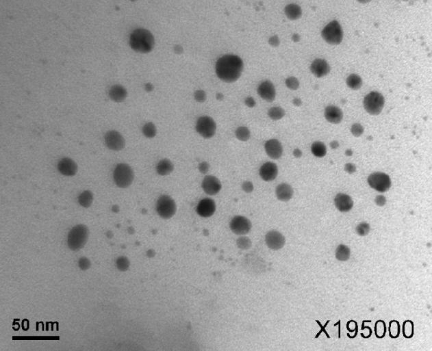
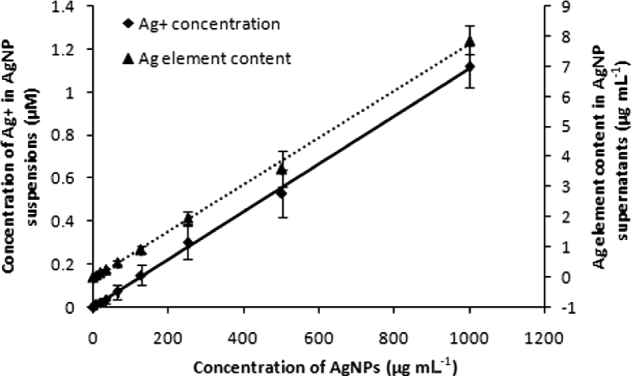
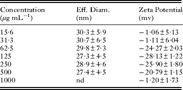
The hydrodynamic diameter and zeta potential of AgNP dilutions are listed in Table 1. Dynamic light scattering (DLS) measurements showed that the average size of an AgNP is around 30 nm. The data indicated that dilution had little effect on particle size and zeta potential in the concentration range from 62·5 to 500 μg mL−1. TEM showed that smaller particles were also present (Fig. 1). The difference between DLS measurement and TEM observation may be attributed to a bias of the DLS measurements towards larger particles. AAS showed that the proportion of supernatant Ag contents were 0·70–0·90% of the total AgNP mass in suspensions (7·8–1000 μg mL−1). ISE analysis showed that increasing AgNP concentration resulted in elevated Ag+ contents with 0·01 μM at the lower end and 1·12 μM at the upper end (Fig. 2 and Table S1, online version only). But Ag+ contents barely varied as a proportion of the total Ag contents in supernatants (1·50–1·94%).
Silver nanoparticles (AgNPs) observed under a transmission electron microscope (TEM).

Concentration of silver ions (Ag+) in silver nanoparticle (AgNP) suspensions and Ag element content in AgNP suspernatants.

Particle size and zeta potential of AgNPs (n = 3)

nd, Not detected.
Effects of AgNPs on cercarial activity, morphology and viability
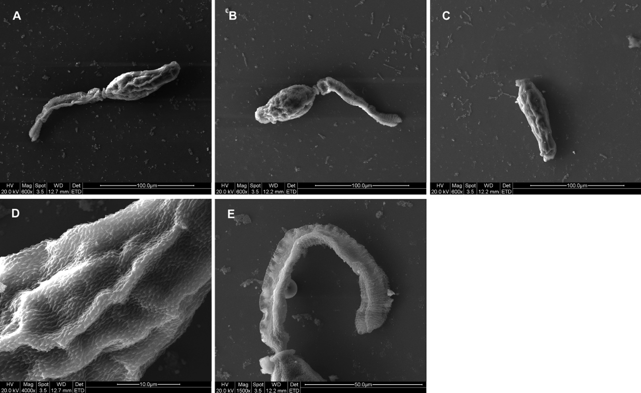
Cercariae were exposed to AgNPs at concentrations ranging from 15·6 to 1000 μg mL−1. SEM examination showed the cercariae exposed to AgNPs with intact teguments covered with spines and no tegumental deformation was found (Fig. 3). However, agitated behaviour such as cercarial corporal contraction and violent tail swinging movements occurred at a concentration of 125 μg mL−1 and above (Supplementary Video S1, online version only). The occurrence and frequency of agitated behaviour was shown to be dependent on AgNP concentration.
Scanning electron microscopy (SEM) of cercariae exposed to 500 μg/ml silver nanoparticles (AgNPs). (A) Cercaria in distilled water served as control (600 × magnification). (B) Cercaria exposed to AgNPs (600 × magnification). (C) AgNPs-induced cercarial tail loss (600 × magnification). (D) Intact tegument with spines covering the body region of cercaria exposed to AgNPs (4000 × magnification). (E) Intact tegument of tail of cercaria exposed to AgNPs (1500 × magnification).

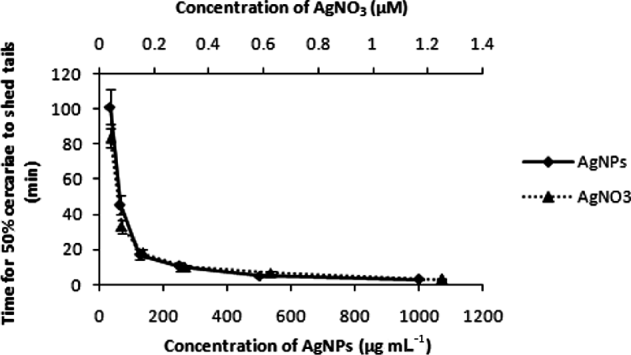
Concentrations of AgNPs above 31·25 μg mL−1 induced cercarial tail shedding. In this study, the time for 50% of the cercariae to shed their tails was measured and expressed as the median effective time (ET50). ET50 was plotted against AgNP concentration in Fig. 4. It can be clearly seen that ET50 shortened dramatically with the increase of AgNP concentration.
Time for 50% of cercariae to shed tails (ET50) in the presence of silver nanoparticles (AgNPs) or silver nitrate (AgNO3).

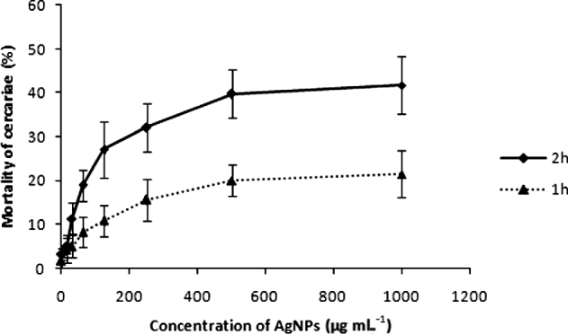
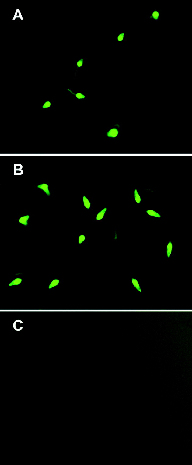
Despite manifest agitated behaviour, the majority of cercariae could be stained by FDA after treatment of AgNPs at all concentrations for up to 2 h (Fig. 5). Notably, even most of the AgNP-treated cercariae that lost their tails were still also able to be stained, suggesting maintenance of viability (Fig. 6B). With the increase of AgNP exposure time, a minor part of the cercariae would sink to the bottom of the container and could not be stained by FDA, indicating loss of viability.
Cercarial mortality after AgNP exposure for 1 h and 2 h.

Assessment of cercarial viability by fluorescein diacetate (FDA) staining. (A) Viable cercariae in distilled water fluoresce bright green at an excitation wave length of 490 nm. (B) Cercariae that lost their tails due to exposure to silver nanoparticles (AgNPs) (250 μg mL−1) for 30 min were still able to be stained by FDA. (C) Cercariae rendered unviable by treatment with 10 μM silver nitrate (AgNO3) for 10 min were unable to be stained by FDA.

Observations on cercariae exposed to AgNO3 were conducted for comparison. At concentrations up to 1·25 μM, AgNO3 elicited cercarial tail loss in a dose-dependent manner, in a pattern closely resembling AgNPs (Fig. 4). AgNO3 at concentrations above 2·5 μM did not induce cercarial tail loss and agitated behaviour, but led to acute toxicity to cercariae as evidenced by impaired cercarial motility and even loss of viability. See Fig. 6C for the absence of FDA staining in cercariae treated with 10 μM AgNO3.
Effects of AgNPs on cercarial infectivity and fecundity
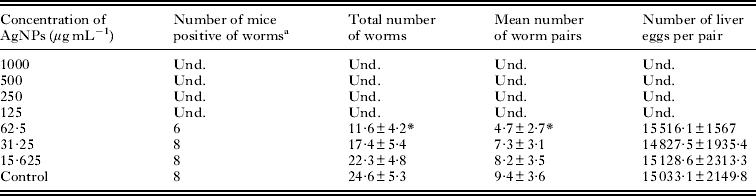
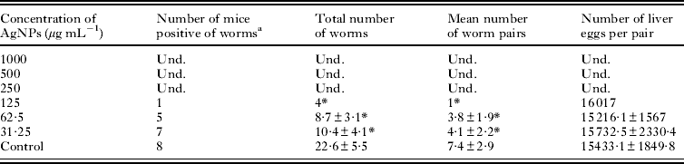
Two separate experiments were conducted to investigate whether AgNPs might affect cercarial penetration and subsequent development into schistosomes in the host. In the first experiment, cercariae were incubated with AgNP suspensions for 30 min and the suspensions containing cercariae were then transferred onto the shaved abdominal skin of anaesthetized mice. Results showed that no adult worms and eggs were detected in mice treated with cercariae that had been exposed to AgNPs at concentrations of 125 μg mL−1 and above (Table 2). However, no significant difference was found in the number of liver eggs per worm pair among different groups, in which adult worms were detected, indicating that AgNP treatment did not affect the worms' fecundity. As we observed that cercariae maintained their viability in the presence of AgNPs despite tail loss, a second experiment was conducted to see whether tail-less cercariae were still infective and fecund. AgNP suspensions containing cercariae were transferred onto the shaved abdominal skin of anaesthetized mice as soon as all cercariae had lost their tails, which was visually confirmed by microscopic examination. Interestingly, despite a decreased worm burden, adult worms and eggs were still found in mice treated with cercariae that had been exposed to AgNPs at concentrations up to 125 μg mL−1 (Table 3). This result suggests that cercariae without tails can still penetrate into mice, develop into adult worms and are able to breed, in spite of a decreased infection rate.
Total number of worms and worm pairs from mice infected with Schistosoma japonicum cercariae treated with AgNPs for 30 min

Und., undetectable.
a 8 mice in each group were infected with S. japonicum cercariae.
* The difference between experimental group and control group was statistically significant (P < 0·05).
Total number of worms and worm pairs from mice infected by Schistosoma japonicum cercariae rendered tailless by AgNPs

Und., undetectable.
a 8 mice in each group were infected with S. japonicum cercariae.
* The difference between experimental group and control group was statistically significant (P < 0·05).
Effect of AgNPs on cercarial secretion
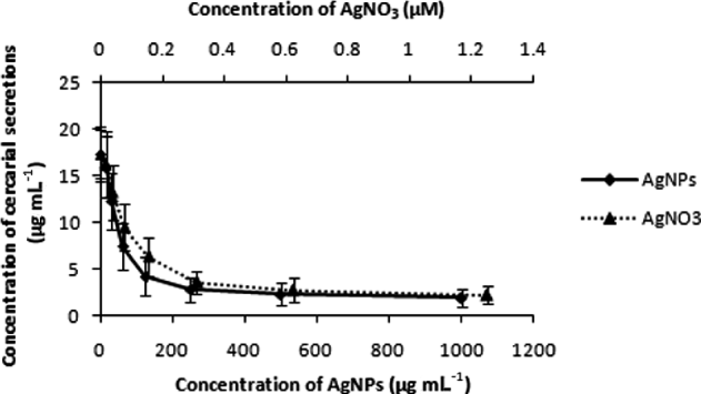
Protein contents in the supernatants of both AgNP-and AgNO3-treated cercarial suspensions displayed a dose-dependent decrease, with similar patterns, suggesting reduced cercarial secretion (Fig. 7).
Effects of silver nanoparticles (AgNPs) and silver nitrate (AgNO3) on cercarial secretion.

Influence of Ag+ antagonism on AgNP cercarial effects
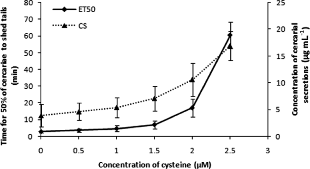
N-acetylcysteine, a specific Ag+ blocker, dose dependently antagonized cercarial effects of 500 μg mL−1 of AgNPs. See Fig. 8 for the increase in both ET50 for tail shedding and cercarial secretions. N-acetylcysteine at 3 μM even completely abolished AgNP-induced cercarial behavioural changes.
Time for 50% of cercariae to shed tails (ET50) and cercarial secretion induced by 500 μg mL−1 of silver nanoparticles (AgNPs) in the presence of N-acetylcysteine.

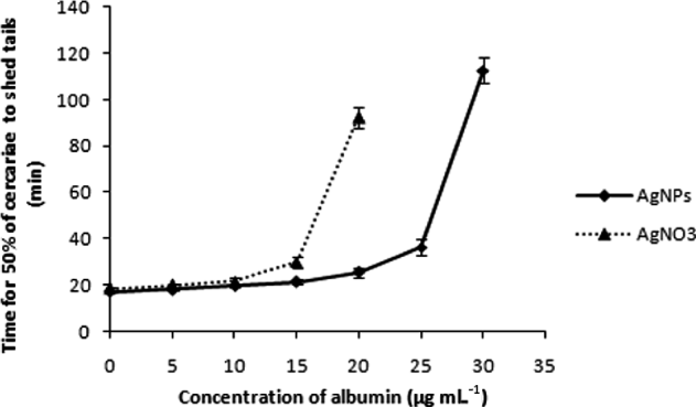
The tail-shedding ET50 value at a concentration of 125 μg mL−1 of AgNPs was very close to that of 0·16 μM of AgNO3 (Fig. 4). Therefore, the tail-shedding ET50 was still used as an index to evaluate the influence of albumin on the cercarial effects of 125 μg mL−1 of AgNPs in comparison with 0·16 μM of AgNO3. As can be seen in Fig. 9, albumin dose dependently extended the tail-shedding ET50 either in the presence of AgNPs or Ag+, suggesting albumin's antagonism of silver. Interestingly, 125 μg mL−1 of AgNPs that contained 0·16 μM of Ag+ by ISE measurement appeared to be more resistant to albumin's antagonism compared with 0·16 μM of AgNO3.
Influence of albumin on cercarial tail loss induced by 125 μg/ml of AgNPs and 0·16 μM of silver nitrate (AgNO3).

DISCUSSION
Human schistosome cercariae infect their hosts by penetration of skin. Upon contact with the host, the cercariae are exclusively stimulated by host chemical signals, i.e. unsaturated free fatty acids (FAs) on the skin surface, and perform a sequence of behavioural responses (phases of host invasion): attachment and enduring adherence to the skin, tail shedding, secretion of acetabular gland contents and penetration (Haeberlein and Haas, Reference Haeberlein and Haas2008). Interrupting the penetration process of cercariae is considered to be an important and feasible strategy for prevention of schistosome infection. Efforts have been made to develop drugs for this purpose (Birrie et al. Reference Birrie, Balcha, Erko, Bezuneh and Gemeda1998).
In this study we investigated the effects of AgNPs on the activity, viability, infectivity and fecundity of the cercariae of the human parasite, S. japonicum. Our major findings were that AgNPs reduced the infectivity of S. japonicum cercariae in a dose-dependent manner and AgNPs at concentrations of 125 μg mL−1 and above completely blocked cercarial infection after 30 min of exposure. Despite loss of infectivity, however, the majority of cercariae maintained viability in the presence of all tested concentrations of AgNPs for up to 2 h. A remarkable cercarial reaction elicited by AgNPs (31·25 μg mL−1 and above) was the rapid occurrence of tail shedding, as well as evident agitated behaviour. Interestingly, despite a decreased infectivity, tail-less cercariae in low AgNP concentrations could still be viable and infective, suggesting that the keeping of tails is not essential for cercarial infectivity. Cercarial secretion, which is crucial for cercarial penetration of host skin, was also reduced in the presence of AgNPs in a dose-dependent way, which might in part explain the decrease in cercarial infectivity. A small portion of cercariae was found to be dead after prolonged AgNP exposure, which we assume might have resulted from water intolerance due to tail loss rather than AgNP toxicity.
Silver nanoparticles are hydrophobic and are not stable in aquatic suspension. In our work, we had expected deposition of AgNPs out of suspension. Indeed, element analysis showed that only a very small portion of the total Ag mass (0·70–0·90%) was still present in suspension at the end of the longest cercarial exposure period (2 h), whereas the viable cercariae were mostly seen near the surface of the water. This discrepancy prompted us to postulate that it might be Ag+ that made the major contribution to the cercarial effects of AgNPs, as silver nanoparticles have been reported to stably release Ag+ in water suspensions (Liu and Hurt, Reference Liu and Hurt2010). ISE confirmed an elevated Ag+ content with increasing AgNP concentrations (0·01–1·12 μM). The cercarial reactions of AgNP suspensions were also found to be dose-dependently attenuated by N-acetylcysteine, a specific Ag+ ligand that could reduce the availability of Ag+. We further made control observations of AgNO3 as a source of Ag+. A dose-dependent occurrence of cercarial responses was also observed with AgNO3 below a concentration of 1·25 μM, in a pattern closely similar to that of AgNPs. Furthermore, ISE data revealed that Ag+ contents in our test AgNP suspensions corresponded nearly perfectly to the control concentrations of AgNO3. Taken together, a major role of Ag+ was strongly indicated in the cercarial effects of AgNP suspensions in this work. Higher concentrations of AgNO3 (above 2·5 μM) were toxic to cercariae, resulting in impaired motility and rapid death without occurrence of tail loss, whereas no acute cercarial toxicity was observed with AgNPs at a maximum concentration of 1000 μg mL−1. Yet, this difference may simply be due to the fact that the Ag+ content in a suspension of 1000 μg mL−1 of AgNP did not reach 2·5 μM. However, the AgNP suspension did act differently from the AgNO3 solution in some way. Namely, AgNP suspensions showed greater resistance to the antagonism of albumin than did the AgNO3 solution. This difference might be explained, at least in part, by AgNPs serving as a reservoir constantly replenishing with fresh Ag+. In summary, the apparent AgNP concentrations used in our work are rather high. However, we believe that what was really at work was only a small portion of the total silver mass – Ag+ and small Ag particles in the supernatant which accounted for less than 1% of the total silver mass. A limitation herein is that, due to condition constraints, changes in Ag and Ag+ contents in the AgNP suspensions were only measured once at 2 h into cercarial exposure rather than being kinetically followed throughout the period. However, we still believe that the data presented here are very revealing concerning the relation between the particle form and ion form of Ag in AgNP suspension. Note an interesting observation that the proportion of Ag+ in the total supernatant Ag barely varied (1·50–1·94%) despite the increase of supernatant Ag contents with AgNP concentrations.
As for the possible mechanisms of action of Ag+, our work as well many literature reports may have provided some clues. Ultrastructural studies showed that schistosome cercariae possess surface chemosensory papillae with ciliated nerve endings as special receptors for chemical signals (Cavalcanti et al. Reference Cavalcanti, Araújo, Paiva, Silva, Barbosa, Silva, Brayner and Alves2009). Chemical attractants in human skin such as FAs can bind to the chemosensory papillae sites and stimulate cercarial acetabular gland secretion which is the key for cercarial host penetration (Stirewalt, Reference Stirewalt1959). Short and Cartrett (Reference Short and Cartrett1973) reported that Ag+ preferentially binds to the papillae, especially to the ciliated papillae on the cercarial surface. King and Higashi (Reference King and Highashi1992) also suggested that Ag+ can bind to the papillae sites on the cercarial surface and thereby inhibit FA-induced cercarial acetabular gland secretion. Our work confirmed the presence of Ag+ in the AgNP suspensions and showed a reduction of FAs-induced cercarial secretion therein, which could be reversed by N-acetylcysteine. This indicates an interruption either of receipt of the FAs signal or of the secretion mechanism by Ag+ released from AgNPs. Despite a very strong indication of involvement of Ag+ in the cercarial effects of AgNPs, which is in accord with latest research opinion on Ag+ and AgNPs by some other workers (Xiu et al. Reference Xiu, Zhang, Puppala, Colvin and Alvarez2012), the role of silver nanoparticles in our work still cannot be ruled out since considerable amounts of silver in particle form was still found by AAS in the supernatants of AgNPs at the end of cercarial exposure.
Drugs like praziquantel or miltefosine are also reported to induce schistosome cercariae tail separation (Andrews, Reference Andrews1978; Liang et al. Reference Liang, Wang, Dai, Li and Tao2010; Eissa et al. Reference Eissa, El Bardicy and Tadros2011) and concurrent uncoordinated contraction of body and tail muscles, similar to our observations. However, cercariae exposed to the drugs showed marked loss of the glycocalyx, and thinning of the tegument leading to external protrusion or surface blebbing with focal loss of spines from the tegument (Eissa et al. Reference Eissa, El Bardicy and Tadros2011), which was not seen with AgNP treatment in our work, suggesting different mechanisms. We postulated that AgNP-induced tail shedding might be attributable to Ag+ acting on a specific receptor site on the cercarial surface, which belongs to the ending of the nervous system. However, it was reported that cercarial tail-loss induced by praziquantel was not associated with the signal recognition and transmission of cercariae (Xiao et al. Reference Xiao, Yue, Mei and Zhang1987).
The focus and emphasis of the present study was on investigating AgNPs' cercarial effects and not much consideration was put on the practical application level at the moment. Issues may exist that can negatively affect the feasibility and practicality of AgNPs for schistosome control in the field. For instance, organic matter, particularly that of proteinaceous nature, in natural waters can neutralize the potency of silver in any form, as evidenced by the albumin antagonism in our work. Another problem is that silver, either in the form of ions or particles, is a biocide and can also impact on other organisms in the ecosystem (Navarro et al. Reference Navarro, Piccapietra, Wagner, Marconi, Kaegi, Odzak, Sigg and Behra2008). However, we believe that proper design of an application strategy and products can be realized that can diminish the above problems or even make them irrelevant. Schistosomes and their intermediate hosts are natural links in the ecosystem and it may not be successful to try to eliminate them from Mother Nature just for the sake of the human being. Such efforts made decades ago have proved a failure in some countries. A schistosome control strategy in the field that emphasizes reducing parasite contamination and exposure for a limited time-period without leaving long-lasting impact on the environment might be more practicable and environmentally friendly. Under this notion, rather than being thrown irretrievably into the environment, AgNPs can be incorporated into some retractable vehicles floating in or on the surface of water which release Ag+ that temporarily decontaminate local water where human activity is taking place. This strategy might be particularly suitable for decontaminating schistosome cercariae because viable cercariae tend to swim near the surface of water. Since the AgNPs can be retracted, they will not leave permanent damage on the local aquatic ecosystem. The released Ag+ can be rapidly diluted by flowing water or neutralized by aquatic organic matter.
In conclusion, Ag+ may contribute largely to the effects of AgNPs on schistosome cercariae. Yet, the possible contribution of nano dimensions of AgNPs could not be ruled out, as remarkably unusual physicochemical properties and biological activities of various nano-sized materials including AgNPs have been demonstrated.
ACKNOWLEDGEMENTS
We thank Professor Yonglong Li (Huazhong University of Science and Technology, China) for his helpful comments on the manuscript. This work was supported by grants from the National Natural Science Foundation of China (81000741/H1906) and the programme of ‘Schistosome Prevention and Control’ from the Health Department of Hubei Province, China (XF2010-25).