Introduction
First described by Meltzer and Kelemen in 1959, central skull base osteomyelitis is a rare but life-threatening disease.Reference Meltzer and Kelemen1 The central skull base is made up of the temporal, sphenoid and occipital bones. Central skull base osteomyelitis is most common in diabetics, immunocompromised individuals and the elderly. The usual symptoms are various cranial nerve palsies and headache.Reference Patmore, Jebreel, Uppal, Raine and McWhinney2
There are three types of central skull base osteomyelitis: (1) necrotising otitis externa extending to the central skull base; (2) central skull base osteomyelitis that presents after resolution of necrotising otitis externa; and (3) central skull base osteomyelitis as a primary presentation. It most frequently presents as an extension of necrotising otitis externa; however, cases also present without any clear proceeding lateral infection.Reference Clark, Pretorius, Byren and Milford3, Reference Cavel, Fliss, Segev, Zik, Khafif and Landsberg4 This type 3 presentation is the most difficult diagnostic challenge.
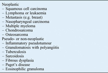
The history, examination and imaging features of central skull base osteomyelitis can mimic malignancy, including chordoma, metastases and inflammatory disorders such as pseudotumour and granulomatosis with polyangiitis,Reference Sharma, Deshmukh, Shaikh and Dabholkar5, Reference Ozgen, Oguz and Cila6 adding to the difficulty in diagnosis.
The most common pathogens in central skull base osteomyelitis are Pseudomonas aeruginosa, Staphylococcus aureus and fungal infection.Reference Chandler, Grobman, Quencer and Serafini7, Reference Johnson and Batra8 Tissue sampling is the ideal method to confirm the diagnosis, and for microbiological control of treatment, but this can be hazardous in an ill patient at presentation. If the imaging can provide a diagnosis, then antibiotic treatment can be instituted without the need of a general anaesthetic and deep biopsies of the clivus.
The imaging findings in central skull base osteomyelitis reported in the literature are soft tissue infiltration and bone erosion. It has been reported that magnetic resonance imaging (MRI) is better than computed tomography (CT) in its ability to assess subtle dural enhancement and the medullary cavity of bone, and so is the preferred method of imaging.Reference Adams and Offiah9 Findings on MRI are clival hypointensity and soft tissue infiltration on T1-weighted images, with hyperintensity and post-contrast enhancement of the central skull base on T2-weighted images.Reference Chang, Fischbein and Holliday10 It has also been reported that with treatment the abnormalities found on MRI improve but do not fully return to normal.Reference Grandis, Curtin and Yu11 Computed tomography is better at eliciting bone erosion and after treatment the abnormalities do not normalise.Reference Grandis, Curtin and Yu11 In reality, both modalities are required to attain as much information as possible. In addition, most of the abovementioned findings are useful to stage the disease rather than to solve the issue of differential diagnosis.
Materials and methods
We identified six patients with central skull base osteomyelitis and compared their CT and MRI scans with those of patients with other skull base pathology.
The medical records and imaging studies of the patients identified as having central skull base osteomyelitis were retrospectively reviewed. Cases were identified over the course of five years, from 2009 to 2014. Five males and one female, aged 64–90 years (mean, 79 years), were identified.
All patients had undergone CT and MRI prior to biopsy, culture testing and treatment. Magnetic resonance imaging included axial, coronal and sagittal T1-weighted scans, T2-weighted scans with fat saturation, and post-contrast T1-weighted scans. Patients also underwent high-resolution CT scanning of the temporal bones and skull base with soft tissue and bone windows. Images were assessed with regard to skull base marrow signal intensity, the presence of abnormal soft tissue and the signal intensity of any abnormal soft tissue.
Results
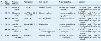
The central skull base osteomyelitis patients presented with headache and/or otalgia, and three patients had suffered from otorrhoea. All patients had cranial neuropathy by the time they presented, with VIIth and VIIIth cranial nerve damage present in two patients, and a variety of IVth, Vth, VIth, IXth and Xth cranial nerve damage in others (Table I).
Clinical characteristics of central skull base osteomyelitis patients

Pt no = patient number; y = years; M = male; CN = cranial nerve; tds = three times a day; IV = intravenously; wk = weeks; bd = twice a day; mth = months; EAC = external auditory canal; F = female
Three patients had a history of previous otorrhoea and had undergone treatment for presumed otitis externa or otitis media; the other three patients had no history of otitis externa or media. Four patients had underlying diabetes mellitus, one patient was undergoing chemotherapy and one patient was on immunosuppressive therapy following a renal transplant. All patients were apyrexial, with no documented fever. All central skull base osteomyelitis patients had raised inflammatory markers (white cell count, C-reactive protein or erythrocyte sedimentation rate).
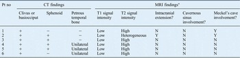
The most consistent CT finding in the central skull base osteomyelitis patients was regional destruction of the bony cortex of the clivus, which was noted in all six patients (Table II). Sphenoid bony involvement was observed in four patients and petrous temporal bone involvement was observed in three patients. Only two patients had unilateral temporal bone, basiocciput and sphenoid bone involvement. In the presentation scans of the central skull base osteomyelitis patients, the extent of bony regional destruction was less than expected based on the extent of involvement observed in the MRI scans. In contrast, the extent of bony involvement on CT in the malignant patients was more in keeping with the extent of disease observed on MRI scanning. On review of the central skull base osteomyelitis patients' post-treatment scans, the MRI scans showed improvement but the abnormalities on CT remained the same. The differences between CT and MRI in terms of disease involvement and extent thus decreased with time.
Imaging findings in central skull base osteomyelitis patients

Computed tomography tends to underestimate the extension of skull base infection when compared to MRI. *With regard to contrast-enhanced T1-weighted MRI, enhanced fascial planes mean that the extension of central skull base osteomyelitis is more difficult to define and anatomy appears ‘restored’. Pt no = patient number; CT = computed tomography; MRI = magnetic resonance imaging; + = involved; − = uninvolved; N = no; Y = yes
The most consistent MRI finding in the central skull base osteomyelitis patients was regional or diffuse clival hypointensity on T1-weighted images relative to normal fatty marrow, which was noted in all patients. There were also signs of pre- and para-clival soft tissue infiltration, with the obliteration of normal fat planes and frank soft tissue masses in all patients. Signal intensity on T2-weighted images of the clivus was high in five patients and heterogeneous in one patient. However, the most interesting finding was observed following the administration of intravenous contrast. The enhancement of the fascial planes showed restored anatomy that was almost in keeping with that of the uninvolved skull base. This restoration of anatomy was not seen in the scans of malignant processes used for comparison. The post-contrast enhancement of malignancy highlights the destruction of anatomical planes as opposed to improving their appearance.
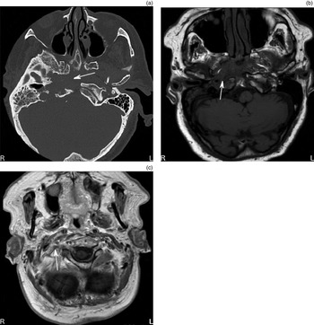
Figures 1b, 1c and 1d show involvement of the clivus and petrous temporal bone on MRI. There is low signal intensity on the T1-weighted images. The post-contrast T1-weighted image shows the restoration of fascial planes in relation to the unaffected (right) side.
(a) Axial computed tomography image on bone setting showing erosive bone loss of the anterior cortex of the clivus (arrow). (b) Axial, short tau inversion recovery sequence magnetic resonance imaging (MRI) scan highlights middle-ear infection (arrow). (c) Axial, T1-weighted MRI scan shows a loss of bone marrow in the clivus and a loss of infratemporal fat (arrow), before the intravenous administration of gadolinium as contrast medium. (d) Axial, contrast-enhanced, T1-weighted MRI scan reveals a restoration of anatomy as the fascial planes are enhanced (arrow). R = right; L = left

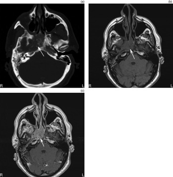
Differentiation of central skull base osteomyelitis and malignant processes can be difficult. In our series, malignant lesions tended to have a high signal on post-contrast T1-weighted images. This differs to the central skull base osteomyelitis findings, where, on post-contrast T1-weighted images, better delineation of the anatomy was observed because of the reasonably intact fascial planes and spaces. This means that the extent of the malignant lesion is harder to identify post-contrast when compared with the low signal lesion identifiable on T1-weighted pre-contrast images, as shown in Figure 2.
(a) Axial computed tomography image on bone setting showing central skull base bone erosion (arrow). (b) Axial, T1-weighted magnetic resonance imaging (MRI) scan shows lack of bone marrow and fat (arrow). (c) Axial, contrast-enhanced, T1-weighted MRI scan reveals a restoration of anatomy as the gadolinium enhances fascial planes (arrow). R = right; L = left

Figure 3 shows a nasopharyngeal carcinoma deposit affecting the clivus and petrous apex. The post-contrast image (Figure 3c) deserves direct comparison with Figures 1d and 2c. The malignant process is easily identifiable.
(a) Axial computed tomography image on bone setting shows central bone destruction of clivus and petrous apex (arrow) associated with nasopharyngeal carcinoma. (b) Axial, T1-weighted magnetic resonance imaging (MRI) scan shows soft tissue mass at site of bone destruction (arrow). (c) Axial, contrast-enhanced, T1-weighted MRI scan shows diffuse enhancement of tumour mass and persistent destruction of anatomy (arrow). R = right; L = left

Figure 4 shows a further example of a malignant process in the skull base. A metastatic deposit of breast cancer is shown extending from the petrous temporal bone to the central skull base. The bony destruction noted in the bone setting of CT scans compares equivocally to post-gadolinium T1-weighted MRI scans. The post-contrast images also show high signal uptake of the lesion, clearly identifying the margins of disease.
(a) Axial computed tomography image on bone setting showing destruction of the temporal bone adjacent to the jugular foramen (arrow). (b) Axial, post-gadolinium, T1-weighted magnetic resonance imaging scan showing heterogeneous enhancement of the destructive lesion (arrow), which is a metastatic deposit of breast cancer. R = right; L = left

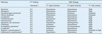
For comparison, we looked at diseases reported as having osteolytic tendencies that are therefore often included in the differential diagnosis of central skull base osteomyelitis (Table III).
Differential diagnoses of central skull base osteomyelitis

Table IV shows the key imaging points to consider when assessing skull base pathology.
Imaging findings in malignant process of central skull base

*Computed tomography destruction correlates with MRI findings in terms of volume; †CT and MRI appearance is often multifocal; ‡CT tends to underestimate extension of skull base involvement when compared to MRI. CT = computed tomography; MRI = magnetic resonance imaging
Discussion
Although imaging, patient history, examination and serum markers of inflammation may be sufficient to commence treatment for central skull base osteomyelitis, a tissue sampling procedure is often required for a definitive diagnosis of this condition. Only three of our patients underwent biopsy, and on each occasion the specimen was taken from the postnasal space, requiring general anaesthetic. Computed tomography guided fine needle aspiration of pre-clival tissue has been described.Reference Clark, Pretorius, Byren and Milford3 Procedures that entail obtaining tissue from open craniotomy or sphenoidotomy are also performed to aid diagnosis. In addition, some rely on tissue sampling from the external auditory canal, and consider the findings in conjunction with the clinical picture before commencing treatment.
Other imaging techniques can help support the diagnosis of skull base osteomyelitis. Technetium-99m methylene diphosphonate scintigraphy may localise the focus of necrotising otitis externa and osteomyelitis within the central skull base because of increased uptake of radiotracer in these areas.Reference Singh, Al Khabori and Hyder12 This technique has low specificity as the findings correlate with increased osteoblastic activity, which may also be observed in inflammatory, neoplastic and post-surgical conditions.
Radioisotope studies are limited by poor anatomical resolution. Changes also lag behind clinical improvement, which limits technetium-99m scintigraphy in the assessment of treatment response. Some of the limitations of technetium-99m scintigraphy can be overcome with the use of alternative radioisotopes. Gallium-67 citrate binds to leucocytes and forms a complex with lactoferrin. It is positive in soft tissue and bone infections. Moreover, it can be used to assess treatment response as it rapidly reverts to normal with disease resolution. Anatomical localisation can be further improved with positron emission tomography or single photon emission CT (‘SPECT’) indium-111 white blood cell studies.
• Central skull base osteomyelitis is a rare but life-threatening disease that can be difficult to differentiate from malignancy
• At presentation, magnetic resonance imaging (MRI) shows greater disease involvement than computed tomography (CT) in central skull base osteomyelitis
• Malignancy usually shows equal involvement
• In central skull base osteomyelitis (unlike malignancy), contrast-enhanced MRI reveals tissue planes
• Combining MRI (enhanced and non-enhanced) and CT findings can enable differentiation between central skull base osteomyelitis and malignancy
• This allows treatment to start before biopsy, and may remove the need for biopsy
The new findings described in this paper of mismatches between CT and MRI scans at disease presentation and regarding the normalisation of the fascial plane after enhancement can be used to indicate a diagnosis of central skull base osteomyelitis, rather than malignancy or other skull base diseases, and may allow early treatment to be commenced without the need for potentially hazardous tissue biopsy.
As this paper describes a relatively small series of cases, it is not clear whether these findings are present in all cases of central skull base osteomyelitis. To confirm or refute these novel findings, a blinded trial would need to be carried out.
Conclusion
Central skull base osteomyelitis is a life-threatening condition that can be misdiagnosed as malignancy. In the acute presentation of central skull base osteomyelitis, there is a mismatch between the MRI findings (which appear to show more extensive disease) and the level of bone destruction on CT scanning. In general, malignancy shows similar levels of involvement and destruction on both CT and MRI. The MRI findings in central skull base osteomyelitis reveal fascial plane ‘normalisation’ after contrast. In an ill patient with raised inflammatory markers, these findings provide enough evidence to make a diagnosis and allow empirical lifesaving treatment with antibiotics. It appears that the differences between MRI and CT in central skull base osteomyelitis cases lessen as a result of treatment and time.
The imaging appearances lag behind clinical response. Neither CT nor MRI are reliable methods for evaluating clinical response in central skull base osteomyelitis. In cases of malignancy, the effect of time on imaging is dependent on the type of malignancy and treatment undertaken.