Case presentation
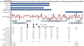
A 5-month-old male patient was admitted for a fever that had persisted for 5 days. The patient showed symptoms of bilateral conjunctiva injection and red lips from 3 days before admission. Meanwhile, polymorphous exanthema had also appeared on his trunk. On the day of admission, erythematous swelling on his soles began to appear since morning, whereas there were no indications of lymphadenopathy or an overt focus of infection. The patient did not have unusual findings in his family or personal medical history. His vital signs upon admission were a blood pressure of 90/50 mmHg, a pulse rate of 140/minute, a respiratory rate of 40/minute, and a body temperature of 37.7°C. The results from the laboratory test performed upon admission indicated leucocytosis (15,730 cells/mm3), elevated C-reactive protein (10.6 mg/dl), and prolongated prothrombin time (20.2 seconds) (Fig 1). The results of all viral studies and blood culture were negative.
Clinical course. aPTT=activated partial thromboplastin time; CRP=C-reactive protein; FDP=fibrin degradation product; FFP=fresh-frozen plasma, INF=infliximab 5 mg/kg; IVIG=intravenous immunoglobulin 2 g/kg; LMCA=left main coronary artery; MethylPDS=intravenous methylprednisolone pulse therapy; PT=prothrombin time; RCA=right coronary artery; WBC=white blood cell count.

The patient’s fever persisted even after admission, befitting the diagnostic criteria for Kawasaki disease. Accordingly, intravenous immunoglobulin (2 g/kg) and high-dose aspirin (100 mg/kg/day) were administered. Echocardiography was performed on the 1st day of hospitalisation, which confirmed the normal cardiac function and normal coronary arteries of the patient.
On the 4th day of hospitalisation, the fever persisted even 36 hours after the initial intravenous immunoglobulin administration and, as a result, intravenous immunoglobulin (2 g/kg) was administered again. In the follow-up laboratory test, platelet levels decreased to 119,000 cells/mm3, and prothrombin time and activated partial thromboplastin time measurements were prolonged further to 302 and 142 seconds, respectively (Fig 1). Aspirin administration was discontinued, and fresh-frozen plasma (10 ml/kg×3 times) and antithrombin-III (2000 IU/day×3 days) were administered as treatment for disseminated intravascular coagulation.
On the 6th day of hospitalisation, the coagulopathy improved after fresh-frozen plasma and antithrombin-III administration, and the platelet count also increased (Fig 1); however, the fever (38.7°C) returned even after the second intravenous immunoglobulin administration, and the elevated C-reactive protein level persisted. Meanwhile, Kawasaki disease symptoms such as skin rash, bilateral conjunctival injection, red lips, and erythematous swelling on soles persisted. The patient remained an intravenous immunoglobulin non-responder, and as a result methylprednisolone pulse therapy (30 mg/kg/day) was initiated. On the 2nd day of methylprednisolone administration, all Kawasaki disease symptoms such as skin rash, conjunctival injection, red lips, and erythematous swelling on soles improved and the fever also subsided to below 37°C. On the 3rd day of methylprednisolone administration, that is, the 8th day of hospitalisation, however, a single episode of fever was noted (38.7°C). Another episode of fever (38.5°C) recurred on the next day, which prompted methylprednisolone pulse therapy for a total of 5 days.
On the 11th day of hospitalisation, the patient had a mild fever (37.8°C), but the C-reactive protein level reduced. Although coagulopathy had been normalised, thrombocytosis was observed (Fig 1). Administration of high-dose aspirin (50 mg/kg) was again initiated.
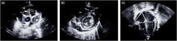
An echocardiography performed on the 12th day of hospitalisation indicated diffuse bilateral coronary artery dilatations (Fig 2a). The proximal right coronary artery was dilated to 7 mm (z score 18.24), and the proximal left main coronary artery was dilated to 7 mm (z score 13.95). Mild pericardial effusion was observed, but the cardiac function was normal (Fig 2b and 2c). The electrocardiography showed no abnormality. A high fever of 39°C was observed, and the C-reactive protein level was elevated. On the basis of the observation that Kawasaki disease persisted without remission, infliximab (5 mg/kg), a tumour necrosis α inhibitor, was administered. On the 13th day of hospitalisation, however, the fever returned even after infliximab administration. On the afternoon of the 13th day of hospitalisation, the patient died after experiencing a sudden cardiac arrest. An autopsy was not performed.
Echocardiography on the 12th day of hospitalisation. ( a ) Diffuse bilateral coronary artery dilatations were observed. The proximal right coronary artery (RCA) was dilated to 7 mm (z score 18.24). The left anterior descending artery (LAD) and the left circumflex artery (LCx) were also dilated. ( b and c ) Mild pericardial effusion (PE) was observed. The mid RCA and the mid LAD were also dilated. Ao=aorta; LV=left ventricle; PA=pulmonary artery.

Discussion
Disseminated intravascular coagulation refers to a syndrome wherein the coagulation cascade is activated by various diseases, thereby creating blood clots throughout the circulatory system, which causes abnormal bleeding following the depletion of clotting factors and the inhibition of normal clotting. Disseminated intravascular coagulation usually occurs following severe infection, malignant tumour growth, or obstetric complications, and sometimes occurs after severe trauma.Reference Levi and Ten Cate 1
Although the exact aetiology of Kawasaki disease is unknown, Kawasaki disease is believed to be systemic vasculitis that infiltrates medium-size arteries accompanied by inflammatory hypercytokinaemia. Inflammatory hypercytokinaemia may trigger a hypercoagulable state and may cause disseminated intravascular coagulation; moreover, vasculitis-induced endothelial injury in Kawasaki disease can activate coagulation, which theoretically can cause disseminated intravascular coagulation to occur as a complication of Kawasaki disease.Reference Sakurai, Takatsuka, Onaka, Takada and Nishino 2
Clinically significant disseminated intravascular coagulation occurring as a complication of Kawasaki disease, however, is very rare and its exact prevalence is still unknown. In the 2007–2008 Japanese Kawasaki disease nationwide survey published by Nakamura et al,Reference Nakamura, Yashiro and Uehara 3 disseminated intravascular coagulation was reported as a complication in 0.08% of all Kawasaki disease patients, and the number of reported cases were few, with only five cases having been reported to date;Reference Usui, Ishii and Akaike 4 – Reference Lee, Jeon, Yang and Pyun 8 one of those cases involved a Kawasaki disease patient with confirmed Yersinia pseudotuberculosis type 4a infection,Reference Usui, Ishii and Akaike 4 whereas another case involved the onset of immune haemolysis, serum sickness, and disseminated intravascular coagulation following high-dose intravenous immunoglobulin administration.Reference Comenzo, Malachowski, Meissner, Fulton and Berkman 5 In the remaining three cases, disseminated intravascular coagulation of an unknown cause had resulted as a complication of Kawasaki disease.Reference Huang, Sun and Huang 6 – Reference Lee, Jeon, Yang and Pyun 8
Among the published Kawasaki disease cases with disseminated intravascular coagulation of unknown cause, most were refractory cases that did not respond to single intravenous immunoglobulin treatment. Among the three cases, only one case exhibited an improvement in Kawasaki disease after a single intravenous immunoglobulin administration.Reference Huang, Sun and Huang 6 In one other case, symptoms persisted while intravenous immunoglobulin was being administered; however, an improvement in patient symptoms was observed after another round of intravenous immunoglobulin administration.Reference Parvathy, Manuel, Criton and Rajesh 7 Meanwhile, in another case, symptoms persisted even after two rounds of intravenous immunoglobulin administration, with an improvement achieved only after the administration of corticosteroids.Reference Lee, Jeon, Yang and Pyun 8 In the present case, a corticosteroid was administered as the fever persisted even after repeat intravenous immunoglobulin administration, and infliximab, a tumour necrosis factor α blocker, was administered because of recurrence of fever. The fever, however, continued to persist. Eventually, a giant aneurysm appeared as a complication, and the patient died suddenly without being treated.
Niwa et alReference Niwa, Aotsuka, Hamada, Uchishiba, Terai and Niimi 9 reported that Kawasaki disease patients who also experience thrombocytopaenia are more susceptible to coronary artery aneurysms and myocardial infarction compared with other Kawasaki disease patients; moreover, in a study by Nofech-Mozes et al,Reference Nofech-Mozes and Garty 10 which reviewed 30 Kawasaki disease patients who also developed thrombocytopaenia, thrombocytopaenia was observed to be more prevalent in the young age group and female patients, whereas it also increased the risk of coronary artery aneurysms and myocardial infarction. Although it is believed that this can be attributed to thrombocytopaenia, which appears as an outcome of platelet consumption from more marked intravascular coagulation caused by endothelial injury and hypercytokinaemia in Kawasaki disease patients with more severe vasculitis, additional studies are needed.
The authors of the present study report their experience, in which all available treatments, including intravenous immunoglobulin, methylprednisolone, and infliximab, were administered to a 5-month-old male Kawasaki disease patient; however, the case was refractory, and the patient subsequently developed a giant coronary artery aneurysm and eventually died. As an autopsy was not perfomed, the cause of death could not be clearly established.
According to a literature review, when disseminated intravascular coagulation appears during the acute phase in Kawasaki disease patients, Kawasaki disease can result in refractory and critical outcomes, and thus more careful observation and treatment are required.
Acknowledgements
None.
Financial Support
This article received no specific grant from any funding agency or from commercial or non-for-profit sectors.
Conflicts of Interest
None.