Introduction
Carcinoma cuniculatum, also referred to as epithelioma cuniculatum,Reference Aird, Johnson, Lennox and Stansfeld1, Reference Kao, Graham and Helwig2 is a rare variant of low-grade squamous cell carcinoma (SCC).Reference Bendelac, Grossin, Sigal, Crickx, Godeau and Bocquet3, Reference Schwartz4 At histology, carcinoma cuniculatum is characterised by a proliferative deep malpighian infiltration with aspects of normal cell maturation and foci of cellular atypia. The term ‘cuniculatum’ derives from the linear, branched and volvulated, rabbit burrow-like crypts and sinuses found in this tumour due to the proliferative deep malpighian infiltration.Reference Aird, Johnson, Lennox and Stansfeld1
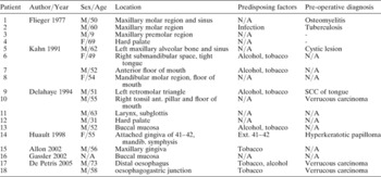
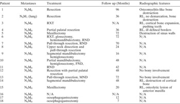
First described in the English literature by Aird and co-workers in 1954,Reference Aird, Johnson, Lennox and Stansfeld1 carcinoma cuniculatum may affect any cutaneous area of the body. It most commonly occurs with elective localisation in the skin of the plantar surface of the foot,Reference Ho, Diven, Butler and Tyring5 followed by the toes,Reference Coldiron, Brown and Freeman6 and the heel2 and only rarely has it been described in the mucosa of the upper aerodigestive tract (Tables I and II).Reference Flieger and Owinski7–Reference De Petris, Lewin and Shoji13
Data from the 18 cases of carcinoma cuniculatum of the upper aerodigestive tract published in the english language literature

N/A = no information available; Ext = extraction; SCC = squamous cell carcinoma; ant = anterior; mandib = mandibular
Data from the 18 cases of carcinoma cuniculatum of the upper aerodigestive tract published in the english language literature

N0 = no lymph nodal disease; M0 = no distant metastasis; N1 = nodal disease present; M1 = distant metastasis present; RXT = radiotherapy; RL = radiolucent; RND = neck dissection; MND = modified radical neck dissection; SCC = squamous cell carcinoma; N/A = no information available
Carcinoma cuniculatum of the skin generally appears as a slow growing painful, non-healing exophytic verruciform mass, but an endophytic growing phase can also be observed particularly at the level of the mucous membrane.
Carcinoma cuniculatum is locally invasive, and metastases to regional lymph nodes have been only rarely described.Reference Schwartz4, Reference Kahn, Blez, Gasser, Weill-Bousson, Vetter and Champy8, Reference Delahaye, Janser, Rodier and Auge9, Reference Van Geertruyden, Olemans, Laporte and Noel14 Local resection is the only definitive treatment, which is generally performed without regional lymph node dissection. To our knowledge, laryngeal localisation has previously been reported in only one case.Reference Delahaye, Janser, Rodier and Auge9
We present the second case of carcinoma cuniculatum of the larynx with special emphasis on the clinical, histopathological and therapeutic aspects of this rare tumour entity.
Case report
A 72-year-old Caucasian man, with a 50-year heavy smoking history, presented to the Section of Otorhinolaryngology of the Department of Surgical Sciences and Organ Transplantations of the University of Cagliari, in September 1999, with recurrent dysphonia lasting six months. He complained of dysphagia for solids, odynophagia and increasing inspiratory dyspnoea with stridor for one week.
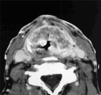
Flexible and rigid laryngoscopy revealed a reddish submucosal glottic–supraglottic swelling. An emergency tracheostomy was performed because of sudden worsening of the dyspnoea. Computed tomography (CT) of the neck demonstrated extensive involvement of the larynx and prelaryngeal structures by a neoplasm with the addition of multiple abscesses, starting from the hyoid bone along with wide destruction of the thyroid cartilage (Figure 1). There was no cervical lymphadenopathy.
Axial CT shows an extensive involvement of the thyroid cartilage, pre-epiglottic space and prelaryngeal tissues by multiple abscesses.

Direct microlaryngoscopy with the aid of rigid telescopes, showed the presence of an open abscess at the level of the right hemilarynx. After CO2 laser removal of the right false vocal fold, it was possible to expose many fragments of the thyroid cartilage that was extensively destroyed by an apparent inflammatory process with purulent chondritis, and no solid tumour was detected. Multiple biopsies histologically reported it as a chronic non-specific inflammatory process. Microbiology performed on the purulent material from the cavity showed Pseudomonas aeruginosa and the patient was treated with an intensive course of intravenous ceftazidime 2 g and ciprofloxacin 750 mg every 12 hours.
As two weeks of antibiotic therapy did not lead to any clinical improvement on the basis of the clinicopathological data and after an appropriate counselling with the patient, a supracricoid laryngectomy with crico-hyoidopexy extended to the prelaryngeal muscles was performed.
All specimens sent for frozen section were negative for carcinoma showing only a pseudoepitheliomatous hyperplasia. Piperacillin-tazobactam 4.5 g every 12 hours was administered for two weeks and the post-operative course was uneventful. The tracheostomy and nasogastric feeding tube were removed after 23 and 25 days, respectively.
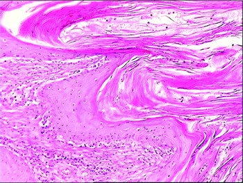
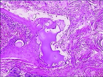
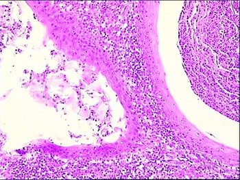
Definitive histopathological evaluation of the entire specimen showed multiple, complex branching keratin-filled cysts surrounded by well-differentiated squamous epithelium (Figure 2). The locally aggressive tumour showed deep invasion into the cartilage (Figure 3) and focal, minimal cytologic atypia were present. A peri- and intratumoural prominent lymphocytic, plasmacytic and neutrophilic infiltration was also seen (Figure 4). Different abscesses were found into the larger keratin masses. The definitive histological diagnosis was carcinoma cuniculatum, and no additional therapy was given. A strict follow up was planned and the patient was free of disease at 70 months after surgery.
Carcinoma cuniculatum: Histological section exhibits keratin-filled crypts surrounded by well-differentiated squamous epithelium (H&E; ×100).

Carcinoma cuniculatum: The neoplastic cells show deep invasion into the cartilage (H&E; ×100).

Carcinoma cuniculatum: Intratumoral abscess (H&E; ×250).

Review of the literature
A review of the literature was performed with the aid of PubMed and Medline searchs. To our knowledge, only 14 cases of carcinoma cuniculatum of the oral cavity,Reference Allon, Kaplan, Manor and Calderon11, Reference Gassler, Helmke, Schweigert, Hassfeld, Otto and Flechtenmacher12 two of the oesophagus,Reference De Petris, Lewin and Shoji13 one of the sinonasal mucosaReference Kahn, Blez, Gasser, Weill-Bousson, Vetter and Champy8 and one case of the larynxReference Delahaye, Janser, Rodier and Auge9 have been reported up to now.
Discussion
Carcinoma cuniculatum represents a rare variant of well-differentiated SCC, which occurs predominantly on the skin at the ball of the soles.Reference Kao, Graham and Helwig2 The aetiology of this tumour in the skin is not well defined, but trauma and chronic irritation have been considered as possible triggering factors.Reference Kraus and Perezmesa15–Reference Zielonka, Goldschmidt and de Fontaine20 Some aspects, such as cytoplasmic vacuolation and binuclear cells, suggest the possibility of viral infections by human papillomavirus, HPV 6 -11 -16 -18, that precede the tumour growthReference Kao, Graham and Helwig2, Reference Eisenberg, Rosenberg and Krutchkoff16–Reference Petersen, Sjolin, Rosman and Lindeberg18, Reference Zielonka, Goldschmidt and de Fontaine20 as in verrucous carcinoma. The correlation of carcinoma cuniculatum to human papillomavirus infection in the aerodigestive tract has been also consideredReference Eisenberg, Rosenberg and Krutchkoff16 and alcohol and tobacco consumption has been proposed as predisposing factors (Table I).Reference Allon, Kaplan, Manor and Calderon11, Reference De Petris, Lewin and Shoji13
The tumour is characterised by an endophytic as well as an exophytic growth pattern with sharp limits and marked hyperkeratosis, parakeratosis and corneous strands intervalled by hyperacanthosic and papillomatous components at various stages of development.Reference Kahn, Blez, Gasser, Weill-Bousson, Vetter and Champy8 As in the skin, also in the aerodigestive tract the deep branched and volvulated rabbit burrow-like formations and keratin-filled crypts and sinuses are peculiar. Polypoid fibroepithelial structures are present where it is possible to distinguish corneal cystic inclusions into their fibrous axis, sometimes super-infected, that correspond to the galleries or the fistulae macroscopically observed.Reference Kao, Graham and Helwig2 The basal membrane is preserved apart from some areas crossed by invasive foci.Reference Delahaye, Janser, Rodier and Auge9 The surrounding area of the tumour is infiltrated by inflammatory cells, mainly lymphocytes, histiocytes, eosinophils and plasma cells, infiltrate is produced, which can organise to form abscesses or micro-abscesses and chronic suppuration.Reference Kao, Graham and Helwig2, Reference Schwartz4, Reference Kahn, Blez, Gasser, Weill-Bousson, Vetter and Champy8, Reference Delahaye, Janser, Rodier and Auge9, Reference Zielonka, Goldschmidt and de Fontaine20 This peculiar histological scenario was characteristic of our case, with the subsequent narrowing of the airway and the need for tracheostomy.
At CT with contrast medium the extensive destruction of the thyroid cartilage with the presence of multiple abscesses with increased perilesional enhancement was present. Destruction of adjacent anatomic structures, such as the nasal floor, sinus walls, and mandibular canal, has been described in the literature.Reference Allon, Kaplan, Manor and Calderon11 In our patient, a chondritis of the laryngeal framework cartilage, with an abscess of the thyroid cartilage, was also hypothesised and only the definitive histology from the specimen after the partial laryngectomy ruled it out.
The only case of carcinoma cuniculatum localised to the larynx reported in the literature refers to a patient whose tumour involved the whole right hemi-larynx with subglottic extension. The patient was cured after a total laryngectomy and bilateral neck dissection without any evidence at histology of metastases.Reference Delahaye, Janser, Rodier and Auge9
Definitive diagnosis is very difficult and requires careful clinical and instrumental examination completed by deep biopsy for histopathology. Multiple biopsies are often necessary to establish the correct diagnosisReference Delahaye, Janser, Rodier and Auge9, Reference Martin, Dalac and Lambert19, Reference Lever and Schaumberg-Lever21 and a long length of time between the onset of the neoplasia and the definitive diagnosis is not infrequent.Reference Kao, Graham and Helwig2, Reference Bendelac, Grossin, Sigal, Crickx, Godeau and Bocquet3 In our case, even when multiple biopsies were performed the results were inadequate to obtain the correct diagnosis and this was possible only after examination of the entire specimen.
At histology carcinoma cuniculatum of the larynx must be differentiated from verrucous carcinoma, well-differentiated SCC and papillary SCC. Verrucous carcinoma, which exhibits similar features, shows an exophytic as well as endophytic growth pattern on a broad front with locally aggressive pushing margins,Reference Shanmugaratnam and Sobin22 but dissociated invasive growth with conspicuous deep duct-like formations and keratin-filled sinuses are absent. Well-differentiated SCC demonstrates cytological signs of malignancy, a brisk mitotic rate, and even atypical mitotic figures, whereas papillary SCC shows an exophytic papillary component with the histological features of papillary carcinoma in situ.Reference Shanmugaratnam and Sobin22
Carcinoma cuniculatum of the upper aerodigestive tract is considered a low-grade tumour and prognosis is usually good, as confirmed by the uncommon lymph node metastatic involvement, and there is general agreement that surgery is the treatment of choiceReference Schwartz4, Reference Kahn, Blez, Gasser, Weill-Bousson, Vetter and Champy8, Reference Delahaye, Janser, Rodier and Auge9, Reference De Petris, Lewin and Shoji13, Reference McKee, Wilkinson, Corbett, Davey, Sauven and Black23 without neck dissection (Table II), while radiotherapy has been considered contraindicated because of the risk of transformation into an anaplastic carcinoma.Reference Schwartz4, Reference Van Geertruyden, Olemans, Laporte and Noel14 The hypothesis of an inflammatory process led us to perform a supracricoid laryngectomy extended to the prelaryngeal muscles, which were extensively involved by the suppurative process. Even though a diagnosis of carcinoma cuniculatum was established definitively from the histology, the absence of any involvement of surgical margins by the tumour suggested that no further treatment was required.
Conclusions
Carcinoma cuniculatum of the larynx is an extremely rare variant of SCC. Histological evaluation often results in the tumours being underestimated and a wrong diagnosis of a benign papillomatous lesion, pseudoepitheliomatous hyperplasia or chronic inflammation being made instead. Carcinoma cuniculatum must be considered as a distinct ‘clinicopathological entity’, as a consequence, the close cooperation between the clinician, the radiologist and the pathologist is essential for the correct diagnosis of these tumours as regards to the correct classification and therapy. In the present case the prevalent inflammatory reaction with multiple micro-abscesses, the destruction of the thyroid cartilage and surrounding tissues, and the negativity of the biopsies obtained before surgery all contributed to the diagnosis of purulent chondritis. The conservative laryngectomy carried out on the basis of the pre-operative diagnosis, appeared to be therefore the best option for the patient.