Non-technical Summary
The new fossil eryopid amphibian Stenokranio boldi, a temnospondyl closely related to Eryops, is described here. The new crocodile-like amphibian is based on well-preserved cranial and postcranial material from ca. 300 million-year-old fluvio-lacustrine deposits of the Permo-Carboniferous (Gzhelian/Asselian) in the Saar–Nahe Basin of southwestern Germany. Phylogenetic analysis identifies Stenokranio as a sister taxon to Eryops. Stenokranio was among the largest predators of the Saar–Nahe Basin. Due to its semiaquatic lifestyle, Stenokranio was able to scour the river and lake shores for prey, but most likely fed on aquatic vertebrates. Stenokranio was part of a faunal assemblage of aquatic, semiaquatic, and fully terrestrial vertebrates, such as sarcopterygian and actinopterygian fishes, xenacanthid sharks, a dvinosaurian temnospondyl, and various other tetrapods (“lepospondyls”, diadectomorphs, and synapsids). This corresponds broadly to the vertebrate community from Permo-Carboniferous rocks in North America that are approximately the same age.
Introduction
Eryopids are widespread nonmarine temnospondyl amphibians that are especially well known from large representatives of the genus Eryops Cope, Reference Cope1878, from the latest Carboniferous and early Permian rocks of the United States (Cope, Reference Cope1882; Case, Reference Case1911; Miner, Reference Miner1925; Sawin, Reference Sawin1941; Pawley and Warren, Reference Pawley and Warren2006; Werneburg et al., Reference Werneburg, Lucas, Schneider, Rinehart, Lucas, Schneider and Spielmann2010). Eryopids were a conspicuous component of the North American early Permian tetrapod assemblage, which included aquatic temnospondyls and terrestrial diadectids, edaphosaurids, and sphencacodontids. Eryopids had a wide geographical distribution on northern Pangea, from the well-known occurrences of North America up to the eastern margin of Europe, with several European localities, including Germany (Werneburg, Reference Werneburg1987; Boy, Reference Boy1990; Schoch and Hampe, Reference Schoch and Hampe2004; Witzmann, Reference Witzmann2013; Witzmann and Voigt, Reference Witzmann and Voigt2014), France (Werneburg, Reference Werneburg1997; Werneburg and Steyer, Reference Werneburg and Steyer1999), Czech Republic (Werneburg, Reference Werneburg1993), Poland (Meyer, Reference Meyer1860a), and Russia (Gubin, Reference Gubin1983).
This work focuses on eryopid remains found in fluvio-lacustrine deposits of the latest Carboniferous–earliest Permian (Gzhelian/Asselian) Remigiusberg Formation at the Remigiusberg quarry near Kusel, Saar–Nahe Basin, southwest Germany, in 2013–2018 (Voigt et al., Reference Voigt, Fischer, Schindler, Wuttke, Spindler and Rinehart2014, Reference Voigt, Schindler, Thum and Fischer2019). The incomplete, but excellently preserved material, including two skulls, is interpreted to represent a new species and new genus. The new eryopid of the Saar–Nahe Basin is one of the stratigraphically oldest eryopids of Europe and among the oldest eryopids in the world. The closest in age is the European temnospondyl Onchiodon thuringiensis Werneburg, Reference Werneburg2007, from the earliest Asselian of the Thuringian Forest Basin, central Germany (Werneburg, Reference Werneburg2007), but this form shows many differences from the new eryopid. Apart from the anatomical description and taxonomic and phylogenetic analyses of the new eryopid from SW Germany, this contribution stimulates the reevaluation of eryopid paleoecology as a whole.
Geological setting and age
All of the eryopid material described herein comes from the Remigiusberg quarry near Kusel, Rhineland–Palatinate, SW-Germany (Fig. 1). The Remigiusberg quarry belongs to the continental Carboniferous–Permian Lorraine–Saar–Nahe Basin, which is one of the largest intramontane basins of the European Variscides (Schäfer, Reference Schäfer1986). Because the French part of the basin fill is mainly covered by younger sediments, regional geologists often talk merely of the Saar–Nahe Basin referring to an area of about 40 × 120 km in SW Germany where Carboniferous–Permian rocks almost continuously crop out (Boy et al., Reference Boy, Haneke, Kowalczyk, Lorenz, Schindler, Stollhofen and Thum2012). The Saar–Nahe Basin accumulated an up to a 10,000-m-thick succession of volcano-sedimentary rocks between early late Carboniferous (Bashkirian) and supposed middle to late early Permian (Artinskian–Kungurian) time (Schneider et al., Reference Schneider, Lucas, Scholze, Voigt and Marchetti2020; Menning et al., Reference Menning, Glodny, Boy, Gast, Kowalczyk, Martens, Rößler, Schindler, von Seckendorff and Voigt2022).
Simplified geological map and lithostratigraphic subdivision of the post-Moscovian part of the Carboniferous–Permian volcano-sedimentary succession of the Saar–Nahe Basin (adapted from Stapf, Reference Stapf1990, Boy et al., Reference Boy, Haneke, Kowalczyk, Lorenz, Schindler, Stollhofen and Thum2012; correlation of formation boundaries with the chronostratigraphic timescale based on Schneider et al., Reference Schneider, Lucas, Scholze, Voigt and Marchetti2020). The type locality of Stenokranio boldi n. gen. n. sp. in the Remigiusberg Formation at the Remigiusberg quarry near Kusel is marked by a star.

The Remigiusberg quarry is a large, active, open-cast mine, producing subvolcanic rock for the production of road and railroad gravel. Subvolcanic rock of the quarry is derived from a sill-like early Permian (Asselian–Sakmarian) intrusion into mainly siliciclastic rocks of the latest Carboniferous to earliest Permian (Gzhelian–Asselian) Remigiusberg Formation. Up to 40 m of fluvio-lacustrine and deltaic sediments of the Remigiusberg Formation currently exposed at the Remigiusberg quarry show a complex interbedding of fluvio-deltaic conglomerate, sandstone, and mudstone, with lacustrine limestone and volcanic tuff beds as minor components. Lithostratigraphic subdivision of the succession is based on seven limestone units ranging 20–150 cm in thickness. The limestone units are referred to the Theisbergstegen and Haschbach lake levels of the middle part and to the Friedelhausen lake level of the basal upper part of the Remigiusberg Formation (Boy and Schindler, Reference Boy and Schindler2000; Fröbisch et al., Reference Fröbisch, Schoch, Müller, Schindler and Schweiss2011; Boy et al., Reference Boy, Haneke, Kowalczyk, Lorenz, Schindler, Stollhofen and Thum2012; Voigt et al., Reference Voigt, Fischer, Schindler, Wuttke, Spindler and Rinehart2014, Reference Voigt, Schindler, Thum and Fischer2019; Fig. 2). Radioisotopic dates from volcanic tuff beds of the Remigiusberg and immediately overlying Altenglan formations suggest that the sediments exposed at the Remigiusberg quarry cover the Carboniferous–Permian boundary with a minimum age of 298.7 ± 0.4 Ma (Burger et al., Reference Burger, Hess and Lippolt1997; Boy et al., Reference Boy, Haneke, Kowalczyk, Lorenz, Schindler, Stollhofen and Thum2012; von Seckendorff, Reference von Seckendorff2012; Voigt et al., Reference Voigt, Schindler, Tichomirowa, Käßner, Schneider and Linnemann2022).
Lithostratigraphy of the Remigiusberg Formation at the Remigiusberg quarry near Kusel and detailed section of the lower Theisbergstegen lake level at the type locality of Stenokranio boldi n. gen. n. sp.

The described eryopid material from the Remigiusberg quarry comes from the lower Theisbergstegen lake level (sensu Boy et al., Reference Boy, Haneke, Kowalczyk, Lorenz, Schindler, Stollhofen and Thum2012) and is here referred to the base of the middle part of the Remigiusberg Formation (Fig. 2). One of the eryopid specimens (holotype; Fig. 2) was preserved in a dark grayish unbedded carbonaceous mudstone (unit 3; Fig. 2) with abundant intraformational pebbles interpreted to represent mudflow deposition in a shallow subaquatic lacustrine paleoenvironment. The other described eryopid specimen (paratype; Fig. 2) was preserved in greenish phytoturbated massive mudstone (unit 4; Fig. 2) of a supposed lake shoreline paleoenvironment. More detailed information on the litho- and biofacies of the Remigiusberg Formation at the Remigiusberg quarry are given in Voigt et al. (Reference Voigt, Schindler, Thum and Fischer2019, p. 229–232).
Materials and methods
This work is based on two eryopid specimens discovered at the southwest German Remigiusberg quarry near Kusel, Rhineland–Palatinate, in autumn 2013 (paratype) and spring 2018 (holotype). Both specimens are represented by disarticulated bone material consisting of the skull with remains of the mandibles (holotype) and an incomplete skull, mandibles, and anterior postcranial material (paratype), respectively. The remains seem to be derived from partially to largely fully decomposed skeletons that were transported into a marginal lacustrine paleoenvironment. The holotype specimen is closely associated with non-eryopid, probably microsaur bone remains.
Preparation of the specimens was carried out mechanically by one of us (LR) and Georg Sommer from the Naturhistorisches Museum Schloss Bertholdsburg Schleusingen (NHMS). Photographs were taken with a Nikon D5100. Drawings were prepared from an A3-photograph and with a ‘camera lucida’ at a Motic binocular.
The new eryopid specimens are stored at the Urweltmuseum GEOSKOP/Lichtenberg Castle near Kusel (UGKU) but are owned by the state of Rhineland–Palatinate according to the local heritage protection law and, thus, inventory numbers refer to the Natural History Museum Mainz/State Collection of Natural History of Rhineland–Palatinate (NHMMZ/LS).
Repositories and institutional abbreviations
CM, Carnegie Museum of Natural History, Pittsburgh, Pennsylvania; CMNH, Cleveland Museum of Natural History, Cleveland, Ohio; FMNH, Field Museum of Natural History, Chicago, Illinois; MCZ, Museum of Comparative Zoology of the Harvard University, Cambridge, Massachusetts; MHNA, Museum of Natural History, Autun, France; MMG, Museum für Mineralogie und Geologie, Dresden, Germany; NHMMZ/LS, Natural History Museum Mainz/State Collection of Natural History of Rhineland–Palatinate, Germany; NHMS, Naturhistorisches Museum Schloss Bertholdsburg, Schleusingen, Germany; NMMNH, New Mexico Museum of Natural History, Albuquerque, New Mexico; UGKU, Urweltmuseum GEOSKOP/Lichtenberg Castle near Kusel, Germany.
Systematic paleontology
Tetrapoda Jaekel, Reference Jaekel1909
Amphibia Linneaus, Reference Linnaeus1758
Temnospondyli von Zittel, Reference von Zittel1888
Eryopidae Cope, Reference Cope1882
Diagnosis
Synapomorphies (after Sawin, Reference Sawin1941; Romer, Reference Romer1947; Milner, Reference Milner1989, Reference Milner, Taylor and Larwood1990; Boy, Reference Boy1990; Werneburg and Steyer, Reference Werneburg and Steyer1999; Schoch and Hampe, Reference Schoch and Hampe2004; Werneburg, Reference Werneburg2007; Werneburg and Berman, Reference Werneburg and Berman2012; Schoch and Milner, Reference Schoch, Milner and Sues2014): (1) enlarged choana medially wide; (2) ectopterygoid, palatine and vomer only with two or three fangs (without subsequent smaller teeth); (3) lacrimal reaches anteriorly to the naris or septomaxilla; (4) enlarged posterior width of skull (pSw/Sl = 0.92–1.10); (5) posterior part of the cultriform process widened (partly); (6) interclavicle of adults proportionally small and broadly ovate in outline; (7) ilium with vertically directed dorsal process, which is anteroposteriorly widened dorsally.
Stenokranio new genus
Types species
Stenokranio boldi n. gen. n. sp.
Diagnosis
As for type species by monotypy.
Etymology
Greek στενός (stenos) for narrow, κρανίο (kranio) for skull.
Remarks
None.
Stenokranio boldi new species
Figures 3–11, 12.1–12.3, 13–15, 17, 18.1, 19.1
Stenokranio boldi n. gen. n. sp., skull roof in dorsal view, holotype NHMMZ/LS PW 2019/5025, with the anterior view of the right premaxilla; note the low and nearly equal height of the teeth.

Stenokranio boldi n. gen. n. sp., skull roof in dorsal view, holotype NHMMZ/LS PW 2019/5025. (1) Interpretative drawing with atlas; (2) reconstruction. atl = atlas; d = dentary; f = frontal; ior = intraorbital ridge; j = jugal; jr = jugal ridge; l = lacrimal; ld = lamina descendens; lf = lacrimal furrow; m = maxilla; md = mandible; n = nasal; ol = occipital lamella; p = parietal; pm = premaxilla; po = postorbital; pp = postparietal; prf = prefrontal; prr = prefrontal-rostral ridge; psr = parietal-supratemporal ridge; pt = pterygoid; ptf = postfrontal; ptr = postorbital-tabular ridge; q = quadrate; qj = quadratojugal; Sl = skull length; sm = septomaxilla; sq = squamosal; st = supratemporal; t = tabular; tfl = tabular flange; v = vomer.

Stenokranio boldi n. gen. n. sp., holotype, NHMMZ/LS PW 2019/5025. (1) Palate in ventral view, with both mandibles, atlas, and external tetrapod bones (see Fig. 15); (2) teeth between pterygoid and vomer; (3) tooth-like palatal denticles on vomer; (4) left stapes (in the lower right part of the image) with stapedial foramen.

Stenokranio boldi n. gen. n. sp., palate in ventral view, with both mandibles, atlas, and external tetrapod bones (see Fig. 15), holotype NHMMZ/LS PW 2019/5025. (1) Interpretative drawing; (2) reconstruction. a = angular; ar = articular; atl = atlas; ch = choane; copr = coprolith; d = dentary; dph = diadectid phalangal; eo = exoccipital; f = frontal; fpqa = quadratojugal foramen accessory; il = ilium; l = lacrimal; md = mandible; mf = meckelian fenestra; n = nasal; o = orbit; p = parietal; pc = cultriform process of parasphenoid; plt = palatine tooth; pp = postparietal; ppl = palatal plates; prf = prefrontal; ps = parasphenoid; pt = pterygoid; q = quadrate; qj = quadratojugal; r = rib; se = sphenethmoid; Sl = skull length; sq = squamosal; stf = stapedial foramen; stp = stapes; v = vomer; vt = vomerine tooth.

Stenokranio boldi n. gen. n. sp., skull with mandibles and anterior postcranial skeleton, paratype NHMMZ/LS PW 2019/5022. (1) Dorsal skull roof with left mandible, shoulder girdle, and anterior axial skeleton; (2) palatal skull in ventral view, right mandible in labial view, shoulder girdle, and ribs. Sl = skull length.

Stenokranio boldi n. gen. n. sp., interpretative drawing of skull with mandibles and anterior postcranial skeleton, paratype NHMMZ/LS PW 2019/5022. (1) Dorsal skull roof with left mandible, shoulder girdle, and anterior axial skeleton; (2) palatal skull in ventral view, right mandible in labial view, shoulder girdle, and ribs. a = angular; ch = choane; cl = clavicle; cor 3 = coronoid 3; cth = cleithrum; d = dentary; dup = distal uncinate process; ect = ectopterygoid teeth; f = frontal; ic = intercentrum; icl = interclavicle; j = jugal; l = lacrimal; m = maxilla; md = mandible; n = nasal; na = neural arch; p = parietal; pl = palatine; plc = pleurocentrum; pm = premaxilla; po = postorbital; prf = prefrontal; psp = postsplenial; pt = pterygoid; ptf = postfrontal; pup = proximal uncinate process; q = quadrate; qj = quadratojugal; r = rib; sa = surangular; sc = scapulocoracoid; se = sphenethmoid; Sl = skull length; sp = splenial; sq = squamosal; st = supratemporal; t = tabular; v = vomer.

Stenokranio boldi n. gen. n. sp., skull in various views, paratype NHMMZ/LS PW 2019/5022. (1) Skull roof in posterodorsal view, with left mandible and left clavicle; note the dorsal strutting pattern with large longitudinal ridges; (2) dermal sculpture of the dorsomedian skull roof; (3) sphenethmoid with longitudinal, ventral ridge (holotype NHMMZ/LS PW 2019/5025); (4) skull in posterior view, note the nearly uncompacted natural skull shape, and the compact sphenethmoid below the pineal foramen; (5) large ectopterygoid fang pair pierce the first or second rib without fracture; (6) wide choana and palatine with large fang, and vomer with fang pair and intensive denticulation as well as teeth of premaxilla and maxilla. ch = choane; ect = ectopterygoid teeth; m = maxilla; pc = cultriform process; pif = pineal foramen; pl = palatine; pt = pterygoid; rib = rib; se = sphenethmoid; Sl = skull length; v = vomer.

Stenokranio boldi n. gen. n. sp., interpretative drawing of skull with mandibles and anterior postcranial skeleton, paratype NHMMZ/LS PW 2019/5022. (1) Dorsal skull roof with left mandible; the arrow indicates the suture between premaxilla and maxilla; (2) reconstruction of dorsal skull roof; (3) reconstruction of ventral palatal skull; (4) palatal skull in ventral view, with, ribs, intercentrum, and cleithrum; (5) enlarged quadrate condyle in ventral view. a = angular; ch = choane; cl = clavicle; cor 3 = coronoid 3; cth = cleithrum; d = dentary; dup = distal uncinate process; ec = ectopterygoid; ect = ectopterygoid teeth; f = frontal; fpq = quadratojugal foramen; ftr = frontal transverse ridge; ic = intercentrum; j = jugal; l = lacrimal; m = maxilla; md = mandible; n = nasal; ntr = nasal transverse ridge; p = parietal; pif = pineal foramen; pjr = prefrontal–jugal ridge; pl = palatine; plt = palatine tooth; pm = premaxilla; po = postorbital; ppr = postfrontal-parietal ridge; prf = prefrontal; prr = prefrontal-rostral ridge; psp = postsplenial; pt = pterygoid; ptf = postfrontal; ptr = postorbital–tabular ridge; pup = proximal uncinate process; q = quadrate; qb = quadrate boss; qj = quadratojugal; r = rib; sa = surangular; se = sphenethmoid; Sl = skull length; sm = septomaxilla; sp = splenial; sq = squamosal; st = supratemporal; syt = symphyseal teeth; t = tabular; v = vomer; vt = vomerine tooth.

Measured distances of the reconstructed skull roof from Stenokranio boldi n. gen. n. sp. aSw = anterior width of skull at level of maxilla-premaxilla sutures; Hl = postorbital midline length of skull from level of posterior margins of orbits; Hw = postorbital width of skull between lateral margins of supratemporals; INw = minimum internarial width; IOw = minimum interorbital width; Jw = transverse width of jugal at maximum lateral lacrimal extent of orbit; Lal = maximum length of lacrimal; Law = maximum transverse width of lacrimal; mSw = midlength width of skull at midlength level of orbits; Ol = maximum length of orbit; POl = preorbital midline length of skull from level of anterior margins of orbits; Pol = maximum posterior length of postorbital from posteriormost extent of orbit; Pow = maximum transverse width of postorbital at contribution to orbital margin; pSw = maximum posterior width of skull at level of posterolateral margins of cheeks; Sl = midline skull length; Thl = length of tabular horn region between levels of posterior tabular corner and occipital midline margin; Ww = maximum transverse width of cheek from lateral margin of supratemporal anterior to otic notch.

Two different eryopid mandibles from the Remigiusberg quarry. (1–3) Right mandible of Stenokranio boldi n. gen. n. sp., in labial view, note the sharp carina on the anterior teeth (2), length 232 mm, paratype NHMMZ/LS PW 2019/5022;the small arrow indicates the suture between surangular and coronoid 3; (4, 5) left mandible of a probable eryopid in lingual view, length 213 mm, NHMMZ/LS PW 2019/5020 (former: POL-F 2012-001), after Witzmann (Reference Witzmann2013). a = angular; art = articular; cor 3 = coronoid 3; d = dentary; mf = meckelian fenestra; pa = prearticular; psp = postsplenial; sa = surangular; sp = splenial; Mdh = mandibular height; Sph = surangular height.

Stenokranio boldi n. gen. n. sp., interpretative drawing of neural arches. (1) Atlas in lateral view, holotype NHMMZ/LS PW 2019/5025; (2) anteriormost neural arch in posterolateral view; (3) two neural arches in lateral and anterolateral view; (4, 5) neural arches in lateral view, (2–5) paratype NHMMZ/LS PW 2019/5022. atl = atlas; fpc = facets for pleurocentra; poz = postzygapophysis; prz = prezygapophysis; rds = roughened dorsal surface; spp = spinose process; trp = transversal process.

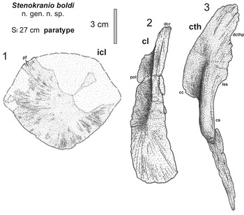
Stenokranio boldi n. gen. n. sp., interpretative drawing of shoulder girdle bones, paratype NHMMZ/LS PW 2019/5022. (1) Interclavicle in dorsal view; (2) left clavicle in dorsal view; (3) left cleithrum in lateral view. cc = cleithral crest; cl = clavicle; cs = cleithral shaft; cth = cleithrum; dcr = dorsal clavicular rod; dcthp = dorsal cleithral process; icl = interclavicle; lss = suprascapular lamina; pf = pectinate fringe; pol = posterior lamina of clavicle.

Stenokranio boldi n. gen. n. sp., interpretative drawing of both scapulocoracoids, paratype NHMMZ/LS PW 2019/5022. (1) Right scapulocoracoid from medial view; (2–5) left scapulocoracoid (2) in medial view, (3) in posterior view, (4) in lateral view, (5) in anterior view. fcor = coracoid foramen; fgl = glenoid foramen; fsgl = supraglenoid foramen; gl = glenoid fossa; igb = intraglenoid buttress; igf = infraglenoid fossa; igs = infraglenoid recess; lsr = lateral supraglenoid ridge; sc = scapulocoracoid; sctor = scapular torus (scapular blade); sgb = supraglenoid buttress; sgf = supraglenoid fossa; Sl = midline skull length; ssf = subscapular fossa.

Foreign tetrapod bones from the palatal skull of Stenokranio boldi n. gen. n. sp. (compare Figs. 5 and 6.1), holotype NHMMZ/LS PW 2019/5025. (1) Interpretative drawing of a possible microsaur skeletal remains with the pelvis in lateral view, ribs, ?humerus, and mandible; (2) photo and interpretative drawing of a diadectomorph phalanx, with cross section (diagonal-line fill). act = acetabulum; ar = articular; d = dentary; hum = humerus; il = ilium; is = ischium; pu = pubis; r = rib.

Stenokranio boldi n. gen. n. sp., reconstruction of the new eryopid. (1) Dorsal skull roof; (2) palatal skull; (3) life restoration of the whole animal (artwork by Frederik Spindler, Kipfenberg). ap = anterior palatal depression; bp = basal plate of parasphenoid; ch = choane; cp = cultriform process; ec = ectopterygoid; eo = exoccipital; f = frontal; fpq = quadratojugal foramen; ior = intraorbital ridge; j = jugal; jr = jugal ridge; l = lacrimal; lf = lacrimal furrow; m = maxilla; n = nasal; ol = occipital lamella; p = parietal; pl = palatine; pm = premaxilla; po = postorbital; pp = postparietal; ppr = postfrontal-parietal ridge; prf = prefrontal; prr = prefrontal-rostral ridge; ps = parasphenoid; psr = parietal-supratemporal ridge; pt = pterygoid; ptf = postfrontal; ptr = postorbital-tabular ridge; q = quadrate; qj = quadratojugal; se = sphenethmoid; Sl = midline skull length; sm = septomaxilla; sq = squamosal; st = supratemporal; t = tabular; v = vomer.

Comparison of eryopid skull roofs in dorsal view. (1) Stenokranio boldi n. gen. n. sp., from this paper. (2) Glaukerpeton avinoffi, after Werneburg and Berman (Reference Werneburg and Berman2012). (3) Eryops megacephalus, after Sawin (Reference Sawin1941). (4) Onchiodon thuringiensis, after Werneburg (Reference Werneburg2007). (5) Onchiodon labyrinthicus, after Boy (1991). (6) Actinodon frossardi, after Werneburg (Reference Werneburg1997). f = frontal; j = jugal; l = lacrimal; m = maxilla; n = nasal; p = parietal; pm = premaxilla; po = postorbital; pp = postparietal; prf = prefrontal; ptf = postfrontal; q = quadrate; qj = quadratojugal; Sl = midline skull length; sm = septomaxilla; sq = squamosal; st = supratemporal; t = tabular.

Comparison of eryopid skulls in palatal view. (1) Stenokranio boldi n. gen. n. sp., from this paper (2) Glaukerpeton avinoffi, after Werneburg and Berman (Reference Werneburg and Berman2012). (3) Eryops megacephalus, after Sawin (Reference Sawin1941). (4) Onchiodon thuringiensis, after Werneburg (Reference Werneburg2007). (5) Onchiodon labyrinthicus, after Boy (1991). (6) Actinodon frossardi, after Werneburg and Steyer (Reference Werneburg and Steyer1999). ch = choane; ec = ectopterygoid; eo = exoccipital; m = maxilla; pl = palatine; pm = premaxilla; ps = parasphenoid; pt = pterygoid; q = quadrate; se = sphenethmoid; v = vomer.

Holotype
NHMMZ/LS PW 2019/5025 (formerly: UGKU 2564), consisting of the skull with skull roof and palate, together with remains of the mandibles (skull length [elsewhere = midline skull length] 24.7 cm).
Paratype
NHMMZ/LS PW 2019/5022 (formerly: UGKU 1998), consisting of the greater portion of skull roof, parts of the palate with palatine, choana and several pairs of fangs, and the mandible in lateral view (skull length 27 cm), together with a few bones of the anterior postcranium.
Diagnosis
Autapomorphies: (1) small posterior skull width (pSw/Sl = 0.92) and posterior half of skull with nearly longitudinally straight margins; (2) postparietals and tabulars forming a longitudinally short bony strip; (3) ectopterygoid very wide, its most posterior part and neighboring pterygoid equal in width.
Synapomorphies with some of the eryopids.—(1) Density of sculpture pattern quantified as the number of pits per in2 on frontal + jugal in relation to skull length range between 0.64 and 1.42, and bones of normal thickness, in contrast to the thinner bones in Glaukerpeton Romer, Reference Romer1952; (2) small width of skull table between lateral margins of supratemporals (Hw/Sl = 0.42–0.43), in contrast to Glaukerpeton and Actinodon Gaudry, Reference Gaudry1866; (3) equal internarial and interorbital width, in contrast to Glaukerpeton, in which the internarial width is smaller; (4) occipital margin of skull roof is relatively straight and only slightly concave, only shared with Onchiodon thuringiensis; (5) septomaxilla is completely sculptured, shared with Onchiodon Geinitz, Reference Geinitz1862, and in contrast to Eryops and Glaukerpeton with a smooth anterior portion of the bone; (6) no interfrontal, in contrast to Eryops; (7) no lateral line sulci, in contrast to Glaukerpeton and Actinodon; (8) palatine is very wide, only shared with Glaukerpeton; (9) fang pair of ectopterygoid and vomer of equal size (in contrast to Onchiodon, in which ectopterygoid fangs are smaller), and consist of two teeth (in contrast to Glaukerpeton with three ectopterygoid teeth); (10) straight dorsal margin of the surangular process and the participation of the posterior half of coronoid 3 in this process with the same height; surangular process is relatively low in comparison to the maximum height of the mandible, in contrast to the second eryopid from Remigiusberg; (11) dentary with approximately 48–50 marginal tooth positions, in contrast to the lower number in the second eryopid from Remigiusberg; (12) homodont marginal dentition of mandible and maxilla, with the size of the teeth generally small and gradually decreasing from rostral to abrostral; parasymphyseal teeth are similar in size to the adjacent dentary teeth; this stands in contrast to the second eryopid from Remigiusberg, Eryops, and Onchiodon with a rather heterodont dentition of larger teeth; (13) low coracoid region, angle between supraglenoid buttress and anterior margin of scapular blade is 90° or slightly greater, in contrast to Glaukerpeton and Onchiodon labyrinthicus Geinitz, Reference Geinitz1862.
Occurrence
Remigiusberg quarry at the northeastern rim of the Remigiusberg (387685 E, 5487644 N, UTM 32U, WGS 84; UGKU L-21), ~1 km northeast of Haschbach, Kusel County, western Rhineland–Palatinate, Germany (Voigt et al., Reference Voigt, Fischer, Schindler, Wuttke, Spindler and Rinehart2014, Reference Voigt, Schindler, Thum and Fischer2019; Fig. 1). Type horizon is a mudstone of the lower Theisbergstegen lake level, middle part of the Remigiusberg Formation, base of Rotliegend, Gzhelian–Asselian boundary, latest Carboniferous or earliest Permian (Figs. 1, 2).
Comparative description
The two specimens are similar in size with skull lengths of 24.7 cm (holotype) and 27 cm (paratype), respectively. Their possession of shared characters such as the very similar skull roof proportions (Table 1), posteriorly notched orbits, and the similar type of dermal sculpturing indicate that they belong to the same taxon.
Comparative measurements of adult eryopid skulls (important values in bold; after Boy, Reference Boy1990; Werneburg, Reference Werneburg1997, Reference Werneburg2007; Werneburg and Berman, Reference Werneburg and Berman2012).

General skull morphology.—The dermal sculpture of the dorsal skull roof corresponds to the relatively coarse sculpture pattern known from most eryopids (Werneburg and Berman, Reference Werneburg and Berman2012; Table 2). The dermal sculpture of the dorsal skull roof consists of a reticulated pattern of small pits and valleys separated by narrow ridges (Figs. 3, 9.1, 9.2). The nasal shows much more radially directed ridges on the smaller holotypic skull roof (Fig. 3) than on the larger paratypic skull with a close reticulate system (Fig. 9.1). The density of the sculpture pattern is quantified as the number of pits per in2 (6.452 cm2) on the frontal and jugal, which are typically well-preserved bones in eryopid skulls, and as a proportion of those counts to skull length. These intraspecific indices range between elements and specimens of Stenokranio n. gen. between 0.64 and 1.42, which are very similar in Onchiodon and Eryops specimens. The dermal sculpture of the dorsal skull roof in similar-sized Glaukerpeton specimens is of much finer sculpture pattern with indices from 2.6 to 4.0 (Werneburg and Berman, Reference Werneburg and Berman2012).
Ranges of density counts of dermal sculpture pits and valleys of frontal and jugal in relation to the skull length (in cm) given separately and combined for eryopids and grouped by genus, species, and maturity (partly after Werneburg and Berman, Reference Werneburg and Berman2012); p = number of dermal skull pits or valleys per in2 (= 6.452 cm2), mainly from frontal and jugal at midlength level of orbits.

Most eryopid skulls exhibit a dorsal strutting pattern with large ridges, as in Eryops and Onchiodon, which increased the mechanical stability of the skull (Sawin, Reference Sawin1941; Boy, Reference Boy1990; Werneburg, Reference Werneburg2007). A paired, well-developed and large longitudinal ridge extends from the lateral portion of the tabular and supratemporal to the postorbital. The ridge then runs on the post- and prefrontal and the median part of the nasal to the premaxilla, where it forms a median wall to the naris (Fig. 17.1). This pair of longitudinal ridges is consistent in both skulls. Additional transverse ridges may occur between the longitudinal ridges on parietals, frontals, and nasals. These transverse ridges are differently pronounced and most completely developed on the paratype skull roof (Fig. 10.1). Depressions are present between these ridges. Also, the ridges on the jugal are variable. A Y-shaped ridge is developed on the anterior part of the jugal in the holotypic skull, whereas a curved ridge extending from the prefrontal is present on the anterior part of the jugal in the paratypic skull.
The degree of skull roof ossification is relatively high, and the bones are normally thick as in other eryopids, but in contrast to the 30–50% thinner skull roof bones of Glaukerpeton.
The combination of both known skulls of Stenokranio n. gen. allowed a tentative reconstruction of the skull roof in dorsal view and of the palate in ventral view (Fig. 17.1, 17.2). The skull is longer than wide. In dorsal view the lateral margin of the skull describes a wide parabolic curve with a broad, bluntly rounded snout and a short postorbital region. The width of the posterior skull is unusually small in comparison with other eryopids (pSw/Sl = 0.92; see Table 1). Therefore, Stenokranio n. gen. has a skull with nearly parallel lateral margins, which is a unique character in eryopids. The postorbital width of the skull measured between the lateral margins of the supratemporals is small (Hw/Sl = 0.42–0.43) in contrast to that of Glaukerpeton and Actinodon. The preorbital skull is elongate (POl/Sl = 0.60–0.61), similar to large skulls of Eryops or Onchiodon thuringiensis. The internarial and interorbital width are equal (INw/Sl = IOw/Sl = 0.24 in the holotype and = 0.26/0.27 in the paratype), as in most eryopids, but in contrast both to Glaukerpeton, in which the internarial width is smaller, and to Eryops megacephalus Cope, Reference Cope1878, with a smaller interorbital width (Table 1). The occipital margin of the skull roof is relatively straight and only slightly concave, as in O. thuringiensis. The quadrate condyles lie distinctly posterior to the occipital condyles. The orbits are large compared to most other eryopids (Ol/Sl = 0.17–0.19). They bear a posterior notch formed by the postorbital.
Growth stage.—Both known specimens of Stenokranio n. gen. are clearly adult animals because (1) the dermal sculpture consists of a reticulated pattern of small pits and valleys separated by narrow ridges; (2) exoccipitals, sphenethmoid (partly) and quadrate are well ossified; (3) vertebrae have ossified inter- and pleurocentra, neural arches with well-developed transverse processes and high spinous processes; (4) ribs present large uncinate processes; (5) scapulocoracoid is well ossified; (6) the skull length of 24–27 cm is large in the family Eryopidae and only Eryops, O. thuringiensis, and Osteophorus (Meyer, Reference Meyer1860a) have larger skulls. However, two characters of the Stenokranio n. gen. specimens indicate that they are early adult, and they did not reach the late adult or senile stage: (1) sphenethmoid is very narrow and probably only partly ossified, and (2) basioccipital is not ossified.
Skull roof (Figs. 4, 7.1, 8.1, 9.1, 9.2, 10.1, 17.1, 18).—The interpremaxillary suture is elongated and accounts for ~13% of the midline length of the skull. This is similar to Glaukerpeton and Eryops megacephalus, whereas this suture is proportionally shorter in the Pennsylvanian Eryops of New Mexico (Werneburg et al., Reference Werneburg, Lucas, Schneider, Rinehart, Lucas, Schneider and Spielmann2010). The alary process of the premaxilla is relatively wide as in Onchiodon labyrinthicus (Boy, Reference Boy1990) but stronger in the Pennsylvanian Eryops. The premaxilla has 14 tooth loci in its ventral tooth arcade.
The rounded oval- to triangular-shaped naris is elongated, as in Glaukerpeton or Eryops megacephalus, comprising 11% of the midline length of the skull. The kidney-shaped septomaxilla is completely sculptured (Fig. 4.1) and is completely located in a dorsal position at the posteromedial part of the naris (shared with Onchiodon). It covered about one-third of the area of the narial opening and excludes the nasal from the naris. The mostly ventrally directed and smooth septomaxilla in Eryops occupies almost the entire naris proper (Sawin, Reference Sawin1941).
The lacrimal is roughly diamond shaped. It is separated from the orbit by an elongated contact between jugal and prefrontal. However, the anterior part of the lacrimal is wide, as in Glaukerpeton or Eryops megacephalus, and participates in the posterolateral narial margin. A short, narrow, anterior–posteriorly directed furrow is visible in this part of the bone and is interpreted as the ductus nasolacrimalis (holotypic skull in Figs. 3, 4.1). It is completely closed in the slightly larger paratypic skull (Figs. 7.1, 10.1). This duct is closed earlier in the late juvenile stage of O. labyrinthicus (Boy, Reference Boy1990, fig. 2). A groove for the lacrimal duct is also known from the lacrimal in Eryops (Sawin, Reference Sawin1941, p. 419). The maxilla has a wide dorsal shelf, especially near the lacrimal–jugal suture and about 40 tooth loci in its ventral tooth arcade. The nasal is considerably elongated and narrow as is typical of eryopids with the exception of O. thuringiensis in which the nasal is anterolaterally wider. The frontal is narrow as in most other eryopids.
The jugal is proportionally wider than in Glaukerpeton or Actinodon but proportionally narrower than in O. thuringiensis. The postorbital is triangular in outline and has an angled (or notched) orbital margin. The postfrontal and prefrontal clearly contact one another as in all eryopids, but their dorsal orbital processes are proportionally much wider than in Glaukerpeton and Actinodon. The prefrontal is anteriorly widened and the postfrontal is posteriorly elongated and wide. Together with the shortened postorbital skull table this leads to the shortening of the supratemporal, which is approximately as long as wide and is one of the proportionally shortest supratemporals in eryopids. Only in Clamorosaurus borealis Gubin, Reference Gubin1983, is the supratemporal much wider than long (RW and FW in preparation).
The parietals extend anterior to the posterior orbital margin. Posteriorly, they nearly reach a common transverse line with the posterior margin of the short supratemporals, which is a unique situation in eryopids. The parietal foramen lies at the transversal line at the posterior end of postfrontals (Figs. 3, 4, 9.2) and is not located in a smooth depression on the parietals as described in O. thuringiensis. Stenokranio n. gen. bears the shortest postparietals and tabulars in eryopids. These bones form a continuous short strip at the slightly concave occipital margin, as in O. thuringiensis. The tabular horn is short (Thl/Sl = 0.06). The occipital lamella is medially bilobed and anteriorly bordered by one or two transverse ridges (Fig. 4.1). The tabular bears a pronounced ventrally directed tabular flange. In accordance with the generally slender cheek (Ww/Sl = 0.26), the squamosal and quadratojugal are relatively narrow. In Onchiodon and Actinodon the cheek is proportionally wider whereas Glaukerpeton has the proportionally narrowest cheeks (Table 1).
In Stenokranio n. gen. the exposure of the quadrate on the occipital surface of the cheek (Figs. 4.1, 10.1) consists of a narrow, dorsally short process that is directed anteromedially between the squamosal and the quadrate ramus of the pterygoid. In Eryops and Glaukerpeton this construction is very similar, but the dorsal portion of the quadrate is anteromedially more elongated whereas in O. thuringiensis it is similarly short but wider. A transverse strip-like dorsal part of the quadrate is known in O. labyrinthicus and Actinodon (Fig. 18). A well-developed, boss-like protuberance occurs at the ventral margin of the dorsal quadrate process in Stenokranio n. gen., Glaukerpeton, and O. labyrinthicus. In Stenokranio n. gen., Onchiodon, and Eryops a quadratojugal foramen near the posterolateral margin of the quadratojugal is visible only in ventral view of the skull (Figs. 6.1, 10.1). In Glaukerpeton, the foramen is visible in lateral and dorsal views of the skull roof. In the Eryops sp. specimen NMMNH P-46379 from the Late Pennsylvanian of New Mexico two small and one larger quadratojugal foramen are visible at the posteroventral margin of the quadratojugal only in dorsal and lateral views of the skull roof (Werneburg et al., Reference Werneburg, Lucas, Schneider, Rinehart, Lucas, Schneider and Spielmann2010).
Lateral line sulci are not present in Stenokranio n. gen. and only known among postlarval forms of Glaukerpeton and Actinodon (Werneburg, Reference Werneburg1997; Werneburg and Berman, Reference Werneburg and Berman2012). However, Warren (Reference Warren2007) argued that at least an enclosed quadratojugal lateral line canal is present in Eryops.
Palate and braincase (Figs. 5–8, 9.3–9.5, 10.3, 10.4, 17.2, 19).—In addition to the skull roof, the palate is informationally complete in both skulls. The pattern of thickened longitudinal ridges on the palatal bones is only partly preserved. A pronounced, narrow ridge starts posterior to the vomerine tusks and borders the choana medially (Figs. 5, 6, 9.5, 10.4, 19.1). Another elevated longitudinal structure starts posterior to the palatine fang pair and continues posteriorly on more than half the length of the ectopterygoid (Figs. 9.5, 10.4, 19.1), as in O. thuringiensis.
Just anterior to the transverse level of the vomerine tusks, the anterior palatal fossae extend mostly on the anterior part of the vomers (Fig. 10.4) and probably on the dental shelf of the premaxilla. The anterior palatal fossae are restricted to the premaxillae in Glaukerpeton because these bones extend posteriorly almost to the level of the vomerine tusks.
The vomer is elongated and wide. The smallest width of both vomers (= interchoanal width) is clearly wider than the interorbital width in Stenokranio n. gen., Eryops, and Onchiodon. In contrast, the interchoanal and interorbital width is equal in Glaukerpeton and Actinodon. The process-like posterolateral corner of the vomer in Stenokranio n. gen., Glaukerpeton, and Onchiodon extends a short distance between the pterygoid and palatine (Figs. 6.1, 10.4), although it is much narrower in the latter, whereas in the Permian and Pennsylvanian Eryops the process is much longer and vermiform. The suture between vomer and palatine is much more elongated than in Glaukerpeton and Actinodon. The palatine is short and very wide and only Glaukerpeton has a similarly shaped palatine. The ectopterygoid is unusually wide (Fig. 10.4, and by reconstruction in Fig. 19.1) in contrast to all other eryopids. Its posteriormost part is equal in width to the neighboring pterygoid. The dentition of all three palatal bones consists of one pair of fangs on each bone (Figs. 6.1, 9.4, 9.5, 10.4). The fang pair of the palatine is the largest, and the fang pairs of the vomer and ectopterygoid slightly smaller and of equal size. Glaukerpeton has three fangs and both species of Onchiodon present one pair of very small teeth on the ectopterygoid.
The choana is shortened and nearly circular (Fig. 9.5). This character of Stenokranio n. gen. is in contrast to that of Glaukerpeton, Actinodon, and Eryops in which the choanae are more elongate. The anterior margin of the choana exhibits a short, narrow, V-shaped notch of variable development lateral to the alveolus of the vomerine tusks as in all eryopids except for O. labyrinthicus.
The pterygoid (Figs. 5, 6, 10.4) has a narrow palatinal ramus as in Glaukerpeton, Eryops, and Actinodon. The anterior third of the the palatinal branch forms a sharp anteromedially directed corner, similar to Eryops and O. labyrinthicus. The transverse flange of the pterygoid in Stenokranio n. gen., Glaukerpeton, and Eryops exhibits a low, angular expansion. In Onchiodon and Actinodon, on the other hand, the entire, free, lateral margin of the pterygoid is greatly expanded into a right-angled projection. The interpterygoid vacuities were filled with mosaics of irregular polygonal bony plates without preserved denticles (Fig. 6).
In Stenokranio n. gen., Glaukerpeton, and Onchiodon the basicranial union is formed by the basipterygoid process of the pterygoid suturally overlapping the ventral surface of the anterolateral corner of the parasphenoidal basal plate (Fig. 6). In Eryops, in contrast, a short, stout, laterally projecting basipterygoid process of the braincase unites with the internal process of the pterygoid in a nearly vertical interdigitating suture. The cultriform process of the parasphenoid is generally narrow (Fig. 6), has a broad triangular base, and it is narrower at midlength. Anteriorly, it becomes a little wider again and abruptly narrows at its distal end in accordance with the posteromedial vomers and it extends a short distance between the midline union of the vomers. The cultriform process in O. labyrinthicus and Eryops is swollen in its posterior half with convex lateral margins. The cultriform process supports a diamond-outlined sphenethmoid in Eryops, Glaukerpeton, and O. thuringiensis. However, the sphenethmoid is much less ossified in Stenokranio n. gen. It is only slightly wider than the cultriform process and bears a longitudinal, ventral ridge (Fig. 6, 9.3). The sphenethmoid is triangular in cross section and massive (Fig. 9.4). The ventral surface of the parasphenoidal basal plate is smooth and lacks a denticle field, but a fine groove for the carotid artery closely parallels the medial margin of the facet for the internal process of the pterygoid (Fig. 6). The basal plate is wide in contrast to Eryops and short as in O. labyrinthicus. The basioccipital probably was not ossified in this growth stage of Stenokranio n. gen., whereas the separate exoccipitals are well ossified.
Numerous denticles are present on the whole vomer, on the palatinal branch of the pterygoid, partly on the palatine, and probably on the ectopterygoid. Some of the denticles on the pterygoid and vomer are of equal size to the posteriormost maxillary teeth (Fig. 6). The parasphenoid is free of denticulation.
The articular condyle of the quadrate is transversely expanded and divided into a pair of condylar facets (Figs. 6.1, 10.5). A narrow, notch-like channel that separates the quadrate condyle from the posterior end of the ventral margin of the quadratojugal is absent in Stenokranio n. gen., Eryops, O. labyrinthicus, and Actinodon, but established in Glaukerpeton and O. thuringiensis.
Visceral skeleton.—The only preserved elements of the visceral skeleton are the stapes (Fig. 6). The footplates are widened and pierced by the stapedial foramen. The shaft is elongated as in Eryops (Sawin, Reference Sawin1941, pl. 6), keel-like thin in ventral/dorsal view, and 1.5–2.0 times wider and flattened in posterior/anterior view. The shaft of the stapes in O. thuringiensis is wider and probably shorter (Werneburg, Reference Werneburg2007, fig. 8b).
Mandibles (Figs. 5–8, 9.1, 10.1, 10.4, 12.1–12.3).—The mandibles of Stenokranio n. gen. are mostly preserved in labial aspect. In the holotypic skull the posterior mandibular bones are badly preserved in lingual view, but the low, oval meckelian fenestra is well established (Figs. 5, 6). The right mandible is completely preserved in labial view (Figs. 7.2, 8.2, 12.1–12.3). It exhibits a morphology that, with few exceptions, is very similar to that of Glaukerpeton and Eryops. One of these shared characters is the straight dorsal margin of the surangular process and the participation of the posterior half of coronoid 3 in that process with the same height. The dorsal surangular process is relatively low in comparison with the maximum height of the mandible (the height of the step of the dorsal surangular process above the level of the most posterior tooth arcade in relation to the mandibular height at this point is 0.23). The mandibles indicate 48–50 marginal tooth positions (Figs. 8.2, 10.1). The marginal teeth of the mandible are small compared to those of the maxilla, and in contrast to the development of a caniniform region in Eryops. All tips of the dentary teeth form a relatively straight line. The pair of parasymphyseal teeth and the marginal teeth on the dentary are of equal size (Fig. 10.1).
Dentition resembles that of other aquatic temnospondyl relatives with a basal labyrinthodont infolding of enamel and dentine resulting in distinct longitudinal grooves. All the marginal teeth are curved lingually (Fig. 12.1–12.3), but the fangs are directed posteriorly. The upper, smooth part of the teeth show mesiodistally aligned, well-developed carinae (Fig. 12.2).
An isolated 21-cm-long mandible (POL-F 2012-001), also found at the locus typicus Remigiusberg quarry of the new eryopid described here, was reported by Witzmann (Reference Witzmann2013) as the stratigraphically oldest eryopoid of the Saar–Nahe basin and tentatively assigned to eryopids. This mandible presents its lingual side (Fig. 12.4, 12.5), whereas the new eryopid Stenokranio n. gen. shows a perfect labially preserved mandible of ~30 cm length (Fig. 12.1–12.3). One hypothesis is that this isolated mandible belongs to the new taxon, Stenokranio n. gen. However, some important differences of the eryopid mandible POL-F 2012-001 are (1) higher step of the dorsal surangular process (the height of the step of the dorsal surangular process above the level of the most posterior tooth arcade in relation to the mandibular height at this point is 0.33); (2) dorsal margin of coronoid 3 is not continuous with the straight dorsal margin of the surangular process; (3) only 35–40 marginal tooth positions; (4) generally longer teeth; (5) wave-like differentiation of mandibular teeth with two or three caniniform regions; and (6) meckelian fenestra is possibly dorsoventrally higher. Thus, the most parsimonious interpretation is that two eryopid taxa were present.
Postcranium (Figs. 7, 8, 10.4, 13–15).—Few bones of the anterior postcranial skeleton are associated with the 27-cm-long skull of the paratypic specimen. The paired neural arches of the atlas lie posterior to the 24.7-cm long holotypic skull. Additional vertebrae as well as ribs and the shoulder girdle are preserved.
The preserved axial elements of Stenokranio n. gen. are essentially identical to those in Eryops based on Moulton's (Reference Moulton1974) detailed description of its vertebral column. The atlantal neural arch is narrow and has a relatively short, dorsally rounded spinous process above the triangular postzygapophyses (Fig. 13.1). The process-like ventral part of the atlas may belong to the centrum, which is similarly illustrated from the stereospondylomorph Korkonterpeton (Werneburg et al., Reference Werneburg, Štamberg and Steyer2020, fig. 10a, b). The preserved neural arches have spinous processes with rugose areas on the flanks (Fig. 13.2–13.5) and represent the most anterior vertebrae. The dorsal surface of the neural arches is roughened, similar to the basal stereospondylomorph Sclerocephalus (Boy, Reference Boy1988; Schoch and Witzmann, Reference Schoch and Witzmann2009a). One pair of the transverse processes bear facets for the pleurocentra on their posterior faces. A few isolated pleuro- and intercentra are preserved but bear few details except for the fact that the intercentra are crescent-shaped as in Eryops (Figs. 8.1, 8.2, 10.4).
Numerous ribs are preserved, some of which are devoid of uncinate processes, whereas others bear one or two processes (distal and proximal). The natural position of these widened anterior ribs is well preserved in a skeleton of Actinodon from Autun in France (Werneburg, Reference Werneburg1997, fig. 2). One rib of the paratype bears a special phenomenon (Fig. 9.4). Both teeth of the ectopterygoid fang pair pierce a rib without fracture. Thus, the bones of the tetrapod-Fossillagerstätte of the Remigiusberg must have passed through a plastic consistency during diagenesis of the sediments.
The shoulder girdle is well preserved in the paratype and includes the interclavicle, both clavicles, cleithra, and scapulocoracoids. Its morphology largely corresponds with that described in detail in Eryops by Pawley and Warren (Reference Pawley and Warren2006).
The interclavicle is slightly wider than long, with pectinate fringes anteriorly (Fig. 14.1; Icll/Iclw = 0.82–0.90). The posterolateral margins form a right angle with a more pointed posterior end. The interclavicle UGKU 3 of an Eryops-like temnospondyl from the Meisenheim Formation of Rockenhausen (Saar–Nahe Basin) differs from that of Stenokranio n. gen. in being much wider than long (Icll/Iclw = 0.70; Witzmann and Voigt, Reference Witzmann and Voigt2014) and its posterior margin forming an obtuse angle. However, the shape of the interclavicle of Stenokranio n. gen. falls within the variability of the three different interclavicles of Eryops figured by Cope (Reference Cope1888, pl. 1, figs. 1, 2), Miner (Reference Miner1925, fig. 15), and Pawley and Warren (Reference Pawley and Warren2006, fig. 3). The clavicula has a narrow ventral blade (Fig. 14.2). The cleithrum (Fig. 14.3), which is longer than the clavicle, has a massive shaft and a long-oval blade.
The right scapulocoracoid (Fig. 15.1) is only preserved from its medial side and shows the complete shape. The left scapulocoracoid (Fig. 15.2–15.5) is visible in all views, but a few anterior and posteroventral parts are missing. The dorsal blade is taller in the mainly larger specimens of Eryops. The angle between the supraglenoid buttress and the anterior margin of the scapular blade varies from 90° on the right to slightly greater than 90° on the left scapulocoracoid of the paratypical Stenokranio n. gen. specimen. Such angles of 90° or slightly greater are typical of most Eryops specimens (Broili, Reference Broili1899; Case, Reference Case1911; Romer, Reference Romer1952; Langston, Reference Langston1953; Pawley and Warren, Reference Pawley and Warren2006; RW, personal observation). One exception exists in Eryops in which the angle between these two structures is less than 90° (Williston, Reference Williston1899, FMNH UR 756), but this may be due to post-mortem distortion. The angle between the supraglenoid buttress and the anterior margin of the scapular blade is less than 90°, and the coracoid region is longer in Glaukerpeton and O. labyrinthicus (Boy, Reference Boy1990, fig. 5C). The dorsoventrally elongated subscapular fossa with both anterior–posterior directed supraglenoid foramen and coracoid foramen is particularly well seen as an oval depression on the narrow anterior side of the scapulocoracoid, while on its medial side it is hidden behind a sharp-angled ridge. The openings of the glenoid foramina can be observed from all four sides of the scapulocoracoid, but the largest depression is developed medially. The glenoid fossa is extended anterior-posteriorly through and demarcated by a bar-like ridge from the dorsally overlying triangular supraglenoid fossa with the lateral supraglenoid ridge and supraglenoid buttress as margins.
Etymology
The species name honors the late Rudolf Bold from Rammelsbach near Kusel who found the holotype and only known specimen of the Remigiusberg sphenacodontid Cryptovenator hirschbergeri Fröbisch et al., Reference Fröbisch, Schoch, Müller, Schindler and Schweiss2011, in 2002 (Fröbisch et al., Reference Fröbisch, Schoch, Müller, Schindler and Schweiss2011).
Foreign tetrapod bones (Figs. 5, 6, 16)
A few bones in the palatal skull of the holotype of Stenokranio boldi n. gen. n. sp. are not assignable to the new eryopid because of their clearly smaller size or their greater robustness. These ‘foreign tetrapod bones’ may belong to a microsaur and a diadectomorph. Both tetrapod groups are known from additional material in the Remigiusberg lake sediments and detailed descriptions are in preparation.
?Microsaur.—The skull fragment is ~12 mm in length and bears a mandible with an ossified articular region and rounded posterior process(es) in dorsal view, and a dentary in lateral view. Its teeth have wide bases. One curved rib is 12 mm long. The probable humerus has rotated proximal and distal parts. In this bone, 7 mm of its length are preserved, but the full length may have been 10 mm. No entepicondylar foramen is preserved. The fully ossified pelvis in lateral view is 8–9 mm high, with a sutured ilium.
Diadectomorph.—A single phalanx with well-formed condyles is recorded. Its length is 11–12 mm.
Remarks
None.
Phylogenetic relationships
Previous work
In the last three decades, several authors have discussed the phylogenetic relationships of eryopids with other temnospondyl clades and yielded different results, however most of them can be assigned to one of two main concepts. In the first main concept, eryopids are the sister group of zatracheids and form the rather terrestrial clade Euskelia together with dissorophoids (Yates and Warren, Reference Yates and Warren2000). This concept is based on phylogenetic hypotheses of Milner (Reference Milner1993) and Schoch (Reference Schoch1997), in which eryopids, zatracheids, and dissorophoids likewise form monophyletic groups, but with different ingroup relationships (eryopids as sister group of dissorophoids in Milner, Reference Milner1993; eryopids as sister group of zatracheids plus dissorophoids in Schoch, Reference Schoch1997). Among others, the shortened postorbital skull table, the sutural connection between parasphenoid and pterygoid in adults, and the proportionally less-elongate interclavicle compared to dvinosaurians and stereospondylomorphs were mentioned as supporting characters. However, Werneburg (Reference Werneburg2007) noted that an abbreviated skull table is not characteristic of all eryopids, and Witzmann et al. (Reference Witzmann, Schoch and Milner2007) pointed to the fact that the proportions of the interclavicle in larval eryopids (Onchiodon) are comparable to those of adult stereospondylomorphs, rendering these synapomorphies doubtful and indicating a closer relationship of eryopids with stereospondylomorphs. This is the quintessence of the second concept, the Eryopiform hypothesis sensu Schoch (Reference Schoch2013), which is based on the Eryopoidea hypothesis of Boy (Reference Boy1990). Indeed, the phylogenetic analyses of Schoch and Witzmann (Reference Schoch and Witzmann2009a, Reference Schoch and Witzmannb) and Schoch (Reference Schoch2013, Reference Schoch2021a, Reference Schochb) found a sister-group relationship of eryopids and stereospondylomorphs. This grouping, named Eryopiformes by Schoch (Reference Schoch2013), is characterized by proportionally large larval interclavicles and a “crocodyliform” skull with the anterior part of the jugal situated well anterior to the orbit (Schoch, Reference Schoch2013; Schoch and Milner, Reference Schoch, Milner and Sues2014).
In contrast to the phylogenetic relationships of eryopids with other temnospondyl clades, the intrarelationships of the group had long been neglected, although a large number of eryopid taxa have been described (Schoch and Milner, Reference Schoch, Milner and Sues2014). In previous works, no more than two or three different eryopid taxa have been considered in phylogenetic analyses (Boy, Reference Boy1990; Schoch and Witzmann, Reference Schoch and Witzmann2009a; Werneburg and Berman, Reference Werneburg and Berman2012; Schoch, Reference Schoch2013, Reference Schoch2021a). This gap was recently closed by the study of Schoch (Reference Schoch2021b) on eryopid intrarelationships and ontogeny, which provided the first comprehensive phylogenetic analysis of almost all known valid eryopid taxa. The analysis found a monophyletic Eryopidae consisting of the successive sister taxa Actinodon frossardi Gaudry, Reference Gaudry1866; Osteophorus roemeri Meyer, Reference Meyer1856; Glaukerpeton avinoffi Romer, Reference Romer1952; Onchiodon labyrinthicus + O. thuringiensis, Clamorosaurus nocturnus Gubin, Reference Gubin1983; Eryops sp. from the Moran Formation, Eryops anatinus Broom, Reference Broom1913; and E. megacephalus. A variant analysis including the incompletely known Onchiodon langenhani Werneburg, Reference Werneburg1989, led to poorer resolution, with O. langenhani nesting between Glaukerpeton avinoffi and Onchiodon consisting of O. labyrinthicus plus O. thuringiensis (Schoch, Reference Schoch2021b).
Modifications to Schoch (Reference Schoch2021b) matrix
We included Stenokranio boldi n. gen. n. sp. in the recent phylogenetic analysis of Schoch (Reference Schoch2021b) to elucidate the phylogenetic position of this new taxon. We deleted Clamorosaurus nocturnus because of some ambiguities in the original description (Gubin, Reference Gubin1983). Clamorosaurus currently is being redescribed by two of the present authors (RW and FW). We also omitted two incompletely known taxa, the immature Onchiodon langenhani (Werneburg, Reference Werneburg1989) and Eryops anatinus (Broom, Reference Broom1913), which is probably also a juvenile specimen. Thus, our analysis is based on a total number of 26 taxa, including eight taxa referred to as eryopids. Balanerpeton woodi Milner and Sequeira, Reference Milner and Sequeira1994, Dendrerpeton helogenes Steen, Reference Steen1934 (Arbez et al., Reference Arbez, Atkins and Maddin2022; Dendrysekos helogenes sensu Schoch and Milner, Reference Schoch, Milner and Sues2014), and Cochleosaurus bohemicus (Fritsch, Reference Fritsch1876) served as operational outgroups. We deleted six characters from the original matrix of Schoch (Reference Schoch2021b) because of unclear definitions and/or partial redundancies. This refers to Schoch (Reference Schoch2021b) characters #10, #15, #47, #57, #62, and #66.
We modified five characters of Schoch (Reference Schoch2021b). Character #12 (#11 in the present study) as follows: “nasal (lateral margin): straight, longitudinal (0); stepped, with lateral excursion (1); or straight, oblique (2)” (revision based on Gee, Reference Gee2022). Character #13 (#12 in the present study): “lacrimal (length): approximately as long as nasal (0), or shorter than nasal (1).” Character #26 (#24 in the present study): “jugal (anterior expansion): jugal does not reach level of anterior orbital margin in adults (0); jugal extends past orbit but does not reach level of anterior prefrontal margin (1); jugal reaches level of anterior prefrontal margin (2).” Character #29 (#27 in the present study): “vomerine tusks: anteromedial or medial to choana (0) or well anterior to choana (1).” Character #60 (#56 in the present study): “jugal width lateral to the orbit: wider than orbit (0) or markedly narrower (1).”
Two characters affecting the morphology of the postorbital and choana, respectively, were replaced by newly defined characters: #23 of Schoch (Reference Schoch2021b) (#21 in the present analysis): “Postorbital: percentage of postorbital length to length of posterior skull table, measured in the midline from the level of the posterior orbital margin to the posterior end of postparietals. Postorbital part equal or more than 50% (0); less than 50% (1);” and #31 of Schoch (Reference Schoch2021b) (#29 in the present study): “Choana (ratio length to width): ratio length to maximum width of choana between 2 and 3 (0); ratio smaller than 2 (1); ratio larger than 3 (2).”
We added four new characters, leading to a total of 70 characters. Character #67: “Width of interpterygoid vacuities through skull width on the level of orbital midlength larger or equal to 0.5 (0) or smaller (1).” Character #68: “length of interpterygoid vacuity divided by its width: larger or equal to 1 (0), or smaller than 1 (1).” Character #69: “distance of posterior choanal margin to anterior margin of interpterygoid vacuities (measured sagittally) less than half the length of the choana (0) or about half the length or more (1).” Character #70: “ratio skull length (measured from the tip of premaxilla to posterior end of postparietals) through posterior width of skull at level of posterolateral margins of cheeks: larger than 1 (0); smaller or equal to 1 (1).”
Numerous character states in the matrix of Schoch (Reference Schoch2021b) had to be recoded for the present analysis, either because of scoring errors or because of the reformulation of certain characters. In the following, the rescored characters and the affected taxa are listed. Characters #19, #24, and #29 are ordered, all other characters are unordered. The numbering of characters refers to the present study; the numbers given in square brackets refer to the original numbering in Schoch (Reference Schoch2021b), if different. The complete list of characters and the character-taxon matrix are given in Supplementary Information 1 and 2.
Dendrerpeton helogenes
#2-1; #7-1; #17-1 [#19]; #24-1 [#26] (Arbez et al., Reference Arbez, Atkins and Maddin2022).
Balanerpeton woodi
#11-1 [#12]; #12-1 [#13] (Schoch and Milner, Reference Schoch, Milner and Sues2014).
Cochleosaurus bohemicus
#7-0/1; #14-1 [#16]; #17-1 [#19]; #24-1 [#26]; #38-1 [#40] (Sequeira, Reference Sequeira2004).
Micromelerpeton credneri Bulman and Whittard, Reference Bulman and Whittard1926
#11-1 [#12]; #12-1 [#13]; #65-1 [#71] (Schoch and Milner, Reference Schoch, Milner and Sues2014).
Acanthostomatops vorax (Credner, Reference Credner1883)
#11-2 [#12]; #24-1 [#26]; #27-1 [#29]; #29-1 [#31]; #65-1 [#71] (Schoch and Milner, Reference Schoch, Milner and Sues2014).
Iberospondylus schultzei Laurin and Soler-Gijón, Reference Laurin and Soler-Gijón2001
#18-1 [#20]; #24-1 [#26]; #29-2 [#31] (Laurin and Soler-Gijón, Reference Laurin and Soler-Gijón2006).
Actinodon frossardi
#2-1; #21-1 [#23]; #65-1 [#71] (Werneburg, Reference Werneburg1997; Werneburg and Steyer, Reference Werneburg and Steyer1999).
Osteophorus roemeri
#2-1; #11-1 [#12] (Meyer, Reference Meyer1860a).
Glaukerpeton avinoffi
#2-1; #11-1 [#12]; #24-2 [#26]; #29-1 [#31]; #65-1 [#71] (Werneburg and Berman, Reference Werneburg and Berman2012).
Onchiodon labyrinthicus
#2-1; #11-1 [#12]; #29-1 [#31]; #44-0/1 [#46] (Boy, Reference Boy1990).
Onchiodon thuringiensis
#2-1; #21-0 [#23]; #24-2 [#26]; #29-1 [#31] (Werneburg, Reference Werneburg2007).
Eryops megacephalus
#2-1; #11-1 [#12]; #12-1 [#13]; #21-0 [#23]; #24-2 [#26]; #29-1 [#31]; #44-1 [#46] (Sawin, Reference Sawin1941; Schoch and Milner, Reference Schoch, Milner and Sues2014).
Eryops sp. from the Moran Formation
#11-1 [#12]; #12-1 [#13]; #21-0 [#23]; #24-2 [#26]; #29-1 [#31]; #44-1 [#46] (Werneburg, Reference Werneburg2007; Schoch and Milner, Reference Schoch, Milner and Sues2014).
Sclerocephalus stambergi Klembara and Steyer, Reference Klembara and Steyer2012
#2-?; #11-0/1 [#12]; #12-0 [#13]; #21-1 [#23]; #24-1 [#26]; #65-? [#71] (Klembara and Steyer, Reference Klembara and Steyer2012).
Sclerocephalus bavaricus Branco, Reference Branco1887
#12-1 [#13]; #17-1 [#19]; #27-? [#29]; #29-? [#31]; #44-? [#46] (Boy, Reference Boy1988; Schoch and Witzmann, Reference Schoch and Witzmann2009a).
Sclerocephalus haeuseri Goldfuss, Reference Goldfuss1847
#11-1 [#12]; #12-1 [#13]; #17-1 [#19]; #18-0/1 [#20] (Boy, Reference Boy1988; Schoch and Witzmann, Reference Schoch and Witzmann2009a).
Sclerocephalus concordiae Schoch and Sobral, Reference Schoch and Sobral2021
#11-1 [#12]; #12-1 [#13]; #17-1 [#19]; #65-1 [#71] (Schoch and Sobral, Reference Schoch and Sobral2021).
Glanochthon angusta Schoch and Witzmann, Reference Schoch and Witzmann2009b, and G. latirostre (Jordan, Reference Jordan1849a)
#12-0/1 [#13]; #17-1 [#19]; #27-1 [#29]; #29-2 [#31] (Schoch and Witzmann, Reference Schoch and Witzmann2009b).
Intasuchus silvicola Konzhukova, Reference Konzhukova1956
#9-0; #17-1 [#19]; #18-1 [#20]; #29-2 [#31] (Werneburg et al., Reference Werneburg, Štamberg and Steyer2020).
Melosaurus uralensis Meyer, Reference Meyer1857
#12-1 [#13]; #17-1 [#19]; #21-1 [#23]; #29-2 [#31] (Meyer, Reference Meyer1860b; Hartmann-Weinberg, Reference Hartmann-Weinberg1939).
Cheliderpeton vranyi Fritsch, Reference Fritsch1877
#2-1; #17-1 [#19]; #21-1 [#23]; #54-0/1 [#58]; #55-1 [#59] (Werneburg and Steyer, Reference Werneburg and Steyer2002).
Archegosaurus decheni Goldfuss, Reference Goldfuss1847
#17-1 [#19]; #27-0 [#29]; #29-2 [#31]; #65-1 [#71] (Witzmann, Reference Witzmann2006).
Platyoposaurus stuckenbergi (Trautschold, Reference Trautschold1884)
#12-1 [#13]; #17-1 [#19] (Gubin, Reference Gubin1991).
Australerpeton cosgriffi Barberena, Reference Barberena1998
#1-0; #12-1 [#13]; #17-1 [#19]; #29-2 [#31]; #49-0 [#52]; #50-2 [#53] (Eltink and Langer, Reference Eltink and Langer2014; Eltink et al., Reference Eltink, Dias, Dias-da-Silva, Schultz and Langer2016).
Results using modified, updated matrix
The analysis was conducted with PAUP 3.1/MacClade 3.0 (Swofford, Reference Swofford1991; Maddison and Maddison, Reference Maddison and Maddison1992) in the heuristic mode with branch swapping (TBR) options, using random addition sequence replicates (number of replicates = 1000). The analysis yielded 56 most parsimonious trees. The tree length is 172, the consistency index CI = 0.4419, and the retention index RI = 0.7405. The resulting strict consensus tree is shown in Figure 20. The strict consensus of the eryopid intrarelationships is illustrated in Figure 21. Additionally, a parsimony bootstrap analysis with heuristic search under the same setting was performed using 200 bootstrap replicates.
Phylogenetic position of the Eryopidae within temnospondyls. Strict consensus tree of 56 most parsimonious trees. The intrarelationships of the Eryopidae are shown in Figure 21. 1 = Eryopiformes; 2 = Stereospondylomorpha.

Intrarelationships of the different species of Eryopidae. Strict consensus tree of 56 most parsimonious trees. Supporting characters are mapped on nodes, with synapomorphies represented by black and homoplasies by white rectangles. The numbers refer to the characters listed in Supplementary Information 1.

The present analysis found a monophyletic Eryopidae (bootstrap 80%) as the sister group to Stereospondylomorpha, thus supporting the Eryopiformes hypothesis. However, in contrast to previous analyses, the position of Actinodon frossardi is not resolved. This taxon may fall outside Eryopidae as a basal stereospondylomorph. This unusual position is supported by one unambiguous synapomorphy: the interclavicle being longer than half the skull length (#43-1). Nevertheless, the bootstrap analysis found Actinodon frossardi as the basalmost eryopid, as revealed by the analysis of Schoch (Reference Schoch2021b), albeit poorly supported (bootstrap support of 49%). The Eryopidae (with Actinodon excluded) is supported by two unique derived characters: the posterolaterally expanded lateral suture of the lacrimal (#13-1) and the length of the posterior skull table measuring 0.4–0.6 times the width (#19-3). Five further derived, but non-unique characters are: the frontal being shorter than the nasal (#16-1, shared with Cochleosaurus Fritsch, Reference Fritsch1885; Intasuchus Konzhukova, Reference Konzhukova1956; Melosaurus Meyer, Reference Meyer1857; Cheliderpeton Fritsch, Reference Fritsch1877; Archegosaurus Goldfuss, Reference Goldfuss1847; Platyoposaurus Lydekker, Reference Lydekker1889; and Australerpeton Barberena, Reference Barberena1998); the interorbital distance being wider than the orbital width (#17-1; shared with Acanthostomatops Kuhn, Reference Kuhn1961, and Actinodon); a tightly sutured basicranial articulation in adults (#36-1, shared with Sclerocephalus concordiae and Australerpeton); the straight, posterodorsally directed shaft of the ilium (#50-2, shared with Acanthostomatops and Australerpeton); and the short lacrimal (#58-1, reversal in O. labyrinthicus, shared with S. bavaricus, S. haeuseri, S. concordiae, Glanochthon Schoch and Witzmann, Reference Schoch and Witzmann2009b, and Cheliderpeton).
The three basalmost eryopids, Osteophorus roemeri, Glaukerpeton avinoffi, and Onchiodon labyrinthicus, form an unresolved polytomy. The clade consisting of Onchiodon thuringiensis, Stenokranio boldi n. gen. n. sp., Eryops megacephalus, and Eryops sp. from the Moran Formation is supported by the following three apomorphies (bootstrap 82%): the long postorbital (#21-0, shared with Iberospondylus Laurin and Soler-Gijón, Reference Laurin and Soler-Gijón2001, and sterospondylomorphs except for S. stambergi, Melosaurus, and Cheliderpeton); the fully ossified neurocranium (#53-1, shared with S. concordiae and reversal in Stenokranio n. gen.); and the slender interpterygoid vacuities (#67-1, shared with Platyoposaurus).
The next clade, comprising Stenokranio boldi n. gen. n. sp. and the two species of Eryops, possesses three apomorphies (bootstrap 66%): the lacrimal being shorter than the nasal (#12-1, shared with Balanerpeton Milner and Sequeira, Reference Milner and Sequeira1994, Micromelerpeton Bulman and Whittard, Reference Bulman and Whittard1926, and stereospondylomorphs except for S. stambergi, Intasuchus, Cheliderpeton, and Archegosaurus [both Glanochthon species are polymorphic in this respect]); the ectopterygoid fangs being similar to palatine fangs (#66-0, reversal with respect to Onchiodon thuringiensis and O. labyrinthicus); and the slender skull (#70-0, shared with Dendrerpeton Owen, Reference Owen1853, Cochleosaurus, Micromelerpeton, Iberospondylus, and all stereospondylomorphs except S. concordiae and the genus Eryops—represented here by E. megacephalus and the still undescribed species from the Moran Formation (Werneburg, Reference Werneburg2007; Schoch and Milner, Reference Schoch, Milner and Sues2014; Schoch, Reference Schoch2021b)—possesses one unambiguous synapomorphy, the septomaxilla without dorsal exposure (#62-1) (bootstrap 74%).
Summary of phylogenetic relationships
Although the Eryopidae is well supported as a clade, it is striking that those taxa that were found as the most basal representatives of the group by Schoch (Reference Schoch2021b) may be either a basal stereospondylomorph (in the case of Actinodon) or form a polytomy at the base of the Eryopidae (Osteophorus and Glaukerpeton with Onchiodon labyrinthicus). An interesting result of the present study is that Onchiodon is either polyphyletic or paraphyletic, with O. labyrinthicus being in a more basal position and O. thuringiensis being the sister group to Stenokranio n. gen. and Eryops. If this grouping is correct, then the small ectopterygoid fangs, regarded as an autapomorphy of the genus by Werneburg (Reference Werneburg2007) and Schoch (Reference Schoch2021b), evolved in parallel in O. labyrinthicus and O. thuringiensis. However, we refrain from removing O. thuringiensis from the genus Onchiodon, awaiting a more comprehensive analysis of eryopids that includes new descriptions of the eryopids from Russia (Werneburg and Witzmann, in prep.) and the Intrasudetic Basin (Werneburg, Reference Werneburg1993). The large number of homoplastic characters of eryopids with stereospondylomorphs and especially with the zatracheid Acanthostomatops is striking and can be attributed to a high degree of parallel evolution, especially in the similar construction of a widened skull with a relatively elongated snout (as in stereospondylomorphs and zatracheids) and the shortened pectoral girdle (as in zatracheids).
Paleoecology
The Stenokranio n. gen. skulls and preserved postcrania fit the known eryopid bauplan. Eryopid skeletons are conservative in their general proportions (Pawley and Warren, Reference Pawley and Warren2006), therefore, assumptions that Stenokranio n. gen. was similar in appearance to Eryops megacephalus (Case, Reference Case1911), and Onchiodon thuringiensis Werneburg, Reference Werneburg2007, are plausible. Stenokranio n. gen. was a medium-sized temnospondyl (skull size at least 27 cm) whose adults probably reached a length of up to 150 cm, thus it was within the size range of Onchiodon (Werneburg, Reference Werneburg2007).
The large fangs and numerous smaller teeth clearly indicate a carnivorous diet. Based on the rostral morphology protocols of Busby (Reference Busby and Thomason1995, fig. 10.2) the rostrum of Stenokranio n. gen. was alligator-like and may indicate generalist feeding, especially as recent studies show temnospondyls were capable of more than one feeding strategy (e.g., Fortuny et al., Reference Fortuny, Marcé-Nogué, Steyer, de Esteban-Trivigno, Mujal and Gil2016, Konietzko-Meier et al., Reference Konietzko-Meier, Gruntmejer, Marcé-Nogué, Bidzioch and Fortuny2018). The mesiodistally developed carinae on the upper tooth parts (Fig. 12.2) together with the closely packed teeth seem best suited to hold slippery prey, to initiate penetration, to propagate cracks in hard tissue, and to cut through the food item (Rinehart and Lucas, Reference Rinehart and Lucas2013; Fortuny et al., Reference Fortuny, Marcé-Nogué, Steyer, de Esteban-Trivigno, Mujal and Gil2016). Capturing was most likely made by some lateral movement of the head or an aggressively forward strike of the whole body (Witzmann, Reference Witzmann2005) since the lack of a significant neck limited a single thrust of the head forward (Rinehart and Lucas, Reference Rinehart and Lucas2013). But the wider jaw and weaker symphysis of Stenokranio n. gen. would not have allowed the violent side-to-side shaking that modern crocodilians use to subdue prey (Walmsley et al., Reference Walmsley, Smits, Quayle, McCurry, Richards, Oldfield, Wroe, Clausen and McHenry2013). As with other eryopids (Schoch, Reference Schoch2009a), prey may have included predominantly aquatic animals such as fish, freshwater sharks, and amphibians, but terrestrial tetrapods such as synapsid edaphosaurs and sphenacodontids were not excluded (Werneburg, Reference Werneburg2007; Flies et al., Reference Flies, Bakker, Flis, Hass and Cook2019). Moreover, cannibalism (Bakker, Reference Bakker1982; Flies et al., Reference Flies, Bakker, Flis, Hass and Cook2019), as documented in other fossil temnospondyls such as Mastodonsaurus Jaeger, Reference Jaeger1828 (Schoch and Seegis, Reference Schoch and Seegis2016), Sclerocephalus (Schoch, Reference Schoch2014), and branchiosaurids (Werneburg, Reference Werneburg1989; Witzmann, Reference Witzmann2009), seems likely.
Stenokranio n. gen. shows signs of both terrestrial and aquatic adaptation, as known and debated from other eryopids (e.g., Pawley, Reference Pawley2006; Sanchez et al., Reference Sanchez, Germain, de Ricqlès, Abourachid, Goussard and Tafforeau2010; Fortuny et al., Reference Fortuny, Marcé-Nogué, Esteban-Trivigno, De, Gil and Galobart2011, Reference Fortuny, Marcé-Nogué, Steyer, de Esteban-Trivigno, Mujal and Gil2016; Quemeneur et al., Reference Quemeneur, de Buffrénil and Laurin2013; Konietzko-Meier et al., Reference Konietzko-Meier, Shelton and Sander2016; Carter et al., Reference Carter, Hsieh, Dodson and Sallan2021, Herbst et al., Reference Herbst, Manafzadeh and Hutchinson2022). Its terrestrial adaptions include a massive, highly ossified shoulder girdle (Pawley and Warren, Reference Pawley and Warren2006; Schoch, Reference Schoch2009a), uncinate processes to strengthen the rib cage (Boy, Reference Boy, Schindler and Heidtke2007; Quemeneur et al., Reference Quemeneur, de Buffrénil and Laurin2013), intercentra that indicate terrestriality according to geometric morphometric comparisons (Carter et al., Reference Carter, Hsieh, Dodson and Sallan2021), absence of external lateral line sulci (Boy, Reference Boy1990; Werneburg, Reference Werneburg2007), absence of an ossified branchial system (Witzmann, Reference Witzmann2005), and presence of large tympanic ears with rodlike stapes for receiving high-frequency sound (Pawley and Warren, Reference Pawley and Warren2006). Geometric morphometric analysis of the skull stress during feeding using finite element analysis (FEA) and principal components analysis (PCA) (Fortuny et al., Reference Fortuny, Marcé-Nogué, Esteban-Trivigno, De, Gil and Galobart2011) provides additional evidence of terrestrial feeding capability.
Apparent aquatic adaptations include eyes and nostrils that were dorsally located, as in modern crocodiles, to permit stealthy approach of prey (Case, Reference Case1911; Pawley, Reference Pawley2006); laterally expanded ribs that might be related to swimming locomotion (Cowan, Reference Cowan, Jablonski, Erwin and Lipps1996); and sharp, closely packed teeth that may indicate at least some piscivory. Thus, Stenokranio n. gen. probably was, like Onchiodon (Fortuny et al., Reference Fortuny, Marcé-Nogué, Steyer, de Esteban-Trivigno, Mujal and Gil2016), semiaquatic. This allowed the advantages of a wider food range, travel to new water sources, or the ability to change habitats (e.g., for reproduction [Schoch, Reference Schoch2014]), and for habitat shifts of juveniles and adults (Bakker, Reference Bakker1982; Boy, Reference Boy1990; Witzmann, Reference Witzmann2005).
The habitat of Stenokranio n. gen. was probably the marginal lacustrine paleoenvironment of the Theisbergstegen lake of the Remigiusberg Formation. However, it is possible, though unlikely because of the number of Stenokranio n. gen. finds in the Remigiusberg quarry, that the remains are allochthonous, and its home was more upland (Witzmann and Voigt, Reference Witzmann and Voigt2014). Stenokranio n. gen. is associated with a variety of aquatic, semiaquatic, and terrestrial taxa (Voigt et al., Reference Voigt, Schindler, Thum and Fischer2019), such as sarcopterygian fishes, palaeonisciformes, acanthodians, freshwater sharks (Triodus Jordan, Reference Jordan1849b; Lebachacanthus Heidtke, Reference Heidtke1998; Voigt et al., Reference Voigt, Fischer, Schindler, Wuttke, Spindler and Rinehart2014), “lepospondyls” (lysorophids, “microsaurs,” urocordylids; Boy and Schindler, Reference Boy and Schindler2000), a trimerorhachid-like dvinosaur (Trypanognathus remigiusbergensis Schoch and Voigt, Reference Schoch and Voigt2019), diadectomorphs (Voigt et al., Reference Voigt, Schindler, Thum and Fischer2019), a synapsid edaphosaurid (Remigiomontanus robustus Spindler et al., Reference Spindler, Voigt and Fischer2020), and a synapsid sphenacodontid (Cryptovenator hirschbergeri Fröbisch et al., Reference Fröbisch, Schoch, Müller, Schindler and Schweiss2011). This faunal assemblage is largely in accordance on the genus level with vertebrate communities from the Pennsylvanian–Permian of North America (e.g., Case, Reference Case1915; Romer, Reference Romer1928; Olson, Reference Olson1958; Sander, Reference Sander1987; Johnson, Reference Johnson2011; Shelton et al., Reference Shelton, Sander, Stein and Winkelhorst2013; Davis, Reference Davis2018) and the early Permian of Germany (Werneburg, Reference Werneburg2007; Schneider et al., Reference Schneider, Lucas, Werneburg and Rößler2010). The fossil record also indicates that Stenokranio n. gen. and fellow eryopids occupied their habitats exclusively (Witzmann and Voigt, Reference Witzmann and Voigt2014), which precluded other large temnospondyls such as Sclerocephalus, and vice versa. Coexistence of two carnivorous predators of the same size (Schoch, Reference Schoch2009b) and with the same habitat requirements (Boy, Reference Boy, Schindler and Heidtke2007; Schoch, Reference Schoch2014; Schoch and Milner, Reference Schoch, Milner and Sues2014) seems not to have been permitted by the natural resources. Except for some food specialists, such a distinct taxon separation is known, for example, in modern crocodilians (Peters, Reference Peters, Deckert, Deckert, Freytag, Günther, Peters and Sterba1991). A complementary exclusion criterion might have been varying water levels and derived living conditions in the Remigiusberg environment, depending on dry and rainy seasons of a monsoonal climate (Voigt et al., Reference Voigt, Schindler, Thum and Fischer2019), that required different hunting techniques (Konietzko-Maier et al., Reference Konietzko-Meier, Gruntmejer, Marcé-Nogué, Bidzioch and Fortuny2018). Stenokranio n. gen., as a less-specialized predator, may have been better adapted than the predominantly piscivorous Sclerocephalus (Boy, Reference Boy, Schindler and Heidtke2007; Schoch, Reference Schoch2009a). The latter also would have had to rival the large piscivorous freshwater shark Lebachacanthus (Schoch, Reference Schoch2009a).
Conclusions
Based on the new eryopid specimens, we arrive at seven conclusions. (1) Stenokranio boldi n. gen. n. sp. was found in fluvio-lacustrine deposits of the latest Carboniferous–earliest Permian (Gzhelian/Asselian) Remigiusberg Formation at the Remigiusberg quarry near Kusel, Saar–Nahe Basin, southwest Germany. The holotypic specimen was preserved in a dark grayish unbedded carbonaceous mudstone with abundant intraformational pebbles interpreted to represent mudflow deposition in a shallowly subaquatic lacustrine paleoenvironment. The other described (paratypic) specimen was found in greenish phytoturbated massive mudstone of a supposed lake shoreline paleoenvironment. (2) Stenokranio boldi n. gen. n. sp. clearly belongs to the family Eryopidae with five of the seven diagnosed synapomorphies. Three autapomorphies distinguish Stenokranio n. gen. from all other eryopid genera: the posterior part of the skull is distinctly narrow, and therefore the skull has nearly parallel lateral margins; the postparietals and tabulars form a short bony strip, therefore the parietals reach posteriorly nearly to a common transverse line with the posterior margin of the short supratemporals; and the ectopterygoid is very wide, its most posterior part and the neighboring pterygoid are equal in width. (3) The mandible of Stenokranio boldi n. gen. n. sp. presents six significant differences to an isolated eryopid mandible (POL-F 2012-001), which also was found at the locus typicus Remigiusberg quarry and was reported by Witzmann (Reference Witzmann2013). Therefore, probably two different eryopids are known from Remigiusberg. (4) A few “foreign tetrapod bones” in the palatal skull of the holotype from Stenokranio boldi n. gen. n. sp. may belong to a small microsaur and a middle-sized diadectomorph. These bones possibly belonged to earlier captured prey animals, or were accidentally embedded under the skull of Stenokranio n. gen. (5) A phylogenetic analysis finds a monophyletic Eryopidae with the basal taxa Osteophorus, Glaukerpeton, and Onchiodon labyrinthicus forming a polytomy. The status of Actinodon is not clear—it may either be a basal eryopid or stereospondylomorph. Stenokranio n. gen. is found as a more derived eryopid forming the sister taxon to Eryops. Interestingly, the genus Onchiodon is not monophyletic in the present study, but we refrain from removing O. thuringiensis from the genus Onchiodon, awaiting a more comprehensive description and analysis of the eryopids from Russia and the Intrasudetic Basin (Werneburg, Reference Werneburg1993). The remarkably large number of homoplastic characters in the skull shared by eryopids, stereospondylomorphs and zatracheids may be ascribed to a high degree of parallel evolution of a broad skull with an elongate, crocodile-like snout. (6) Stenokranio boldi n. gen. n. sp. was part of a characteristic faunal assemblage of aquatic, semiaquatic, and terrestrial vertebrates that is also known in higher-taxon compositions from other late Carboniferous–early Permian localities of Germany and North America. (7) Stenokranio boldi n. gen. n. sp. was among the largest predators of Carboniferous–Permian time in the Saar–Nahe Basin. Its semiaquatic lifestyle enabled Stenokranio n. gen. to browse riverbanks and lake shorelines for prey, but it probably hunted aquatic vertebrates. As a generalized feeder, Stenokranio boldi n. gen. n. sp. was perfectly adapted to the changing environmental conditions caused by rainy and dry seasons at the time of Theisbergstegen lake environment.
Acknowledgments
G. Sommer, Schleusingen, is appreciated for skillful preparation of the ventral side of the holotype of the new eryopid. For access to the Remigiusberg quarry and logistic support, we are thankful to the Basalt AG and former and local operation managers O. Schneider and K. Schön, respectively. We appreciate field assistance and other help by T. Bach, W. Conrath, O. Emrich, H.-R. Matzenbacher, and H. Rapin. Fossil excavations at the Remigiusberg quarry are conducted under the permission of the Bureau for the Conservation of Historic Monuments in the state of Rhineland–Palatinate (Generaldirektion Kulturelles Erbe Rheinland-Pfalz). D. Marjanović is thanked for help with the phylogenetic analysis. Financial support for field work and laboratory work was given by the Palatinate Museum of Natural History (Pfalzmuseum für Naturkunde – POLLICHIA-Museum), whose geoscientific branch is the Urweltmuseum GEOSKOP. We appreciate careful and constructive reviews of Bryan Gee and an anonymous reviewer.
Declaration of competing interests
The authors declare that they have no competing interests.
Data availability statement
Data available from the Zenodo Digital Repository: https://doi.org/10.5281/zenodo.8298571.