I. INTRODUCTION
On the technology front, during last 10 years, manufacture of microcomponents and miniaturization of manufacturing equipment/devices have obtained notable development globally, which deal with the manufacturing at miniature (e.g., <10 mm), micro (<1.0 mm), and submicro (0.1–1.0 μm) scale. Various manufacturing methods have been emerging to cover a wide range of materials and applications.Reference Alting, Kimura, Hansen and Bissacco1–Reference Qin, Brockett, Ma, Razali, Zhao, Harrison, Pan, Dai and Loziak3 Microsystem technology is one of the leading technologies of producing microcomponents ranging from nanometers to millimeters.Reference Imbaby and Jiang4, Reference Beeby, Enell, Kraft and White5 X-ray lithography is a conventional microfabrication technique, which is suitable to fabricate polymeric and silicon-based materials.Reference Mappes, Worgull, Heckele and Mohr6, Reference Liu, Liu, Xiong, Chen, Kang, Huang and Tian7 Other fabrication techniques, such as micro-electro-discharge machining (EDM) and laser micromachining provide a wide range of material microcomponents.Reference Sheu8, Reference Foong, Koh, Lim, Chua and Ng9 EDM is limited to conductive materials and laser micromachining produces structures with rough edges. Electroforming is another microfabrication technique used for producing microcomponents from metallic materials.Reference Lee, Chen and Huang10, Reference Lee and Jiang11 Electroforming has a difficulty in producing microcomponents with thickness higher than 0.5 mm. Micrometal injection molding is a technique developed from metal injection molding to fabricate microcomponents from wide range of materials,Reference Gietzelt, Jacobi, Piotter, Ruprecht and Hausselt12–Reference Krug and Evans14 and the whole process includes mixing, injection molding, debinding, and sintering. Soft lithography is a new microfabrication technique emerging few years ago. It was successful in the production of freestanding micromachines parts from metallic as well as ceramic materials.Reference Kim, Jiang and Chang15–Reference Martin and Aksay18 However, these methods are either very expensive because of the resources on which they rely or slow (always take several minutes or hours) in production because of the nature of individual fabrication.
Recently, numerous processes have been developed with a view to apply an external electric field/current to assist powder sintering processes, such as Field-Assisted Sintering Technique, Pulse Electric Current Sintering, and so on.Reference Feng, Hong, Yang and Wang19 Our research group has proposed a novel microforming using the multifield coupling method in the recent years. The multifield sintering process is enabled with a Gleeble 1500D thermal simulation controlled by passing an AC current through the die to generate the necessary temperature in the powders. In addition, a pressure can be applied onto the powders simultaneously to facilitate the consolidation of the powders. The powders were sintered by the electric field, thermal field, and external pressure field in the whole sintering time.
Compared with conventional sintering methods, it is a promising and energy-efficient technique that uses electricity to quickly raise the compact’s temperature and exert pressure simultaneously and that can be turned off immediately after the preset sintering time. In the whole method, the existence of a high electric field can not only maintain and control the process of sintering but also significantly promote diffusion among the atoms and lower the sintering temperature of the system.Reference Feng, Hong, Yang and Wang19 This method is different from other external fields sintering such as Spark Plasma Sintering (SPS) or Pressure-Assisted Fast Electric Sintering.Reference Wang, Chen and Hirai20, Reference Groza, Garcia and Schneider21 The significant differences between this method and other external fields sintering are the heating rates, electric current, and applied pressure. In the former, heating rate as high as 200 °C/s can be achieved, which is greatly higher than SPS (e.g., 5 °C/s). In this study, the heating rate is 50 °C/s, while in SPS, the heating rate was found to have no influence on the final density up to 5 °C/s.Reference Demuynck, Erauw, Van der Biest, Delannay and Cambier22 And the sintering process proposed by us can be finished in a much shorter time than SPS process.
The present study aims at investigation of the effect of particle size and sintering temperature on the densification of microspecimens during the sintering process. The outcome of this study is expected to help provide a better understanding of the sintering technique, especially the effects of sintering temperature and particle size on densification.
II. EXPERIMENTAL
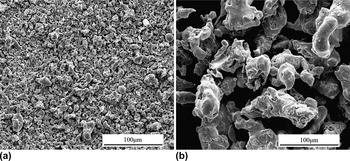
Two kinds of 316L stainless steel powders, (i) with purity 99.9% and median particle size 20 μm and (ii) with purity 99.9% and median particle size 70 μm, manufactured by water atomization from the same company were used as starting powders. The powders were directly subjected to sintering without any further milling step. The chemical composition is given in Table I. Figure 1 shows the morphologies of the two kinds of powders.
SEM micrographs of the starting 316L stainless steel powders: (a) 20 μm; (b) 70 μm.

Chemical composition of 316L stainless steel (wt%).

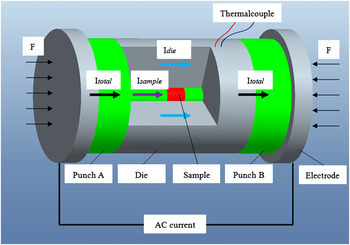
All experiments were carried out with a Gleeble 1500D thermal simulation instrument manufactured by Dynamic Systems Inc. (Poestenkill, NY), which provided a variable AC current through the sample. Moreover, the electric field has low voltage and high current (e.g., >200–400 kA/cm2).Reference Feng, Yang, Hong, Wu and Lan23 A schematic representation of the instrument is shown in Fig. 2.
Schematic of Gleeble 1500D apparatus and current distribution.

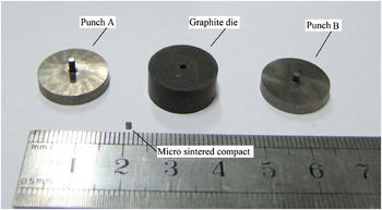
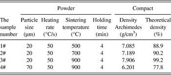
In the microforming/sintering process, the powders are sintered under simultaneous influences of the current, high temperature, and forming pressure. It is a process for the forming of microcomponents for various material systems such as metals, ceramics, and composites. The die and punch setup is shown in Fig. 3. The loose powders with no binder were loaded into a graphite die with outer and inner diameters of 16 and 1 mm, respectively. Both the cemented carbides punches had a diameter of 1 mm and a thickness of 3 mm. A thermocouple was placed on the surface of the die to measure the nominal temperature, as illustrated in Fig. 2. According to previous investigation, due to the excellent electrical and thermal conductivity of the die and powder system, the nominal temperature is slightly lower than the actual temperature of the sample, and thus, a temperature gradient has to be taken into account for the real temperature of the sample.Reference Zadra, Casari, Girardini and Molinari24 The unit was heated by applying AC current under a uniaxial mechanical pressure of 250 MPa in vacuum. During the sintering process, the microsample was first heated to 200 °C and held for 2 min in vacuum (<10−4 Pa) to remove the trapped gases in the compact. Then, it was heated to a preset temperature (T m), which was 500, 700, and 900 °C at a heating rate of 50 °C/s and held for 4 min at T m. After the holding time, the electricity was shut off and the system cooled rapidly from T m to room temperature. The advantages of this technique over conventional ones include lower sintering temperature, shorter forming time, and marked improvements in sintering density. The specimens were named 1#, 2#, 3#, and 4#, respectively. The detailed technological parameters of the experiment are shown in Table II.
The punch and die setup and microsintered compact.

Properties of the powders, processing parameters, and density of the compacts.

Bulk density was determined using the Archimedes principle, and the relative density was calculated using the theoretical density of 316L stainless steel (7.97 g/cm3). Microstructures of cross-sectional surfaces of sintered specimens were observed with a scanning electron microscope (SEM; JSM-5900LV, JEOL, Tokyo, Japan) and a metalloscope (CK-40M, Olympus, Tokyo, Japan).
III. RESULTS
A. Density of the microcompacts
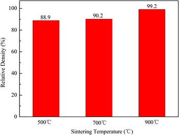
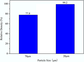
According to the Archimedes measurements, samples 1#, 2#, and 3#, whose starting powder size 20 μm, have got a relative density of 88.9%, 90.2%, and 99.2%, respectively. And under a same sintering condition, sample 3# got nearly full theoretical densification (7.906 g/cm3), but the relative density of sample 4#, whose starting particle size 70 μm, is only 77.8% (Table II).
B. Structural study of the microcompacts
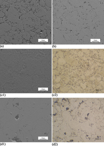
The SEM images and metallographic photographs of 1#, 2#, 3#, and 4# microsintered samples are shown in Fig. 4. Lots of macropores can be observed in Figs. 4(a), 4(b), and 4(d) of samples 1#, 2#, and 4#, respectively. The pores are not all closed circles but are of various shapes including macropores, straight lines, and curves, which indicate that the sintered sample does not densify intensively. Meanwhile, in Fig. 4(c), only a little minor porosity can be observed, resulting in high densification of compact 3#.
High-resolution SEM images and metallographic photographs of microsintered samples.

IV. DISCUSSION
316L stainless steel powders with no binder were sintered under an external electric field, a thermal field, and an external pressure field. The temperature and current are not independent, and thus, the thermal effect of the current (Joule heating) cannot be unambiguously separated from the intrinsic role of the current. There are some other functions of the current, such as current-assisted diffusion (electromigration), cleaning of the contaminated layer, electroplastic effect, cracking of the oxide layer, and local overheating at the contact zone between particles.Reference Jabbar, Couret, Durand and Monchoux25 The coupling fields make an important contribution to the densification of the compacts.
A. Brief introduction to the process of microforming with multifield coupling
The purpose of powder metallurgy was to produce a densified compact from a porous body and to eliminate residual pores and interfaces among particles. The densification process of microforming using the multifield coupling technique is different from the conventional sintering technique. From this research, it seems that for micrometric powder particles, densification is dominated by the plastic deformation, breakage, and fluid flow of the particles under the effect of the applied pressure at relatively low temperature and in a few minutes. However, neck formation and growth is the mechanism of conventional sintering in a long sintering time. To understand the mechanism of the sintering process, a sintering procedure and analyses are needed.
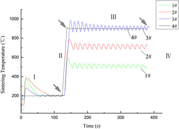
The temperature–time curves of the microcompacts during the sintering process are shown in Fig. 5. Generally, the consolidation process could be divided into four stages based on Fig. 5 (taking compact 4# as an example): (i) preheating period (the compacts were heated to 200 °C from room temperature and held for 2 min); (ii) calefactive period (the compacts were heated to the preset sintering temperature from 200 °C); (iii) sintering period (the compacts were held for a few minutes at the preset sintering temperature); and (iv) cooling period (the current was stopped and the compacts were cooled to room temperature). It should be noted that the division of this method is rather arbitrary and the boundaries between stages are actually unclear and difficult to define; however, such a treatment helps lead to a meaningful analysis and may yield instructive insight into the forming mechanism.
Temperature–time curve of the compacts during the sintering process.

B. Effect of sintering temperature on densification
As shown in Fig. 6, at the same heating rate, the densification degree of the compact was higher when the sintering temperature increased in a certain range. The SEM micrographs are shown in Fig. 4. In our research, when the sintering temperature was up to 1000 °C, all the powders were melt, and no integrity compacts manufactured in that condition.
The effect of sintering temperature on the densification of sintered compacts (20 μm).

As a compact is porous, it contains lots of interfaces. When a current passes through a powder compact, Joule heat will be generated at interfaces due to the contact resistance, which raises the temperature. According to Joule–Lenz’s law, the heat generated by the current passing through the compact is given by:
 $$<![CDATA[$$Q\, = \,I^2 Rt\quad,$$
where Q is the heat generated by the current passing through the compact, I is the current, R is the resistance of the particle or contact resistance, and t is time.
MunirReference Munir26 suggests that the effect of an electric field is dependent on its strength and the value of the effective charge of the diffusing atoms. The results of his studies have been explained in terms of electromigration, where the field modifies the diffusion flux equation by a field-related term, which is expressed by the following equation:
 $$<![CDATA[$$J_i \, = \,{{D_i X_i } \over {RT}}\left( {RT{{\partial \,{\rm{In}}\,X_i } \over {\partial x}} + Fz_i^* E} \right)\quad,$$
where Ji is the flux of atoms of species i, Di is its diffusion coefficient, Xi is its concentration, F is the Faraday constant,  $z_i^* $ is the effective charge of the diffusing atom, E is the electric field, R is the gas constant, T is temperature, and x is the coordinate along which diffusion takes place.
Munir’s research groupReference Quach, Paredes, Kim, Martin and Munir27 also found that the applied pressure is often a result of the additional driving force besides the sintering stress in the sintering procedure, and that at a given temperature, the densification rate is higher when a larger pressure is applied. The function of the applied stress is generally expressed as:
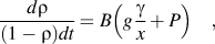 $$<![CDATA[$${{d{\rm{\rho }}} \over {(1 - {\rm{\rho }})dt}} = B\left( {g{{\rm{\gamma }} \over x} + P} \right)\quad,$$
where  ${\rm{\rho }}$ is the fractional density, B is a parameter that includes the diffusion coefficient (of the slowest species) and temperature, g is a geometric constant,
 ${\rm{\gamma }}$ is surface energy, x is a parameter representing a size scale, which is related to particle size, t is time, and P is the effective pressure.
At high pressure, plastic flow is accelerated, which enhances the densification process, as indicated by the Bingham model as followsReference Quach, Paredes, Kim, Martin and Munir27:
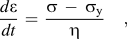 $$<![CDATA[$${{d{\rm{\varepsilon }}} \over {dt}}\, = \,{{{\rm{\sigma }}\, - \,{\rm{\sigma }}_{\rm{y}} } \over {\rm{\eta }}}\quad,$$
where  ${{d{\rm{\varepsilon }}} \over {dt}}$ is the deformation rate of a compact,
 ${\rm{\sigma }}$ is the external stress suffered by a bulk material,
 ${\rm{\sigma }}_{\rm{y}} $ is the yield stress of a bulk material, and
 ${\rm{\eta }}$is the viscosity of a compact.
While the higher the particle temperature, the lower the yield strength is.Reference Guan28 According to Eq. (4), as the contact temperature between particles is high, the yield stresses of the contact area and the viscosity of a compact decrease. So the deformation rate of contact area is greater at higher temperature. Meanwhile, under the applied pressure, the distance between particles decreased, and point contact between particles gradually became surface contact. Parts of atomic group in the powders fill into the macropores by plastic flow. Simultaneously, the particles subjected to shear stress. When the value of shear stress exceeds the yield limit of the particle, fracture and breakage occur easily in its weak position and form some small particles, as shown in Fig. 8.
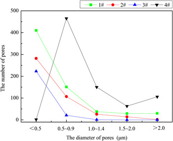
According to the aforementioned sintering kinetics, the higher the sintering temperature and external pressure, the higher the densification degree. But too high a sintering temperature is unfavorable to enhance the densification degree. Comparing the microstructure of the compacts sintered at 500, 700, and 900 °C according to Fig. 4, the number and size of pores in the compacts 1# and 2# are obviously more than 3#. From the effect of sintering temperature on the distribution of pores in sintered compacts shown in Fig. 7, most of the pores in the 3# compact are very small (d ≤ 1 μm), and the number of pores with d ≤ 1 μm is 95% of the total number. In this research, the density of the sintered compact is increasing with the increase of sintering temperature, as shown in Fig. 6. The density of the compact sintered at 900 °C is the highest.
The effect of sintering temperature on the pore distribution of sintered compacts.

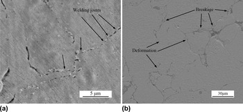
SEM images of compacts with different particle sizes sintered under the same condition (a) welding joints between particles of sample 3#. (b) Particles breakage and deformation of sample 4#.

C. Effect of particle size on densification
As shown in Fig. 5, in the sintering process, the temperature–time curve of microsamples 1#, 2#, and 3# fluctuated around its preset temperature, but curve 4# is almost straight lines.
During the microforming using the multifield coupling process, the electrical field supplied heat power to raise the temperature of the compact and enhance diffusion among particles, simultaneously reducing the yield strength of the material. Under an applied pressure and at a high temperature, point contact between particles gradually became surface contact. This action leads to a decrease in electric resistance and then affects the current flow. Therefore, the multifields matter to each other in several ways.
The particle’s melting point decreases with the decrease in particle size. It is found that the relation between the melting temperature of a powder and its size (particle radius is r) could be represented byReference Zhu29:
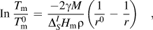 $$<![CDATA[$${\rm{In}}\,{{T_{\rm{m}} } \over {T_{\rm{m}}^0 }}\, = \,{{ - 2{\rm{\gamma }}M} \over {\Delta _S^I H_{\rm{m}} {\rm{\rho }}}}\left( {{1 \over {r^0 }}\, - \,{1 \over r}} \right)\quad,$$
where r 0 is a normal particle radius,  $T_{\rm{m}}^0 $ is the normal particle melting point, r is a fine particle radius, T m is the fine particle melting point,
 ${\rm{\gamma }}$ is interfacial tension between solid particle and liquid, M is molar mass,
 ${\rm{\rho }}$ is density, and
 $\Delta _S^I H_{\rm{m}} $is Moore melting heat.
When r 0 » r, Eq. (5) is simplified to:
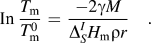 $$<![CDATA[$${\rm{In}}\,{{T_{\rm{m}} } \over {T_{\rm{m}}^0 }}\, = \,{{ - 2{\rm{\gamma }}M} \over {\Delta _S^I H_{\rm{m}} {\rm{\rho }}r}}\quad.$$
At low nominal temperature (measured by the thermocouples installed in the wall of the die), however, the instant temperature at the particle-contacting areas may reach a very high value close to or above the melting temperature of the material. Moreover, due to the small-size effect, the 20 μm particles have a reduced melting temperature compared with 70 μm particles. This is because the arrangement of atoms on the free surface and interfaces (such as grain boundaries, phase boundaries, etc.) is very different from the inner complete lattice of powder particles. Also, the 20 μm particles with high specific surface energy promote the surface melting process.Reference Mondal, Upadhyaya and Agrawal30 Due to the high current and high instant contact temperature, the melting point of small powders may be reached rapidly in stage (iii) of the sintering procedure. Therefore, small particles may evaporate and melt at relatively low nominal temperature because of internal and external factors. In Fig. 8(a), welding joints can obviously be seen between adjacent particles in sample 3#. The liquid phase occurs at an interface due to the contact resistance. The presence of a liquid phase enhances the pore closure of a porous body by capillary force, resulting in fast densification of the compacts.
However, large particles are more prone to fracture at compaction pressure compared to fine particles. As shown in Fig. 8(b), lots of particle deformation and breakage has been found under multifield coupling. The 70 μm particles consume a lot of energy because of deformation and breakage, and as a result, liquid phase hardly appeared on the surface of particles. Besides, the holes between particles [shown in Fig. 4(d)] are too large to be completely eliminated by deformation. From the above-mentioned discussion, a less densified compact has been obtained with 70 μm particles, even at 900 °C.
The particle would consume a large amount of heat when melting happened. At the same time, the liquid phase flows on the surface of solid particles and wets them under the effects of applied pressure and capillary force. Then, the electric resistance of the compact is decreased. According to Eq. (1), the porous compact would gain a smaller quantity of heat, and therefore, the temperature is reduced. The Gleeble machine continuously supplies a variable AC current, the compact gains enough energy, in addition to the latent heat of solidification that is released, and the temperature is raised again. Consuming and gaining of heat repeat alternately, and the temperature–time curve reflects the fluctuation phenomenon directly as shown in Fig. 5 of microsamples 1#, 2#, and 3#. Meanwhile, the fluctuating curve reveals that liquid phase appears on the particle surface indirectly. Viscous liquid flow can promote the rearrangement of powder particles and fill the gaps, to eliminate porosity, rapidly enhancing the compact density. However, the temperature–time curve of 4# compact is about a straight line through the (iii) sintering stage. The density of sample 3# is higher than that of sample 4# under the same sintering condition, as presented in Fig. 9. Smaller sized starting powders can cause the compact to densify at a lower sintering temperature, which is beneficial for reducing grain sizes and therefore improves the properties of the microcompacts.
Densification of compacts with different starting powder sizes.

Conrad’s research resultsReference Stitzer, Smith and Zur Loye31 showed that the rapid mass transfer of liquid phase will become the dominant densification mechanism through the free flow of liquid on the interface of particles. However, the solid phase mass transfer mechanism based on the surface tension effect becomes less important than the liquid phase transfer mechanism.
V. CONCLUSIONS
From the work completed in this study, the following conclusions can be drawn:
Compared with conventional sintering methods, the coupled multifield-activated microforming is an efficient process, and it has lower energy consumption and little impact to the environment. Two kinds of 316L stainless steel powders with different particle sizes (20 and 70 μm) were sintered by microforming using the multifield coupling method by different sintering temperatures (500, 700, 900 °C) at a heating rate of 50 °C/s. To a certain range, the sample with higher sintering temperature always had higher density. When the sintering temperature was reached to 900 °C, a nearly fully densified compact was gained (its relative density is 99.2%). The electric field, thermal field, and external pressure field affect each other mainly due to the electric resistance of the compact. Because melting occurred under a multifield coupling condition, the densification mechanism dominating microsintered compacts with a starting powder size of 20 μm is liquid phase transfer, and a nearly fully densified microcompact was obtained under relatively low temperature. This is proved by the fluctuated temperature–time curves. However, particle deformation, breakage, and solid-state diffusion are the dominant mechanisms during the densification of the microcompact with a starting powder size of 70 μm.
In other words, the process of heating by Joule heat in the microsintering process is faster than conventional sintering. The microcompact can easily gain enough energy to accelerate the densification process by particle deformation, breakage, and melting in a short time and at a lower nominal temperature during the whole process. Further research will concentrate on the investigations of microgears and densification mechanisms of microsintering under multifield coupling.
ACKNOWLEDGMENTS
The authors would like to acknowledge the support from the National Nature Science Foundation of China (No. 50945018) for the conduction of the research reported in this paper.