Introduction
The pattern of growth hormone (GH) secretion, such as GH pulse frequency and amplitude, is of considerable importance for optimal growth. Insulin-like growth factor-I (IGF-I), a 70 amino acid polypeptide, mediates the action of GH on somatic cell growth. The growth axis, like other neuroendocrine systems, functions through the hypothalamic–pituitary and target organ axis interactions. IGF-I has been reported to play a role in the feedback regulation of GH secretion (Muller et al., Reference Muller, Locatelli and Cocchi1999). To that extent, circulating IGF-I concentrations increased, while serum GH levels decreased with age and/or body weight in the pig (Buonomo et al., Reference Buonomo, Lauterio, Caile and Campion1987; Lee et al., Reference Lee, Chung and Simmen1993). The decrease in GH secretion is primarily due to a reduction in the amplitude and frequency of secretory GH pulses (Dubreuil et al., Reference Dubreuil, Pelletier, Petitclerc, Lapierre, Couture, Brazeau, Gaudreau and Morisset1987), which is accompanied by an age-related reduction in the GH response to growth hormone-releasing hormone (GHRH) in growing pigs (Dubreuil et al., Reference Dubreuil, Pelletier, Petitclerc, Lapierre, Couture, Brazeau, Gaudreau and Morisset1987). Similarly in the rat, an age-related decrease in GH response to GHRH was related to a reduction in pituitary somatotroph sensitivity to GHRH (Niimi et al., Reference Niimi, Takahara, Kawanishi and Irino1985). Intracerebroventricular (ICV) administration of IGF-I failed to influence GH secretion in sheep (Fletcher et al., Reference Fletcher, Thomas, Dunshea, Moore and Clarke1995) while intra-pituitary administration suppressed GH secretion. In contrast, ICV administration of a mixture of IGFs suppressed GH secretion (Tannenbaum et al., Reference Tannenbaum, Guyda and Posner1983) and somatomedin-C treatment stimulated somatostatin (SS) release from hypothalamic explants (Berelowitz et al., Reference Berelowitz, Szabo, Frohman, Firestone and Chu1981) in the rat. Thus, the primary site of action of circulating IGF-I concentration on GH secretion is unclear and may, in part, be species dependent.
In addition to its action on somatic cell growth and GH secretion, IGF-I also exerts effects on the reproductive axis. Circulating IGF-I concentrations are elevated at the time of puberty in rodents (Handelsman et al., Reference Handelsman, Spaliviero, Scott and Baxter1987; Crawford and Handelsman, Reference Crawford and Handelsman1996), primates (Mauras et al., Reference Mauras, Rogol, Haymond and Veldhuis1996), ruminants (Roberts et al., Reference Roberts, McCutcheon, Blair, Gluckman and Breier1990) and pigs (Prunier and Louveau, Reference Prunier and Louveau1997). Moreover, IGF-I stimulated gonadotropin-releasing hormone (GnRH) release from hypothalamic explants (Hiney et al., Reference Hiney, Ojeda and Dees1991) and LH from rat pituitary cells in culture (Kanematsu et al., Reference Kanematsu, Irahara, Miyake, Shitsukawa and Aono1991). In the pig, IGF-I-stimulated LH release from pituitary cells in vitro was influenced by stage of the estrous cycle of the pituitary donor (Whitley et al., Reference Whitley, Barb, Utley, Popwell, Kraeling and Rampacek1995). Hiney et al. (Reference Hiney, Srivastava, Nyberg, Ojeda and Dees1996) reported that ICV administration of small doses of IGF-I to immature juvenile and peripuberal rats increased LH and this effect was blocked by immunoneutralization of GnRH. Furthermore, Miller and Gore (Reference Miller and Gore2001) reported that ICV administration of IGF-I in the rat during puberty and reproductive senescence failed to effect GnRH expression, suggesting that IGF-I may play a role in releasing GnRH rather than stimulating gene expression. The LH response to peripheral administration of IGF-I in castrate male sheep with or without estrogen implants was biphasic with the lower doses stimulating and higher doses inhibiting LH secretion (Adam et al., Reference Adam, Findlay and Moore1998). Moreover, a directed effect of IGF-I on basal LH secretion from ovine pituitary cells in culture has also been reported (Adam et al., Reference Adam, Gadd, Findlay and Wathes2000).
Based on the above-cited studies, we hypothesize that circulating IGF-I may act at the level of both the hypothalamus and pituitary to regulate GH and LH secretion. Therefore, three experiments were conducted to test this hypothesis by examining the effect of ICV administration of IGF-I on GH and LH secretion and direct action on the anterior pituitary utilizing pituitary cell culture.
Material and methods
Experiment (EXP I)
Twelve ovary-intact crossbred prepuberal gilts, of 65 ± 6 kg body weight and 140 days of age, were surgically implanted with lateral ICV cannulas using the stereotaxic procedure of Estienne et al. (Reference Estienne, Kesner, Barb, Kraeling, Rampacek and Estienne1990). Animals were individually penned in an environmentally controlled building at a constant temperature of 22°C and artificial 12 : 12-h light : dark photoperiod. Pigs were fed to appetite daily, at 0800 and 1700 h, a corn–soybean meal ration (14% crude protein) supplemented with vitamins and minerals, according to the National Research Council guidelines (NRC, 1998). At 1 week after the last ICV surgery, and 24 h before treatment, all pigs were fitted with an indwelling jugular vein cannula (Barb et al., Reference Barb, Kraeling, Rampacek, Fonda and Kiser1982). On the day of the EXP, pigs were fed at 0700 h and blood sampling started at 0800 h. Blood samples were collected every 15 min from 5 h before to 4 h after ICV injections of 150 μl 0.9% saline (S; n = 4), 25 μg (n = 4) or 75 μg (n = 4) of recombinant IGF-I (GroPrep, Adelaide, Australia) in 150 μl of S. Serum was harvested and stored at −20°C until assayed for GH and LH by radioimmunoassay (RIA). All procedures were approved by the Richard B. Russell Agriculture Research Center Committee on Animal Care and Use.
Experiment (EXP II)
Pituitary glands were aseptically removed from 150-day-old prepuberal gilts (n = 6). Ovaries were examined at sacrifice and all gilts were considered prepuberal because their ovaries were devoid of corpora albicantia and corpora lutea. The anterior lobe was dissected from each pituitary gland and cells were enzymatically dispersed and cultured as previously described (Barb et al., Reference Barb, Kraeling and Rampacek1995). Cells from each pig were cultured separately. Cell viability and number were assessed by counting cells, which excluded trypan blue on a hemocytometer. Cell viability was 96%. Cells were diluted and plated, based on the number of live cells. Cells were plated at 105 cells/ml with a culture medium 60% (vol/vol) Ham’s F-l2 nutrient mix, 30% (vol/vol) Dulbecco’s modified Eagle’s medium (DME); 4500 mg/l glucose (Sigma-Aldrich Corporation, St Louis, MO, USA) and 10% (vol/vol) Fitton-Jackson medium (BGjb; Invitrogen Corporation, Carlsbad, CA, USA) containing 2 mg/ml of BSA, 10 mM HEPES (pH 7.21 100 U/ml of penicillin, 2 mM l-glutamine, 250 ng/ml of amphotericin B (Sigma-Aldrich) and 100 pg/ml of streptomycin (Invitrogen)) to which was added 2% (vol/vol) fetal bovine serum (Sigma-Aldrich), 100 ng/ml of cortisol, 1.0 ng/ml of human insulin, 10 μg/ml of human transferrin (Sigma-Aldrich), 10 pg/ml of glucagon, 100 pg/ml of epidermal growth factor, 200 pg/ml of bovine parathyroid hormone, 400 pg/ml of triiodothyronine (Sigma-Aldrich) and 200 pg/ml of fibroblast growth factor (Collaborative Research, Bedford, MA, USA) in a 24-well cluster plate. Culture medium was changed on day 3 (day of seeding = day 0 of culture) and replaced with serum-free medium as described before (Barb et al., Reference Barb, Kraeling and Rampacek1995), except for using DME containing 1000 mg of glucose/l (Sigma-Aldrich). On day 4 of culture, the medium was discarded, plates were rinsed twice with serum-free medium and cells were cultured in 1 ml fresh serum-free medium containing one of the following treatments: 0.1, 10 or 1000 nM [Ala15]-hGHRH-(1-29)NH2 (Sigma-Aldrich), 0.01, 0.1, 1, 10, or 30 nM IGF-I individually or in combinations with 1000 nM GHRH. Cells were exposed to treatment for 4 and 24 h, during which time medium was harvested and quantified for GH by RIA.
Experiment (EXP III)
Pituitary glands were aseptically removed from 150-day-old barrows (n = 5). Castrate males were utilized to remove the influence of testicular steroids on the pituitary. The anterior lobe was dissected from each pituitary gland and cells were enzymatically dispersed and cultured as previously described above. Cells from each pig were cultured separately and challenged with 10, 100 or 1000 nM GnRH (Sigma-Aldrich, Cat. # L7134) or 0.01, 0.1, 1, 10, 30 nM IGF-I individually or in combinations with 100 nM GnRH. Secreted LH was measured at 4 h after treatment by RIA.
Radioimmunoassays
EXP I and II: Serum and media samples were assayed for GH (Barb et al., Reference Barb, Estienne, Kraeling, Marple, Rampacek, Rahe and Sartin1991) as previously described. Sensitivity of the assays was 0.4 ng/ml and the intra- and inter-assay coefficients of variation were 3.5% and 13%, respectively. Serum and media samples from EXP I and III were assayed for LH (Kesner et al., Reference Kesner, Kraeling, Rampacek and Johnson1987) as previously described. Sensitivity of the assays was 0.15 ng/ml and intra- and inter-assay coefficients of variation were 4.5% and 10.2%, respectively.
Statistical analysis
EXP I: To determine the effect of IGF-I on GH and LH secretion, data were subjected to repeated measures using the PROC MIXED ANOVA procedure of Statistical Analysis Systems Institute (SAS; Littell et al., Reference Littell, Milliken, Stroup and Wolfinger1996). The statistical model included treatment, pig and time. Effects of treatment were tested using pig within treatment as the error term. Time and time × treatment were tested using time × treatment and pig within treatment × time as the error terms, respectively. Differences between treatment means within a time were determined by least-squares contrasts (SAS, 1999).
EXP II and III: Basal secretion (control; C) was the amount of hormone secreted into the culture medium per 105 cells seeded/well in the absence of treatment or a secretagogue. Cells from each pig were cultured separately. For wells that were treated with GHRH, GnRH or IGF-I alone, data were converted to percentage of basal secretion. Data from wells treated with a combination of GHRH or GnRH with IGF-I data were converted to percentage of the hormone response to GHRH or GnRH alone before averaging to minimize differences between wells. To obtain an estimate of variation between control wells, medium GH and LH concentrations for control wells were converted to a percentage of mean basal GH or LH concentration. This was then used to calculate an SE for basal secretion. Converted data were tested for homogeneity of variance using Hartley’s F max test (Gill, Reference Gill1978). Data were then subjected to a one-way ANOVA and differences between means were determined by least-squares contrast (SAS, 1999).
Results
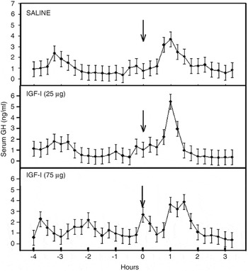
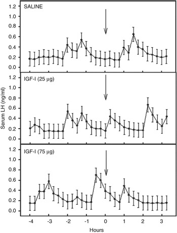
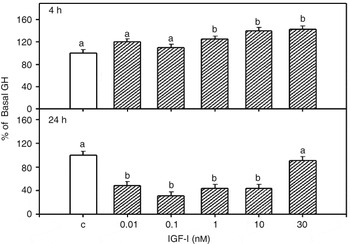
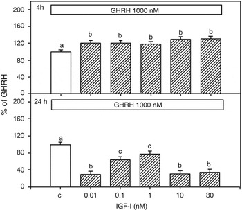
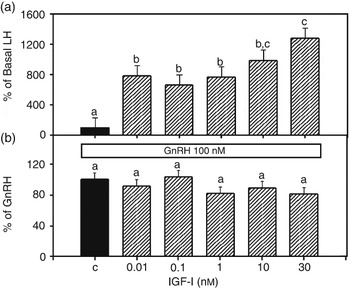
In EXP I, serum GH and LH concentrations were unaffected by ICV administration of 25 or 75 μg of IGF-I (Figures 1 and 2). In EXP II, basal (control; C) GH was 48 ± 2 ng/well at 4 h (n = 6) and 82 ± 5 ng/well at 24 h (n = 6). Relative to C at 4 h, 0.1, 10 or 1000 nM GHRH increased (P < 0.01) GH secretion by 111%, 125% and 150%, respectively. Relative to C 1, 10, 30 nM IGF-I enhanced (P < 0.02) basal GH secretion at 4 h whereas by 24 h all doses, except for the 30 nM IGF-I, suppressed (P < 0.02) GH secretion compared to C wells (Figure 3). All doses of IGF-I in combination with1000 nM GHRH increased (P < 0.04) the GH response to GHRH compared to GHRH alone at 4 h (Figure 4). In contrast, by 24 h all doses of IGF-I suppressed (P < 0.04) the GH response to GHRH alone (Figure 4). In EXP III, basal LH secretion was 5.7 ± 0.7 ng/well (n = 5). Relative to C at 4 h, 10, 100 and 1000 nM GnRH increased (P < 0.0002) LH secretion by 19 ± 2, 19 ± 2 and 18 ± 2-folds, respectively. All doses of IGF-I increased (P < 0.01) basal LH compared to C levels while the LH response to GnRH was unaffected by IGF-I (P > 0.1; Figure 5).
Serum GH concentrations (mean ± s.e.) in prepuberal gilts receiving ICV injection of saline (n = 4), 25 μg IGF-I (n = 4) or 75 μg IGF-I (n = 4) at time 0; arrows indicate time of treatment.

Serum LH concentrations (mean ± s.e.) in prepuberal gilts receiving ICV injection of saline (n = 4), 25 μg IGF-I (n = 4) or 75 μg IGF-I (n = 4) at time 0; arrows indicate time of treatment.

Effects of IGF-I on basal GH secretion from anterior pituitary cells at 4 and 24 h in culture. Values are the mean ± s.e. (n = 6 prepuberal gilts/treatment). Control (C) = basal secretion in the absence of treatment. Data expressed as a percentage change from C. Means with different letters differ (P < 0.02) from control.

Effects of IGF-I on GHRH-stimulated GH secretion from anterior pituitary cells at 4 and 24 h in culture. Values are the mean ± s.e. (n = 6) prepuberal gilts/treatment except for the 1 nM dose at 24 h (n = 5). Control (C) = GH secretion in the presence of GHRH alone. Data expressed as a percentage change from C. Means with different letters differ (P < 0.04).

Effects of IGF-I on basal and GnRH-stimulated LH secretion from anterior pituitary cells at 4 h in culture. Values are the mean ± s.e. (n = 5 barrows). (a) Control (C) = LH secretion in the absence of treatment. Data expressed as a percentage change from C. (b) Control (C) = LH secretion in the presence of GnRH alone. Data expressed as a percentage change from C. Means with different letters differ (P < 0.01).

Discussion
In the present study, we report that ICV administration of IGF-I failed to affect GH secretion in the prepuberal gilt. These findings support the observation of Spencer et al. (Reference Spencer, Bass, Hodgkinson, Edgley and Moore1991 and Reference Spencer, Berry, Hodgkinson and Bass1993) and Harel and Tannenbaum (Reference Harel and Tannenbaum1992a and Reference Harel and Tannenbaum1992b), who were unable to demonstrate a central effect of IGF-I on GH secretion and suggested that IGF-I does not play a role in the negative feedback regulation of GH at the level of the brain. To that extent, Fletcher et al. (Reference Fletcher, Thomas, Dunshea, Moore and Clarke1995) was unable to demonstrate an inhibitory response to IGF-I following either lateral or third ventricle administration or infusion of 5 μg of IGF-I into the third ventricle over a 2 h period. In contrast, in the rat, ICV administration of a mixture of IGFs (Tannenbaum et al., Reference Tannenbaum, Guyda and Posner1983; Harel and Tannenbaum, Reference Harel and Tannenbaum1992b) or somatomedin-C suppressed GH secretion (Abe et al., Reference Abe, Molitch, Van Wyk and Underwood1983). However, the report by Harel and Tannenbaum (Reference Harel and Tannenbaum1992b) demonstrated that only IGF-I in combination with IGF-II suppressed GH secretion. Moreover, the study by Tannenbaum et al. (Reference Tannenbaum, Guyda and Posner1983) utilized a mixture of IGFs that most likely contained IGF-II and may explain the inhibitory affect on GH secretion. This paradox further strengthens the idea of a pituitary site of action of IGF-I on GH secretion.
This is the first report on the pig to demonstrate that IGF-I exerted a biphasic effect on basal and GHRH-induced GH secretion from anterior pituitary cell in vitro. At 4 h, IGF-I stimulated basal GH secretion in a dose-dependent manner and enhanced the GH response to GHRH. However, by 24 h all but 30 nM dose of IGF-I inhibited basal and all doses inhibited the GHRH-induced GH secretion. Although the 30 nM dose is within the physiologic range (Barb et al., Reference Barb, Barrett, Kraeling and Rampacek2001), the failure of IGF-I to affect basal GH secretion at 24 h may, in part, be related to the ligand-mediated down regulation of the receptor, which is dependent on the amount of ligand and time to which the cells are exposed. To that extent, Yamamoto et al. (Reference Yamamoto, Prager, Yamasaki and Melmed1993) reported that exposure of rat GC pituitary cells to IGF-I resulted in a time- and concentration-dependent reduction in cell surface IGF-I receptor binding. Thus, constant exposure to 30 nM might have induced a desensitization of the cell to the inhibitory action of IGF-I.
Our findings agree in part with a study by Berelowitz et al. (Reference Berelowitz, Szabo, Frohman, Firestone and Chu1981) who reported that somatomedin-C had no effect on basal or GH-releasing peptide (RP)-induced GH secretion in rat anterior pituitary cells in culture at 4 h but by 24 h somatomedin-C suppressed both basal and GH-RP-stimulated GH secretion. Several reports have confirmed that the inhibitory action of IGF-I on GH secretion under basal and GHRH-stimulated conditions is due to a reduction in GH mRNA in the rat (Yamashita and Melmed, Reference Yamashita and Melmed1986; Morita et al., Reference Morita, Yamashita and Melmed1987), teleost (Fruchtman et al., Reference Fruchtman, Jackson and Borski2000) and human (Yamashita and Melmed, Reference Yamashita and Melmed1986) anterior pituitary cells in culture, suggesting that IGF-I inhibits transcription. This may explain the delayed inhibitory action of IGF-I on GH secretion observed in the present study.
In addition to IGF-I action on somatic cell growth and GH secretion, several reports have demonstrated that IGF-I acts centrally to affect GnRH release and subsequent LH secretion (for a review, see Daftary and Gore, Reference Daftary and Gore2005). ICV administration of IGF-I failed to affect LH secretion in the present study, although no negative effect on LH secretion was detected. The lack of a central effect on LH secretion may in part be related to maturational changes in hypothalamic sensitivity to IGF-I as proposed by Daftary and Gore (Reference Daftary and Gore2003), or possibly the response to IGF-I may vary due to species, gender, and (or) hormonal status and, lastly, IGF-I may serve as a permissive metabolic signal that may be necessary for activation of the reproductive axis but not as a triggering effect on LH secretion (Hiney et al., Reference Hiney, Ojeda and Dees1991). This could account for the lack of LH response to ICV administration of IGF-I.
We present evidence that IGF-I stimulated basal LH secretion from anterior pituitary cells in culture from castrate males. This was dose dependent and was similar to that reported in male castrate sheep (Adam et al., Reference Adam, Gadd, Findlay and Wathes2000), eel (Huang et al., Reference Huang, Rousseau, Le Belle, Vidal, Burzawa-Ge’rard, Marchelidon and Dufour1998) and mature gilt (Whitley et al., Reference Whitley, Barb, Utley, Popwell, Kraeling and Rampacek1995). The LH response to GnRH was unaffected by IGF-I, suggesting that the action of IGF-I on LH release from anterior pituitary cells in vitro was by direct action, independent of GnRH, rather than by enhanced GnRH-induced LH secretion. In contrast, IGF-I stimulated basal and enhanced the LH response to GnRH in rat anterior pituitary cell culture and this response was reduced by 30% with IGF-I antiserum (Soldani et al., Reference Soldani, Cagnacci, Paoletti, Yen and Melis1994 and Reference Soldani, Cagnacci and Yen1995). The authors suggested that IGF-I may serve as a putative modulator of anterior pituitary LH secretion. This paradox between the current study and those reported by Soldani et al. (Reference Soldani, Cagnacci and Yen1994) may in part be related to species differences.
Conclusions
Under the experimental conditions used in the present study, the results demonstrated a biphasic and time-related effect of IGF-I on basal and GHRH-induced GH release from the anterior pituitary whereas IGF-I only affected basal LH secretion. The role of IGF-I in the feedback regulation of GH and LH secretion at the level of the brain remains to be resolved. Further research is needed to determine the functional role of IGF-I in the regulation of GH and LH secretion in the pig.
Acknowledgments
The authors wish to thank Benny Barrett and Brent Jackson for their technical assistance. The authors wish to thank Dr A. F. Parlow, Harbor-UCLA Medical Center, Torrance, CA, USA for providing the porcine GH antiserum AFP-1021854, porcine GH AFP-10864B and porcine LH AFP-11043B. This research was supported by USDA funds. Mention of a trade name, proprietary product or specific equipment does not constitute a guarantee or warranty by the US Department of Agriculture and does not imply its approval to the exclusion of other products, which may be suitable.