Case Report
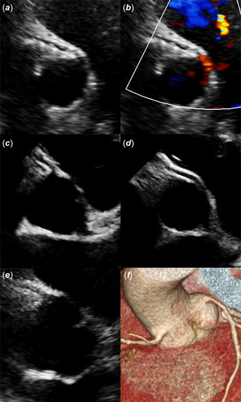
An 8-year-old boy underwent cardiac evaluation for mid-systolic ejection murmur. Physical examination was unremarkable. Electrocardiography, showed a sinus rhythm and incomplete right bundle branch block. 2D echocardiography showed normal biventricular function with competent atrioventricular and semilunar valves, but the anomalous origin of the right coronary artery could be seen (Fig 1a and b). For this reason, a coronary computed tomography was performed and confirmed an anomalous aortic origin of a right coronary artery from the left sinus with inter-arterial course. An exercise treadmill stress test did not show arrhythmias and/or ischemia-induced by exercise. Following the diagnosis of anomalous coronary artery in one sibling, we screened with echocardiography all close relatives which revealed a normal coronary pattern in the 11-year-old brother (Fig 1c) and a similar coronary anomaly in the 15-year-old brother. On medical evaluation, the older brother described himself as asymptomatic. The physical examination and blood pressure were normal. Electrocardiography showed sinus rhythm with normal conduction and normal repolarisation. The echocardiography reported competent valves, normal bi-ventricular dimension, and systolic function. However, an acute take-off origin of the right coronary artery was noticed (Fig 1d). For these reasons, a coronary computed tomography scan was requested to define the origins of the coronary arteries. The coronary computed tomography scan was performed with evidence of an anomalous aortic origin of the right coronary artery with it arising from the left sinus and inter-arterial course. We extended our pedigree investigation to the parents of the children. The mother had a normal origin of the coronary artery at the echocardiographic evaluation (Fig 1e). Coronary anatomy of the father was not evaluable by echocardiography. Coronary computed tomography scan confirms normal coronary anatomy in the father of the children (Fig 1f). We were not able to accomplish a direct pedigree investigation of three-generation. However, the parents of the children had a negative family history for congenital heart disease and/or sudden cardiac death.
Echocardiographic parasternal short axis provides longitudinal views of the proximal portion of the coronary arteries. (a) In the younger brother anomalous origin of the right coronary artery could be seen by 2D image echocardiography and (b) colour Doppler flow. (c) Echocardiography revealed a normal coronary pattern in the 11-year-old brother. (d) In the older brother 2D imaging in the high parasternal short axis view close to the sinotubular junction demonstrates an acute take-off of the right coronary artery. (e) The mother of the children has a normal origin of the right coronary artery as describe by echocardiography. (f) Coronary computed tomography scan confirmed the normal origin of the coronary arteries in the father of the children.

Coronary computed tomography scan findings
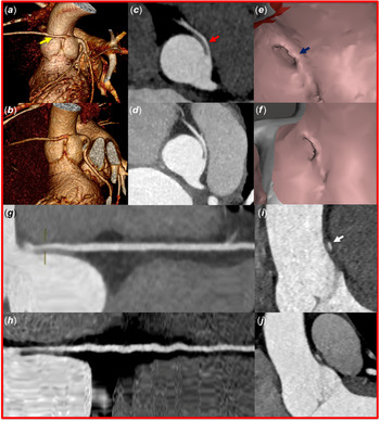
At the computed tomography scan, we analysed specific aspects of the anatomy of the anomalous coronary artery as described in recent publications. Reference Cheezum, Liberthson and Shah1,Reference Harris, Whitehead, Shin, Keller, Weinberg and Fogel2 In particular, number of Ostia and ostial morphology, the asymmetry index defined as the ratio between the maximal and minimal diameter at maximal proximal narrowing point, proximal vessel cross-sectional area percentage narrowing compared to normal distal reference and length of the narrowing, take-off angle, and take-off level of the anomalous coronary artery. Radiological findings are shown in Figure 2a–j. Both patients have anomalous aortic origin of the right coronary artery from the left sinus and inter-arterial course. The anomalous right coronary artery had a separated origin in both two cases. Ostia of the right coronary artery was characterised by an elevated ratio of the vertical-to-horizontal measurements and therefore elliptic morphology. The younger brother had an asymmetry index at maximal proximal narrowing point of 2.9 and the older brother had an asymmetry index of 3.9. The expected asymmetry index value in the normal coronary artery is about 1. Coronary computed tomography scan through a diastolic acquisition evaluated the proximal narrowing of the anomalous coronary artery. The proximal vessel percentage cross-sectional area narrowing compared with a normal distal reference was respectively 67% in the younger and 73% in the older one. Angelini et al describe associated intramural course with proximal stenosis in all 67 patients with anomalous aortic origin of the right coronary artery with inter-arterial course evaluated with cardiac catheterisation and intravascular ultrasonography. In the anomalous coronary artery, the passage of the coronary artery inside the aortic wall (intramural course) becomes complicated by lateral compression. The coronary lateral compression due to dynamic structural change is worse during systole, tachycardia ad exercise. Reference Angelini, Uribe, Monge, Tobis, Elayda and Willerson3 Cardiac computed tomography is a reliable technique for diagnosis of anomalous coronary artery; however, for the dynamic entity of the lateral compression, it cannot precisely quantify the severity of stenosis. Reference Angelini4 The two brothers had an acute take-off angle of the anomalous right coronary artery with an origin right above the sinotubular junction. Familiar clustering of anomalous aortic origin of a coronary artery has already been described. Reference Agrawal, Mery and Sexson Tejtel5,Reference Brothers, Stephens, Gaynor, Lorber, Vricella and Paridon6 Notably, the anomalous coronary patterns in the two brothers are almost identical. Furthermore, we encountered extreme overlapping of the associated anatomic features (as in the vessel take-off angle, ostial morphology, asymmetry index, proximal vessel percentage narrowing, and length of the narrowing).
Coronary computed tomography identified features of the anomalous coronary artery in the two siblings. Figure a-c-e-g-i display the coronary computed tomography features of the younger brother. Figure b-d-f-h-j exhibit the coronary computed tomography features of the older brother. (a–b) Vessel’s take-off level (above the commissure) shown in 3D reformatted images. The yellow arrow points to the origin of the right coronary artery. (c–d) Multiplanar axial computed tomography reconstruction at the level of the coronary artery origin demonstrates the right coronary artery travelling between the aorta and the pulmonary artery. Separate vessel origin (red arrow) can be seen in both the patients. (e–f) Virtual angioscopy post-processing technique is used to visualise the coronary Ostia. The abnormal anomalous coronary Ostia (blue arrow) was characterised by an elevated ratio of the vertical-to-horizontal measurements and therefore elliptic shape. (g–h) Curved multi-planar reformations of the right coronary artery providing an excellent visualisation of the vessel along its entire course. Narrowing of the proximal vessel was detected in both cases. (i–j) Lumen cross-sectional area of the anomalous coronary artery obtained in double oblique view at the level of the anterior aortic commissure. This view is known as the “Angelini/Cheong cut” (white arrow). In the anomalous coronary artery, an elevated asymmetry index defines as the ratio between the maximal and minimal diameter was found.

Discussion
The purpose of the paper was the presentation of two brothers affected by identical anomalous origin of a right coronary artery. The likelihood of finding such a rare anomaly in two brothers is extremely low. Indeed, the prevalence of anomalous aortic origin of a coronary artery is considered between 0.1 and 0.3 %. Reference Cheezum, Liberthson and Shah1 Little is known about familiarity in the anomalous coronary artery. Embryological studies describe a complex development of the coronary artery, with many possible sites at which a mutation might lead to a coronary anomaly. Sans-Coma used the Syrian hamster as an animal model to study the congenital anomalous coronary artery. In the Syrian hamster, there is evidence that the proximal portion of coronary artery pattern with regard to the aorta is influenced by genetic factors. Reference Soto-Navarrete, Arqué and Durán7 Moreover, in the animal model, the frequency of anomalous origin of the coronary artery significantly increase when the aortic valve is bicuspid. Reference Sans-Coma, Arqué, Durán, Cardo and Fernández8 However, the aetiology of this developmental complex consisting of bicuspid aortic valve and anomalous origin of the coronary artery is not established. There is no published evidence about the genetic inheritance of the anomalous coronary artery in humans. Case series are available in the literature of families with relatives affected by anomalous origin of the coronary artery. Reference Brothers, Stephens, Gaynor, Lorber, Vricella and Paridon6 Furthermore, in human, patients with anomalous coronary artery show significant familial clustering of cardiovascular disease. Reference Agrawal, Mery and Sexson Tejtel5 Specific anatomical characteristics (like a slit-like orifice, an acute angle of origin and an intramural segment) concomitant with anomalous aortic origin of a coronary artery seems to be associated with a high risk of sudden cardiac death. Reference Mosca and Phoon9 For these reasons, we decided to present the clinical scenario of these cases and to compare the radiological findings of the coronary computed tomography scans of the two brothers.
Moreover, to our knowledge, this is the first Italian report of an anomalous coronary artery in two siblings.
Conclusion
We report on the diagnosis of anomalous coronary artery in two brothers. Following the diagnosis of anomalous coronary artery in one sibling, we screened immediate family relatives and found the same anomaly in the older brother. These findings might support coronary origin screening in first line relatives of patients with anomalous coronary artery. Patients with anomalous coronary artery in our report were asymptomatic with normal physical examination and electrocardiography. Echocardiography seems an accurate screening tool for this anomaly, particularly in the paediatric population. However, an anatomic diagnosis of anomalous aortic origin of the right coronary artery does not represent an automatic indication for surgical intervention and individualised therapy is mandatory.
Acknowledgements
None.
Financial support
This research received no specific grant from any funding agency, commercial or not-for-profit sectors.
Conflict of interest
None.