



No CrossRef data available.
Article contents
Seasonal variation in Sarcocystis species infections in goats in northern Iraq
Published online by Cambridge University Press: 07 October 2004
Abstract
We investigated the prevalence of sarcocystosis in 826 goats slaughtered in the winter season from November to April in northern Iraq. The prevalence of macrocysts was on average 34%, with only 20% infected animals in November, but 46% in February. The infection rate in 1-, 3- and 6-year-old goats was 4%, 48%, and 83%, respectively. The highest specificity of infection was in the oesophagus (99%) and the lowest in the diaphragm (3%). Grossly, we identified 2 forms of macroscopic sarcocysts, fat and thin, with different morphological characteristics. The prevalence of microcysts was 97% and no effects of age, sex and seasonal variations were observed. Development of microcysts in the small intestine of dogs and cats has also been investigated. The pre-patent period in experimentally infected dogs was 12–14 days and the patent period lasted 64–66 days. A dog shed about 155 million sporocysts, but no sporocysts were shed by cats that had been fed the same infected tissues, thus identifying the microcysts as Sarcocystis capracanis.
Information
- Type
- Research Article
- Information
- Copyright
- © 2005 Cambridge University Press
INTRODUCTION
The protozoon Sarcocystis is one of the most widely distributed in nature and a prevalent parasite in a broad range of vertebrates. Most Sarcocystis species have an obligatory two-host life-cycle, both an intermediate (herbivore) and a definitive (carnivore) host must be present for the completion of the life-cycle (Dubey, Speer & Fayer, 1989).
Goats are intermediate hosts for at least 3 species of Sarcocystis: S. capracanis (Fischer, 1979; Heydorn & Haralambidis, 1982), S. moulei (Moulé, 1888; Neveu-Lemaire, 1912; Levine, 1986; probably identical with S. caprifelis: Dubey et al. 1989), S. caprifelis (El-Rafaii, Abdel-Baki & Selim, 1980), and S. hircicanis (Heydorn & Unterholzner, 1983). Definitive hosts are, respectively, canids, cats, and dogs (Tenter, 1995).
We performed this study with the aims (i) to estimate the prevalence of sarcocystosis in goats slaughtered at the city abattoir of Sulaimany, North Iraq; (ii) to determine whether age, sex and seasonal variation have effects on macroscopic (S. caprifelis) and microscopic, (S. capracanis) sarcocystosis; (iii) to investigate the target-specificity of macrocysts of S. caprifelis in various animal tissues and (iv) to study the developmental stages of S. capracanis in the small intestine of dogs (dog–goat cycle) and cats (cat–goat cycle) experimentally inoculated with microcysts.
MATERIALS AND METHODS
Macroscopical examination
From November 1991 to April 1992, tissue samples from 826 (501 female, 325 male) goats (Capra hircus) slaughtered at the abattoir of Sulaimany, were collected twice per week. All animals appeared healthy before slaughter. The age (estimated by examination of teeth) and sex of each animal were recorded. The oesophagus, diaphragm and skeletal muscles were thoroughly inspected for the presence of Sarcocystis macrocysts. The distinction between macrocysts and microcysts was based on size (see Results section). Specimens containing macrocysts were separated, the cysts excised from the tissue and collected in a Petri dish containing 5 ml of 0·9%, NaCl. Afterwards, the cyst wall was mechanically disrupted and the bradyzoites released from the sarcocysts. One drop (0·05 ml) of homogenized solution was spread on a glass slide, fixed and stained with 1% Giemsa's stain.
Microscopical examination
Tissue sampling
Pieces of oesophagus, diaphragm and skeletal muscle were collected from all goats, examined grossly, and each sample was divided into a smaller portion for the muscle squash and a larger one for pepsin digestion.
Muscle squash
A 0·5 g specimen of each muscle was squashed between a cover-slip and slide and examined unstained at 100× magnification using a bright-field microscope for the detection of microcysts and bradyzoites of Sarcocystis (Singh, Agrawal & Shah, 1990; Dubey et al. 2000).
Pepsin digest of muscles
Twenty g of pooled muscles from the oesophagus, diaphragm and skeletal muscles were incubated for 20 min at 40 °C in 50 ml of acid-pepsin solution (pepsin 2·6 g, NaCl 5 g, 1 M HCl 7 ml plus 993 ml of distilled water). This digest was filtered through a 38 μm sieve, centrifuged at 2000 g for 5 min, and the sediment suspended in 0·5 ml of distilled water (Dubey & Livingston, 1986). A drop of this suspension was examined for the presence of Sarcocystis bradyzoites under the light microscope at 400× magnification. In addition, further drops from the same solution were spread on glass slides, fixed, and stained with 1% Giemsa's stain. A goat was declared ‘negative’ when none of the tissue samples examined yielded microcysts or bradyzoites by both methods.
Histology
All tissue samples were fixed in 10% buffered neutral formalin, embedded in paraffin, sectioned at 6 μm, stained with Haematoxylin and Eosin and microscopically examined for microcysts of S. capracanis (Dubey & Livingston, 1986; Dubey et al. 2000).
Inoculation of dogs and cats with S. capracanis
Seven 3 to 4-week-old dogs, and five 6-week-old cats were obtained at weaning and reared free of Sarcocystis infection. The dogs and cats were housed individually in isolation cages throughout the study, given food and water ad libitum. For 10 days before inoculation, all faeces of all these experimental animals were daily examined, employing the centrifugation–flotation method (Hussein & Warrag, 1985). Four dogs and 4 cats were fed with raw meat containing microcysts of naturally infected goats, but free of macrocysts. Each animal received 25 g of oesophagus and diaphragm for 2 consecutive days. The faeces of each animal were collected daily and incinerated to avoid cross-contamination. Three dogs and 1 cat were left uninfected, housed separately and received clean food and water ad libitum. The faeces of each animal were examined daily for Sarcocystis sporocysts from day zero until day 80 post-inoculation (p.i.), employing the centrifugation-flotation method. The counting of sporocysts was performed by the modified McMaster method.
Two inoculated dogs, 2 inoculated cats, and a non-inoculated dog and cat each were anaesthetized and sacrificed at days 8 or 13 p.i. The small intestine was removed and cut into small portions 5×10×10 mm3. Afterwards, these small tissue blocks were fixed by immersion in 10% buffered neutral formalin, embedded in paraffin, sectioned at 6 μm, stained with Haematoxylin and Eosin and microscopically examined at 400× and 1000× magnifications. Microcysts, sporocysts, and bradyzoites of Sarcocystis were measured using a micrometer eyepiece at 400× magnification, and macrocysts were measured with a standard ruler.
RESULTS
Macrocysts of S. caprifelis
Grossly, 33·6% (278 of 826) of the goats were infected with macrocysts and, as detected with the naked eye, the oesophagus had the highest incidence. It contained macrocysts in 98·9% (275/278) of all infected goats, differing markedly from skeletal muscle which contained macrocysts in 4·3% (12/278) and diaphragm with macrocysts in 2·5% (7/278) of the infected goats. The macroscopic sarcocysts were milky-white and grainy in appearance and were embedded in the muscular tissue (Fig. 1A). Two distinct forms of macrocysts were identified.
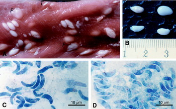
Fig. 1. (A) Oesophagus of a goat heavily infected with fat (F) and thin (T) macrocysts of Sarcocystis caprifelis. Magnification 2×, same scale bar as (B), in cm. (B) Isolated fat macrocysts of S. caprifelis, same magnification as (A). (C) Fat macrocyst of S. caprifelis. After excystment the banana-shaped bradyzoites appear somewhat longer and thicker (range: 11–17 μm×3–6 μm) than those of a thin macrocyst (compare with (D). Giemsa staining, objective 100× oil, final magnification 1000×. (D) Thin macrocyst of S. caprifelis. After excystment the bradyzoites are slightly shorter and more slender (range: 9–13 μm×2–4 μm) than those of a fat macrocyst (C). Giemsa staining.
Fat cysts appeared large, thick and round (Fig. 1A) and were exlusively found in the oesophagus – preferentially in its lower half – with a mean length of 6·2 mm and mean width of 3·8 mm (range: 2–17 mm×2–7 mm, n=50; Fig. 1B). These fat cysts were chambered and fully packed with banana-shaped bradyzoites averaging 14·1 μm×4·5 μm (range: 11–17 μm×3–6 μm, n=25; Fig. 1C).
Thin cysts were small, slender and less frequent than the fat cysts. Thin cysts occurred not only in the oesophagus, but also in diaphragm and skeletal muscles. They were somewhat smaller than fat cysts with a mean length of 3·6 mm and width of 1·1 mm (range: 1·5–7 mm×0·5–2 mm, n=50). Also the bradyzoites of thin cysts were slightly smaller, averaging 12·2 μm×2·9 μm (range: 9–13 μm×2–4 μm, n=25; Fig. 1D).
The prevalence of both fat and thin macrocysts of Sarcocystis greatly increased with the age of the host animal. In young goats (6 to 12-months-old) prevalence was 0% (except in April), but increased to about 80% (5/6) in old goats, aged 6 years and more (see Table 1 for details).
Table 1. Prevalence of Sarcocystis caprifelis macrocysts in goats (This table shows the number of infected goats per number of investigated goats (in parentheses: ratio in %) per month of observation period per age group. From top to bottom the data show the seasonal variation (see text), from left to right they indicate the increase of infection rate with age.)

In male goats the prevalence of Sarcocystis macrocysts was only 17·2% (56/325) of all investigated goats, while in female animals it was 44·3% (222/501). However, this difference must be attributed to the much higher mean age of female goats at slaughter (see Discussion section).
The numbers of goats containing macrocysts (found in oesophagus and/or skeletal muscle or diaphragm) strikingly increased from November to April, but was considerably lower again in November in the next age group: 11·5% of the 6 to 12-month-old goats carried macrocysts in April, i.e. at the end of the cold season, but no macrocysts were detected in the corresponding 1 to 2-year-old goats in November, after the animals had spent the summer outdoors on pasture. Likewise 45·4% of the 2 year olds were infected with macrocysts in April, having spent another winter stabled, but only 11·5% of the 3 year olds contained macrocysts, having been on pasture again. Also 56·2% of the 3 year olds carried macrocysts in April, but only 28·5% of the 4 year olds in November. All of the 4 year olds were infected in April, but only 33·3% of the 5 year olds in November. These data strongly suggest that macrocysts of S. caprifelis are by no means permanent, but transient structures within the muscle. From May to October, during the hot and dry summer season, when all goats are out on pasture and certainly have less close contact to cats, macrocysts were greatly reduced in size or even disappeared so that we were unable to detect them in goats slaughtered in November. Subsequently, they either regrow, increasing with age, or the animals are de novo infected during the cold and wet winter season when, in North Iraq, most goats are stabled.
Microcysts of S. capracanis
Microsarcocysts were found in 97·4% of goats, regardless of age, sex or season (Table 2). By muscle squash, the rate of infection with microcysts was 89·7% (741/826). The microcysts in the oesophagus were elongated, spindle- or spiral-shaped, of mean dimension 529 μm×62 μm (range: 446–645 μm×42–78 μm) with a 3·2 μm thick cyst wall (range: 2·1–4·0 μm). The wall of microcysts was striated, varied in thickness, and had minute finger-like villar protrusions indicating that they were S. capracanis (cf. Fig. 1 in Heydorn & Haralambidis, 1982). Morphologically, microcysts in the skeletal muscle and cardiac muscle were relatively similar to those obtained from the oesophagus, but were of smaller dimensions (mean: 275 μm×52 μm, range: 226–370 μm×36–59 μm) with a thinner cyst wall (2·4 μm, range: 1·9–3·2 μm).
Table 2. Prevalence of Sarcocystis capracanis microcysts in goats (Same design as Table 1. For S. capracanis microcysts there is no seasonal variation and no increase with age. Almost all goats were infected in our material.)

Infection of dogs with S. capracanis
Except for inappetance and mild watery diarrhoea for 1 or 2 days, neither changes in animal behaviour, nor clinical symptoms were observed. Following the pre-patent period of 12–14 days, all inoculated dogs passed free sporocysts in the faeces during the patent period (64–66 days). Under light microscopy it was observed that free sporocysts were passed singly or were clamped together. Sporocysts were ellipsoidal and measured 14·3 μm×9·1 μm on average (range: 13·9–15·5 μm×8·5–9·2 μm). The fully mature sporocysts contained 4 sporozoites and a number of granules that represent residual bodies. The control animals remained clinically normal throughout our study.
The number of sporocysts passed in faeces varied from 20000/g faeces on day 14 p.i., increasing to 470000/g on day 45 p.i. and 80000/g on day 60 p.i., and then declining to zero on day 80 p.i.
DISCUSSION
Our data show a high prevalence of S. capracanis microcysts (97·4%) in goats in northern Iraq. Similar results had previously been found in other geographical locations (Pethkar & Shah, 1982; Erber & Göksu, 1984; Hussein & Warrag, 1985; Dubey & Livingston, 1986; Foreyt, 1989). In the present study the prevalence of microscopic sarcocystosis was not influenced by sex, age, or seasonal variations, which might be explained by the short life-cycle and a possibly quicker rate of maturation of microcysts in comparison to macrocysts. The striated microcyst wall found in S. capracanis sarcocysts was clearly different from microcysts of S. hircicanis, that were not found in the present study.
There is considerable confusion concerning the structure and the synonymity of macroscopic sarcocysts in goats (Dubey et al. 1989, pp. 121–125). After the first report of macrocysts measuring up to 14 mm long and up to 7 mm wide in a Swiss domestic goat (Niederhäusern, 1873), 4 macroscopic Sarcocystis spp. have now been described. Firstly, S. moulei was reported by Moulé (1888) (see also Neveu-Lemarie, 1912), but his description of sarcocysts is too vague for identification. Secondly, S. orientalis (Matschulski & Miskaryan, 1958, cited by Dubey et al. 1989) was observed only in wild mountain goats of Siberia (Capra sibirica), and not in domestic goats (Capra hircus). These sarcocysts were up to 7·5 mm long and up to 2·5 mm wide, which is much smaller than the macrocysts (up to 17 mm long) that we have found in domestic goats. For these reasons, we did not identify the macrocysts shown here (Fig. 1A and B) as S. orientalis. Thirdly, S. ovifelis (Erber & Göksu, 1984) was reported in long-haired goats of Turkey, with sarcocysts up to 4 mm long and up to 2 mm wide. The primary wall of these cysts appeared as a smooth, homogeneous layer, about 1 μm thick and enclosed by a secondary wall (Erber & Göksu, 1984). Such macrocysts were never observed in our material, in which the macrocysts were larger in size and had a much thicker cyst wall of up to 14 μm. Finally, S. caprifelis was first described by El-Rafaii et al. (1980) in domestic goats of Egypt. According to his brief note (1 page of text, no figures), the sarcocysts had ‘a thin or smooth wall with many fibrils, and the infection was successfully transmitted to cats’.
It must be stressed that none of the papers published on macroscopic sarcocystosis in goats contains any figure of a macrocyst. The Fig. 1A and B of this study are – to the best of our knowledge – the first images ever published of macroscopic Sarcocystis in this species. Generally, all well-studied macroscopic Sarcocystis spp. in herbivores other than goats are of felid origin (Tenter, 1995) i.e. in water buffalo S. fusiformis (up to 32 mm long), in cattle S. hirsuta (up to 8 mm long), in sheep S. gigantea (up to 10 mm long). Hence, we suggest that the macrocysts in domestic goat, documented by us are transmitted via cats and need further study.
Morphologically we observed two forms of macrocysts of Sarcocystis in goat muscle samples: Firstly, fat cysts were thick, round in shape and seemed to grow in length and width. They were exclusively found in the oesophagus. These cysts fully coincide with the careful first-ever description of macrocysts in the oesophagus of goat with a round-ovoid shape, up to 14 mm long and up to 7 mm wide (Niederhäusern, 1873). Secondly, thin cysts that were slim and slender and appeared to grow mainly in length, were commonly seen in the oesophagus, diaphragm and skeletal muscles. No data in the literature fit these cysts found in our material. Based on the histological examination, both fat and thin cysts differ not only in shape, wall thickness and wall structure, but also in bradyzoite dimensions. Whether fat and thin macrocysts represent different species or subspecies needs investigation.
With regard to sex, we observed differences in the prevalence of macroscopic sarcocystosis between the investigated male (17·2%) and female (44·3%) goats. This gender-bias is probably based on the simple fact that female goats are slaughtered at an older age (3 years and over) than males (1–2 years).
The most important finding is that our data provide the first evidence of seasonal variation in the prevalence of macroscopic sarcocystosis in goats. This seasonal variation in the rate of infection cannot be attributed to an age factor, because, in November, 85% of all slaughtered goats were 3 years old or older and the infection rate was only 20%, whereas in February somewhat less, namely 77% of all killed goats were 3 years old or older, but the infection rate had more than doubled to 46%. Also, long-term fluctuations in the intensity of infection over several years can definitely be ruled out as a source of artifact in our data, because all goats had been examined within one and the same winter season.
The differences in the proportion of positive cases between the younger goats in April and the next older goats in November may be caused by pasture and living conditions. Temperature has an important role on the viability of Sarcocystis sporocysts in the pasture (Dubey et al. 1989, 2002; Chen, Zuo & Zuo, 1999; Hilda, Oogassawara & Idercio, 2001). Our data on seasonal variation in the prevalence of infection derived from 826 investigated goats can only be explained by the assumption that during the hot and dry season of the year macrocysts disappear or undergo atrophy inside the intermediate host on pasture, but regress in the cold and wet season or by the stabling conditions of the goats in winter. Whether macrocysts are potentially transient structures that can disappear in vivo needs investigation in experimentally infected goats.
We thank Professor Dr S. Ali and Professor Dr M. El-Atar for their useful advices, and Dr J. Rasoul (Sulaimany abattoir) for supply of material. The fine photographical work of I. Koch and M. Schmidt, Universität zu Köln, is highly appreciated. This study was supported by a Clinical Research Grant from the College of Veterinary Medicine, Baghdad, Iraq.
References
REFERENCES
CHEN, X. W.,
ZUO, Y. X. &
ZUO, W. W. (
1999).
Observation on the clinical symptoms and sporocyst excretion in human volunteers experimentally infected with Sarcocystis hominis.
Chinese Journal of Parasitology and Parasitic Diseases
17,
25–27.Google Scholar
DUBEY, J. P. &
LIVINGSTON, C. W. Jr. (
1986).
Sarcocystis capracanis and Toxoplasma gondii infections in range goats from Texas.
American Journal of Veterinary Research
47,
523–524.Google Scholar
DUBEY, J. P.,
SAVILLE, W. J. A.,
LINDSAY, D. S.,
STICH, R. W.,
STANEK, J. F.,
SPEER, C. A.,
ROSENTHAL, B. M.,
NJOKU, C. J.,
KWOK, O. C. H.,
SHEN, S. K. &
REED, S. M. (
2000).
Completion of the life cycle of Sarcocystis neurona.
Journal of Parasitology
86,
1276–1280.CrossRefGoogle Scholar
DUBEY, J. P.,
SAVILLE, W. J. A.,
SREEKUMAR, C.,
SHEN, S. K.,
LINDSAY, D. S.,
PENA, R. W. H. F.,
VIANNA, M. C.,
GENNARI, S. M. &
REED, S. M. (
2002).
Effects of high temperature and disinfectants on the viability of Sarcocystis neurona sporocysts.
Journal of Parasitology
88,
1252–1254.CrossRefGoogle Scholar
DUBEY, J. P.,
SPEER, C. A. &
FAYER, R. (
1989).
Sarcocystosis of Animals and Man.
CRC Press,
Boca Raton, Florida, USA.
EL-RAFAII, A. H.,
ABDEL-BAKI, G. &
SELIM, M. K. (
1980).
Sarcosporidia in goats of Egypt.
Journal of the Egyptian Society of Parasitology
10,
471–472.Google Scholar
ERBER, M. &
GÖKSU, K. (
1984).
Sarcosporidia in goats in Turkey and the differentiation of species. In
Recent German Research on Problems of Parasitology, Animal Health and Animal Breeding in the Tropics and Subtropics (
ed. Markel, H. & Bittner, A.), pp.
21–29.
Verlag Chemie,
Weinheim.
FISCHER, G. (
1979).
Die Entwicklung von Sarcocystis capracanis n. spec. in der Ziege. Veterinary Medicine Thesis,
Freie Universität
Berlin.
FOREYT, W. J. (
1989).
Sarcocystis sp. in mountain goats (Oreamnos americanus) in Washington: prevalence and search for the definitive host.
Journal of Wildlife Diseases
25,
619–622.Google Scholar
HEYDORN, A. O. &
HARALAMBIDIS, S. (
1982).
Zur Entwicklung von Sarcocystis capracanis Fischer, 1979.
Berliner und Münchener Tierärztliche Wochenschrift
95,
265–271.Google Scholar
HEYDORN, A. O. &
UNTERHOLTZNER, J. (
1983).
Zur Entwicklung von Sarcocystis hircicanis n. sp.
Berliner und Münchener Tierärztliche Wochenschrift
96,
275–282.Google Scholar
HILDA, F. J.,
OGASSAWARA, P. S. &
IDERCIO, L. S. (
2001).
Occurrence of cattle Sarcocystis species in raw kibbe from Arabian food establishments in the city of São Paulo, Brazil, and experimental transmission to humans.
Journal of Parasitology
87,
1459–1465.Google Scholar
HUSSEIN, S. H. &
WARRAG, M. (
1985).
Prevalence of Sarcocystis in food animals in Sudan.
Tropical Animal Health and Production
17,
100–101.CrossRefGoogle Scholar
LEVINE, N. D. (
1986).
The taxonomy of Sarcocystis (Protozoa: Apicomplexa) species.
Journal of Parasitology
72,
372–382.CrossRefGoogle Scholar
MOULÉ, L. (
1888).
Des Sarcosporidies et leurs fréquence, principalement chez les animaux de boucherie.
Société des Sciences et Arts de Vitry-le-Francois
14,
3–41.Google Scholar
NEVEU-LEMARIE, M. (
1912).
Les sporozoaires. Part 2. Chapter 1. In
Parasitologie des Animaux Domestiques Maladies Parasitaires Non-bactériennes. p. 198–317.
J. Lamarre et Cie,
Paris.
NIEDERHÄUSERN, D. VON (
1873).
Psorospermien bei der Ziege.
Zeitschrift für praktische Veterinär-Wissenschaften
1,
79–86.Google Scholar
PETHKAR, D. K. &
SHAH, H. L. (
1982).
Prevalence of Sarcocystis in goats in Madhya pradesh.
Indian Veterinary Journal
59,
110–114.Google Scholar
SINGH, K. P.,
AGRAWAL, M. C. &
SHAH, H. L. (
1990).
Prevalence of Sarcocystis capracanis in oesophagus and tail muscle of naturally infected goats.
Veterinary Parasitology
36,
153–155.CrossRefGoogle Scholar
TENTER, A. M. (
1995).
Current research on Sarcocystis species of domestic animals.
International Journal for Parasitology
25,
1311–1330.CrossRefGoogle Scholar

Fig. 1. (A) Oesophagus of a goat heavily infected with fat (F) and thin (T) macrocysts of Sarcocystis caprifelis. Magnification 2×, same scale bar as (B), in cm. (B) Isolated fat macrocysts of S. caprifelis, same magnification as (A). (C) Fat macrocyst of S. caprifelis. After excystment the banana-shaped bradyzoites appear somewhat longer and thicker (range: 11–17 μm×3–6 μm) than those of a thin macrocyst (compare with (D). Giemsa staining, objective 100× oil, final magnification 1000×. (D) Thin macrocyst of S. caprifelis. After excystment the bradyzoites are slightly shorter and more slender (range: 9–13 μm×2–4 μm) than those of a fat macrocyst (C). Giemsa staining.

Table 1. Prevalence of Sarcocystis caprifelis macrocysts in goats

Table 2. Prevalence of Sarcocystis capracanis microcysts in goats
 You have
Access
- 10
- Cited by