Introduction
Primitive neuroectodermal tumours are soft tissue tumours with histologically typical malignant small round cells. These tumours are diagonosed by the detection of translocation t(11;22)(q24;q12) of the EWS gene using reverse transcriptase-polymerase chain reaction or fluorescence in situ hybridization in formalin fixed paraffin embedded tissue. We present the first case to be reported in the tongue of this rare and aggressive tumour.
Case report
A 47-year-old woman presented with a three to four month history of a left-sided sore throat. In the week prior to presentation, a left upper neck swelling had developed. She was a non-smoker with no previous history of illness. She had a family history of maternal uterine cancer and paternal bowel cancer.
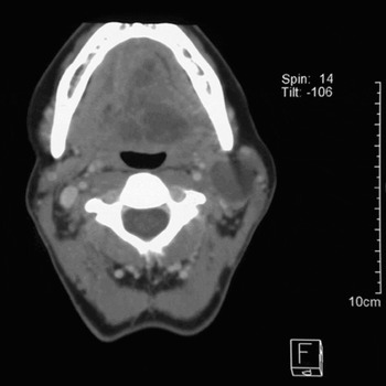
On examination, the patient had a left hypoglossal nerve palsy and a mass in the posterior third of her tongue. Flexible nasoendoscopy showed enlarged lingual tonsils and a normal postnasal space, larynx, hypopharynx and vallecula. There was no evidence of any ulcerative lesions. Palpation of the neck revealed a 4×2 cm mass in the upper region of the sternocleidomastoid muscle and two small lymph nodes in the left posterior triangle.
Panendoscopy and biopsy of the tongue mass was conducted under general anaesthesia. Pharyngoscopy showed a >4 cm, hard, submucosal, base of tongue mass on the left side, with extension into vallecula, left anterior tongue and across the midline. A 1×4 cm left neck node was evident.
Histologically, the biopsy tissue showed squamous mucosa with underlying oedematous and fibrous corium, skeletal muscle, fat, minor salivary glands, and no evidence of tumour. It was decided to perform a repeat biopsy as the mass appeared cystic and non-ulcerative and the patient had no risk factors for primary head and neck cancer.
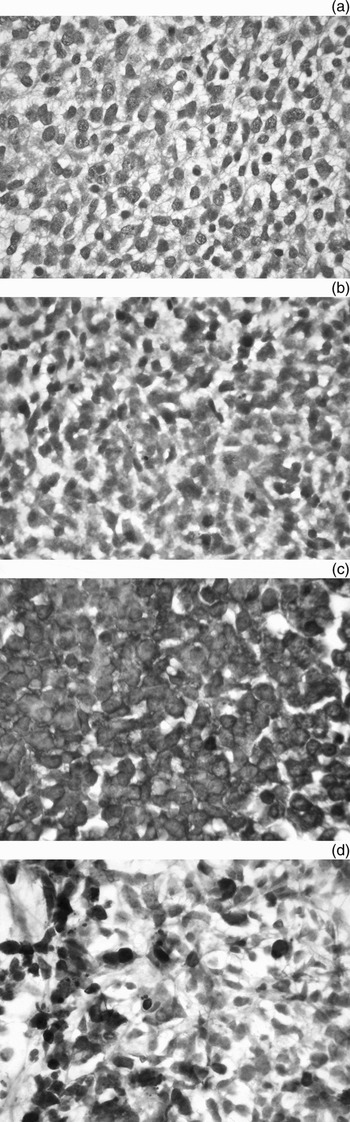
The repeat biopsy showed fragments of tumour admixed with blood clot and necrotic tumour debris. The tumour comprised sheets of tumour cells surviving around delicate blood vessels. The tumour cells had small, mildly hyperchromatic, angulated nuclei, together with a small amount of eosinophilic to clear cytoplasm. The nuclear chromatin was coarse with small nucleoli. No nuclear moulding was seen. There was a myxoid quality to the background stroma.
Immunohistochemical staining showed positive staining within tumour cells for vimentin, Bcl2 (DAKO, dilution 1:5, clone 124) and CD99 (monoclonal antibody that recognizes the HBA71 antigen which is mainly found in Ewing sarcoma and PNET, DAKO, dilution 1:50, clone MIC2-12E7). Focal positive staining with neuron-specific enolase was seen. Immunohistochemical staining for cytokeratin CKAE1/AES (DAKO, dilution 1:50, clone 34betaE12), Cam5.2 (Beckton Dickenson, dilution 1:10), LCA (Leucocyte Common Antigen, DAKO, dilution 1:200), S100 protein (DAKO, dilution 1:600, clone S100), HMB45 (DAKO, dilution 1:100, clone HMB45), chromogranin, synaptophysin, actin, desmin and myoglobin were all negative. A periodic acid–Schiff stain demonstrated moderate amounts of glycogen within the cytoplasm of the tumour cells.
The findings were those of a small, round, blue cell tumour showing mesenchymal differentiation and positivity for Bcl2 and CD99, with glycogen within the cytoplasm. The morphologic findings were most consistent with a primitive neuroectodermal tumour.
Cytogenetic analysis failed to demonstrate metaphases for analysis. Paraffin sections analysed with Fluorescence In Situ Hybridization (FISH) studies of the EWS (gene locus on chromosome 22) gene showed a normal signal pattern (two yellow signals) with EWSR1 (Zymed laboratories, locus 22q12/q13) break apart probe in the majority of sites examined consistent with a normal result.
Electron microscopy
Electron microscopy showed primitive, undifferentiated cells with few cytoplasmic organelles but abundant glycogen. Non-specific intermediate filaments were present but no cytokeratin filaments or myofilaments. Cell junctions were infrequent and rudimentary. Occasional small, dense core granules were noted, but interdigitating cell processes were absent.
Discussion
Peripheral primitive neuroectodermal tumours belong to the Ewing sarcoma family of tumours. These tumours are known to mainly occur in the second decade of life. Histopathologically, these tumours are composed of small, round cells forming rosettes.Reference Dehner1 The diagnosis of primitive neuroectodermal tumour is based on patient history, immunostaining with at least two neural markers, ultrastructural examination and evidence of an abnormal t(11;22)(q24;q12) translocation (being the major feature in the Ewing sarcoma family of tumours).Reference West2
Primitive neuroectodermal tumour in the head and neck region is extremely rare. A recent literature review by WindfuhrReference Windfuhr3 collected a total of 27 patients with clear evidence of primitive neuroectodermal tumour in the head and neck region. We believe the present case of primitive neuroectodermal tumour of the tongue is the first to be reported in the English literature.
Hattinger et al. described a few isolated cases with no detectable rearrangement using an EWSR1 break apart probe but subsequently showing abnormalities with FISH probes for the specific fusion product. In these cases, even if a normal result is reported, a diagnosis of Ewing sarcoma family tumour cannot be excluded.
The lack of rearrangement using the EWSR1 break apart probe led to reconsideration of the diagnosis, and the tumour was sent for a second pathological review. In 5 to 10 per cent of EWS primitive neuroectodermal tumours, it is not possible to demonstrate rearrangements of EWS on 22q12 and ETS related oncogenes.
These tumours are known to have a poor prognosis, with metastases likely by the time of presentation. Surgical resection, along with chemoradiotherapy to treat metastasis or prevent undetected metastasis, has been recommended for these aggressive tumours.Reference Weiss and Goldblum4 Our patient underwent two cycles of chemotherapy (doxorubicin, vincristine, etopside and ifosfamide), followed by radical radiotherapy to the primary site and neck and then two more cycles of chemotherapy. Two months after completion of treatment, there was no sign of residual disease. Surgical resection was not performed due to the large tumour volume, the jugular node metastatic lesions and the goal of function preservation in an otherwise healthy young patient.
Due to the rarity of this diagnosis, guidance for treatment is obtained from experience with the Ewing sarcoma patient group.
Immunostaining and cytogenetics are vital in establishing the diagnosis of peripheral primitive neuroectodermal tumour. Greater availability of these techniques may enable wider recognition of this condition.
Axial computed tomography scan of the neck, showing the tongue mass and the neck nodes.

Tumour histology. (a) H&E, ×100; (b) periodic acid–Schiff stain showing glycogen in the cells; (c) tumour cells staining positive for CD99; (d) tumour cells staining positive for neuron-specific enolase.