In the last 100 years there have been 3 major influenza pandemics: Spanish Flu in 1918, Asian Flu in 1957 and Hong Kong Flu in 1968. These claimed the lives of approximately 50 million, 2 million and 1 million people, respectively. Added to this is the annual death toll of 250,000 to 500,000 people worldwide with a further 3 to 4 million people suffering severe illness. In 1997 the alarming emergence of a new, highly pathogenic subtype, H5N1, which has a 50% mortality rate, provided a major impetus for renewed influenza research. Re-emergence of fatal human influenza A subtype H5N1 disease was reviewed (Peiris et al. Reference Peiris, Yu, Leung, Cheung, Ng, Nicholls, Ng, Chan, Lai, Lim, Yuen and Guan2004). At the same time another subtype, H1N1, has emerged (Neumann et al. Reference Neumann, Noda and Kawaoka2009). This latter subtype causes a relatively mild infection in humans, however it is highly transmittable between people and a new influenza pandemic was declared by the World Health Organization. Development of effective new drugs for the treatment and/or prevention of epidemics and pandemics of influenza is a major sanitary objective. Achievement of this objective is particularly difficult since the influenza viruses are continuously modifying their genomes, thus escaping the conventional vaccine-based treatments. Some widely used anti-influenza drugs such as adamantanes and neuraminidase inhibitors might become rapidly useless due to the emergence of drug-resistant influenza subtypes. Although the neuraminidase inhibitor Tamiflu® is now considered the lead antiviral efficient against H5N1 and H1N1, there were concerns that some resistant mutant strains may soon be around. In vitro, some neuraminidase mutations were found associated with resistance to oseltamivir (Mc Kimm-Breschkin et al. Reference Mc Kimm-Breschkin, Sahasrabudhe, Bilck and McDonald2001, Reference Mc Kimm-Breschkin, Trivedi, Hampson, Hay, Klimov, Tashiro, Hayden and Zambon2003). In July 2009, WHO has been informed by health authorities in Denmark, Japan and the Special Administrative Region of Hong Kong, China of the appearance of H1N1 viruses resistant to the antiviral drug oseltamivir (Tamiflu®) based on laboratory testing (WHO 2009). This situation creates the need for conception of novel effective anti-influenza therapeutics bypassing the appearance of resistances.
Esterified milk proteins display antiviral activities against several different viruses (bacteriophage M13, lactococcal bacteriophages bIL66, bIL67 and bIL170, herpes simplex virus, poliovirus, coxsackie, influenza virus subtype H3N2) (Sitohy et al. Reference Sitohy, Chobert and Haertlé2005, Reference Sitohy, Chobert, Karwowska, Gozdzicka-Jozefiak and Haertlé2006, Reference Sitohy, Billaudel, Haertlé and Chobert2007, Reference Sitohy, Chobert, Dalgalarrondo, Nowoczin, Besse, Billaudel and Haertlé2008, Reference Sitohy, Besse, Billaudel, Haertlé and Chobert2010). Given the current advances in new anti-influenza therapeutics (De Clercq Reference De Clercq2009), it was interesting to test the efficacy of methylated β-lactoglobulin (Met-BLG) also against influenza virus subtype H1N1. The broad specificity and variability of esterified milk proteins is reminiscent of Capoxone in the broadness of its action overcoming the recurrent viral resistances against synthetic antiviral drugs. Additionally, esterified milk proteins are degradable and non-toxic products in contrast with many synthetic drugs.
Materials and Methods
Materials
β-Lactoglobulin (BLG, Protarmor 907 NK) was purchased from Armor Protéines (Saint-Brice-en-Coglès, France) and purified according to Mailliart & Ribadeau Dumas (Reference Mailliart and Ribadeau Dumas1988). BLG was esterified as previously described (Sitohy et al. Reference Sitohy, Chobert and Haertlé2001). MDCK cell lines (derived from the kidney of normal adult female cocker spaniel) were grown in Eagle's minimum essential medium (MEM, Eurobio, Courtabœuf, France) containing 8% fetal calf serum (FCS, Eurobio) and 1% PCS (penicillin 10 000 U, colimycin 25 000 U, streptomycin 10 mg).
H1N1 was obtained from Dr. Christophe Chevalier (Influenza virus group, INRA, Jouy-en-Josas, France) and regenerated in MDCK cell line maintained with MEM containing 1% PCS and 1 μg trypsin/ml (MEM-T). Virus titre was determined by plaque-forming units (pfu).
Hemagglutination (HA) activity
HA activity was assayed with chicken erythrocytes 0·5% suspension according to Donald & Isaacs (Reference Donald and Isaacs1954). All experiments were performed in three replicates and results were expressed as the mean±the standard deviation.
RNA extraction and real time-PCR
H1N1 RNA was extracted from 150 μl of supernatant of infected strains by the GTC-silica technique described by Boom et al. (Reference Boom, Sol, Salimans, Jansen, Wertheim-van Dilen and van der Noordaa1990) and performed using the NucleoSpin® RNA Virus kit (Macherey-Nagel, Düren, Germany) according to the manufacturer's instructions. A real-time (RT)-PCR assay of the most constant region in influenza A virus, the matrix gene, was carried out using primers and probe previously described (Spackman et al. Reference Spackman, Senne, Myers, Bulaga, Garber, Perdue, Lohman, Daum and Suaez2002).
Forward primer: AGA TGA GTC TTC TAA CCG AGG TCG (M+25)
Reverse primer: TGC AAA AAC ATC TTC AAG TCT CTG (M-124)
Probe: Fam-TCA GGC CCC CTC AAA GCC GA-BHQ1 (M+64)
The Invitrogene Superscript III One-Step RT-PCR kit was used with a 25 μl reaction mixture using the following conditions: 1× of kit supplied buffer (dNTP included), 8 mm-MgSO4, 0·3 μm of each primer, 0·1 μm probe, 1 μm T4 gene 32 protein (USB Corporation Cleveland, Ohio USA), 2·5 IU of kit supplied enzyme mixture including Superscript III reverse transcriptase and Platinum Taq (Invitrogene Life Technologies, 2003). Twenty microliters of the reaction mixture was added to 5 μl RNA from one of the supernatant medium or RNA from the standard. The standard curve was obtained from the plasmid pET22b_MHistag with M gene, supplied by INRA (Dr J-C Gaudin, INRA, Nantes, France) and Novagen® and used at concentrations from 5 to 5×10⁴ copies. Influenza virus was reverse transcribed into cDNA (15 min at 49°C and 5 min at 94°C) and amplified by PCR (5 s at 94°C, 30 s at 59°C and 5 s at 72°C) for 45 cycles with an LC instrument (Rotor-Gene 6500, Corbett Research, Mortlake, Australia). RT fluorescent measurements were carried out and the threshold cycle (Ct) value for each sample was calculated by determining the point at which fluorescence exceeded a threshold limit (10 times the baseline standard deviation). A standard curve of the Ct values obtained with a serially diluted external RNA standard was prepared. Ct values obtained from the supernatant samples were plotted on the standard curve, and the number of copies was calculated automatically by the software Sequence Detector (Applied Biosystems). Detector samples were considered negative if their Ct values exceeded 45 cycles.
Influence of Met-BLG concentration on cell viability and hemagglutination activity
The viral hemagglutination activity (HA) and cell viability were assayed in the presence of different concentrations of Met-BLG. MDCK cells in 96-well microtitre-plates (50·000/well) were infected by adding appropriate aliquots of influenza virus suspensions. Aliquots of virus suspension (100 μl) were added to all cell cultures except for cellular and Met-BLG controls, so that virus pfu and cell numbers are equal (1 MOI, multiplicity of infection). Aliquots (100 μl) of Met-BLG dissolved at different concentrations in the same medium were added to the wells containing the infected cells. Infected and mock MDCK cells were incubated at 37°C in the presence of 5% CO2 for different time intervals with MEM-T. At the end of each period, HA was assayed with chicken erythrocytes 0·5% suspension according to Donald & Isaacs (Reference Donald and Isaacs1954) and the viable cells were revealed by neutral red (Sitohy et al. Reference Sitohy, Chobert, Dalgalarrondo, Nowoczin, Besse, Billaudel and Haertlé2008). In a previous study with Vero cells (Sitohy et al. Reference Sitohy, Chobert, Dalgalarrondo, Nowoczin, Besse, Billaudel and Haertlé2008) no cytotoxicity of Met-BLG was observed in all the range of concentrations used. All experiments were performed in triplicate and results were expressed by the mean plus the standard deviation.
Viral replication in presence of Met-BLG
Viral replication in presence of Met-BLG was studied in 96-well micro-plates containing 50 000 MDCK cells in MEM-T infected with H1N1 (0·01, 0·1 and 1 MOI) and incubated at 37°C in the presence of 5% CO2. Plates were incubated for 18 h, then the supernatant was assayed for replicated RNA as previously described (Sitohy et al. Reference Sitohy, Besse, Billaudel, Haertlé and Chobert2010).
Influence of the renewal of esterified BLG
The same protocol as described above was followed except that 50 μg/ml Met-BLG was added to adhered MDCK cells infected by 1 MOI H1N1 at the start of infection. Parallel treatments were performed in which supplementary Met-BLG additions were made as follows: 5 plates were prepared, 1 plate served as a control and received one addition of 50 μg/ml at zero time and the 4 other plates as treatments, which received the same addition as control with some other supplementary additions as described below. The first treatment received a supplementary addition (50 μg/ml) after 24 h. Second treatment received two supplementary additions (50 μg/ml each) after 24 and 32 h. Third treatment received one supplementary addition (50 μg/ml) after 32 h. Fourth treatment received one supplementary addition (100 μg/ml) after 32 h from infection. Aliquots of MEM-T were added to all wells to ensure that the final volume in all studied cell cultures was identical. HA was determined after 2 days. The reduction of this index was taken as an indication of the antiviral action of Met-BLG. All experiments were performed in triplicate and results were expressed by the mean plus the standard deviation.
Influence of time of addition of Met-BLG
Influence of time of Met-BLG addition was determined as described above except that Met-BLG was added to fixed MDCK cells infected by influenza virus (0·1–1·0 MOI) after different times from viral infection, up to 10 h. Antiviral activity was determined by measuring the number of viable cells, viral RNA and HA 24 h after the infection. All experiments were performed in triplicate and results were expressed as the average with the standard deviation.
Virus growth curve
After infection of adhered MDCK cells (96-well micro-plates) at 1·0 MOI in absence and presence of 50 and 100 μg/ml Met-BLG, virus growth was followed by RNA quantification by RT-PCR as well as by HA. Aliquots were withdrawn from each well after 0, 6, 9, 21, 24, 30, 33, 45 and 48 h incubation at 37°C in the presence of 5% CO2. All experiments were performed in triplicate. Results were expressed as the average with the standard deviation.
Electron microscopy
One volume of stock viral suspension was combined with an equal volume of Met-BLG (50 μg/ml) dissolved in distilled water and left at room temperature for 1 h. Another volume of stock viral suspension was combined with an equal volume of distilled water and left in the same conditions to serve as control.
Samples were visualized by a staining technique using 3% sodium phosphotungstate at pH 6·98 (Horne & Wildy, Reference Horne and Wildy1979). Six 300-mesh lattices were used.
The counting of intact and deformed viral particles was performed manually by direct ocular observation by using Electron Microscope JEOL 100C (JEOL Ltd).
Statistical analysis
Obtained data are presented as the mean and standard deviation from at least three independent experiments. For comparison of differences, two-sided t-test for independent samples was used. Statistical treatment showed that the averages were significantly (P<0·05) different.
Results and Discussion
Influence of Met-BLG on viral HA
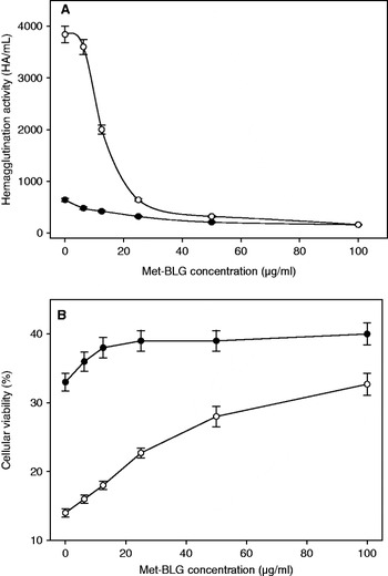
HA of H1N1 was inhibited by applying different concentrations of Met-BLG (Fig. 1A). With a virus titre of 1 MOI, the viral HA was significant and the observed antiviral effect of Met-BLG was concentration-dependent. With lower viral load (0·1 MOI) the viral HA was too weak to assume any readable difference with the treatment (data not shown). A concentration of 17 μg/ml Met-BLG could achieve 50% inhibition of HA after 24 h. Extending the incubation time produced more pronounced antiviral activity as the same concentration inhibited about 61% of HA with an IC50% decreased to 13 μg/ml Met-BLG after 48 h, and made the difference between the viral control and inhibition more marked.
Effect of different concentrations of Met-BLG (0–100 μg/ml) against (A) hemagglutination activity of H1N1 virus (1 MOI) infecting MDCK cells and (B) viability of MCDK cells. Incubation was performed for 24 (•) and 48 h (○). The results are the averages of three replicates with the standard error.

HA is exclusively due to hemagglutinin, a glycoprotein located on the influenza outer coat surface. It is responsible for infection of intact cells through the specific association with the cell receptor, ending with fusion with cell membrane. Recently, it has been reported that salt bridges within hemagglutinin monomers are crucial for its folding and stability (Rachakonda et al. Reference Rachakonda, Veit, Korte, Ludwig, Bottcher, Huang, Schmidt and Herrmann2007). This finding may to some extent help to understand the inhibitory effect of Met-BLG on HA. The large amount of positive charges on this esterified whey protein can disrupt the electrostatic interactions within hemagglutinin subunits, affecting its stability and activity, reducing finally its capacity to fuse and merge with the targeted cell membrane, hence inhibiting its infection potency. In a similar (but electrostatically opposite) way, a tricarboxylate – aurintricarboxylic acid (ATA), a heterogeneous mixture of polymers formed when salicylic acid is treated with formaldehyde, sulphuric acid, and sodium nitrite, is able to bind by electrostatic interactions to proteins containing positively charged residues. ATA was shown to inhibit the cytopathogenicity of HIV (human immunodeficiency virus) in cell culture (Balzarini et al. Reference Balzarini, Mitsuya, De Clercq and Broder1986; Baba et al. Reference Baba, Schols, Pauwels, Balzarini and De Clercq1988).
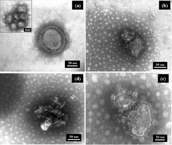
Perturbation of viral surface proteins (particularly hemagglutinin) may be deduced from the observation of electron microscopy image of the virus in absence or presence of Met-BLG. The viral coat looks deformed and rather disrupted in the later case (Fig. 2). The magnitude of the inhibitory effect (expressed by IC50) of Met-BLG is comparable to its previously observed activity against poliovirus type-1 and Coxsackie virus B6 (Sitohy et al. Reference Sitohy, Chobert, Dalgalarrondo, Nowoczin, Besse, Billaudel and Haertlé2008), although these viruses do not have hemagglutinin in their coat structure, indicating the broad specificity of Met-BLG, which may be able to act against a number of components of the virus machinery.
Electron microscopy images of influenza virus H1N1 in its native form (a) and after being in contact with 50 μg/ml Met-BLG at room temperature for 1 h (b–d).

Influence of Met-BLG on the viability of virus-infected MDCK cells
Viability of cells infected by H1N1 (1 MOI) was improved in a concentration-dependent manner thanks to the additions of Met-BLG (Fig. 1B). Maximal cell viability after 24 h was obtained by increasing the concentration of Met-BLG to 20 μg/ml. Cell viability was maintained up to 48 h, but its magnitude was lower than that observed after 24 h. Cell viability was 33% and 14% in the viral control (in absence of Met-BLG) after 24 and 48 h, respectively. In presence of Met-BLG, cell viability was enhanced up to 39% and 33% after 24 and 48 h, respectively. Again, the protective action was more pronounced after 48 h. However, comparing the action of Met-BLG against influenza virus with that against poliovirus type-1 and Coxsackie virus B6 (Sitohy et al. Reference Sitohy, Chobert, Dalgalarrondo, Nowoczin, Besse, Billaudel and Haertlé2008) shows weaker action against H1N1, although the inhibitory effect against HA of the virus was quite evident. This may indicate that HA is not the only factor determining the viral virulence and infectivity. Moreover, further adjustment of the conditions of application of Met-BLG may help to increase its protective effect.
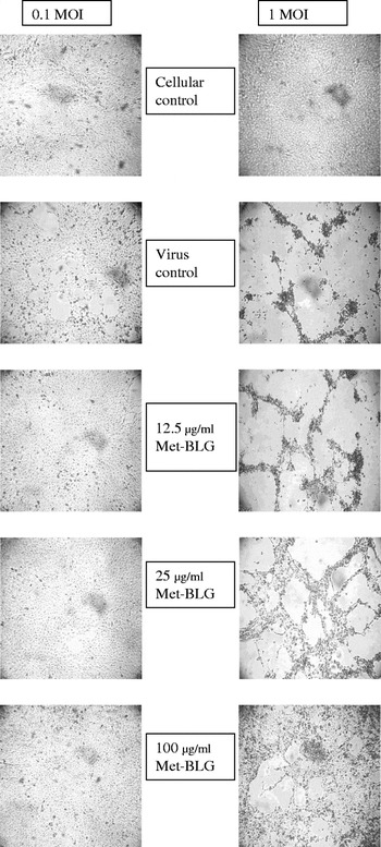
The images of microscopy in Fig. 3 (×40) show that the presence of Met-BLG in the medium of the virus-infected cells could protect them against viral action. The magnitude of this protective effect is dependent on two factors: Met-BLG concentration and MOI. Increase of Met-BLG concentration in the infected cell cultures resulted in higher amounts of viable cells, while the increase of MOI up to 1 decreased the antiviral effect without eliminating it altogether.
Light microscopy images (x 40) of MDCK cells infected with H1N1 virus (0·1 and 1·0 MOI) and treated with different concentrations of Met-BLG after 24 h incubation at 37°C and 5% CO2.

Generally, esterification of proteins neutralizes their negative charges unravelling positive charges, a fact which applies to the well-defined Met-BLG. However, even intensively substituted proteins are still similar in their net charges to native basic proteins existing in nature such as histones, which are able to cross the cellular membranes (Hariton-Gazal et al. Reference Hariton-Gazal, Rosenbluh, Graessmann, Gilon and Loyter2003). Due to its condensed positive net charge, the general effect of Met-BLG may be to interact with the non-structural influenza viral proteins, e.g. NS1 protein, which interact with cellular multifunctional proteins during infection (Murayama et al. Reference Murayama, Harada, Shibata, Kuroda, Hayakawa, Shimizu and Tanaka2007), thus resulting in reduced infection rate and higher cell viability.
Influence of Met-BLG on viral RNA replication
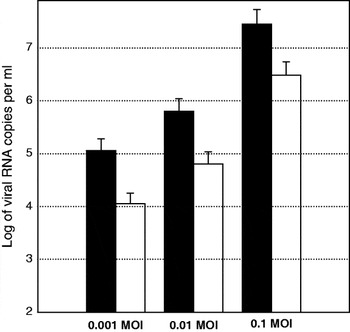
The amount of H1N1 viral RNA replicated in MDCK cells (MOI of 0·01, 0·1 and 1·0) was evaluated in response to treatment with 50 μg/ml Met-BLG (Fig. 4). The number of viral RNA copies decreased by 1 log for 0·1 and 0·01 MOI while it attained a reduction of 0·9 log when using 1 MOI. This slightly diminished effect when using higher MOI may indicate that the amount of Met-BLG should be adapted to the virus concentration. Consequently, additional Met-BLG doses should be used to better inhibit the infection.
Influence of presence of 50 μg/ml Met-BLG on the level of replicated RNA of H1N1 inoculated into MDCK cells at different MOI (0·01, 0·1 and 1) after incubation at 37°C and 5% CO2 for 18 h. In black: without Met-BLG; in white: after addition of Met-BLG. The results are the averages of three replicates with the standard deviation.

The anti-replicating effect of Met-BLG may occur through the interaction with viral proteins during the replicative events or through some RNA–Met-BLG interactions due to different charges of the reacting macromolecules.
Met-BLG might also interact with influenza nuclear proteins (PB1, PB2, PA and NP), which catalyze the transcription of viral RNA (Huang et al. Reference Huang, Palese and Krystal1990; Poole et al. Reference Poole, Elton, Medcalf and Digard2004; Boulo et al. Reference Boulo, Akarsu, Ruigrok and Baudin2007). Since these proteins are normally associated with RNA and undergo systematic dissociation during replication, the positively charged Met-BLG molecules may interfere with this association-dissociation process or compete for the negative charges on the exposed region of single stranded RNA, disturbing the replication.
Influence of Met-BLG on the virus growth curve
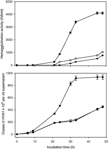
The evolution of growth of H1N1 in MDCK infected cells (1 MOI) was followed through assessing HA and replicated RNA during 48 h of incubation at 37°C and 5% CO2 in the presence of Met-BLG (Fig. 5). HA of virus control was detected after its first cycle of replication when it started to increase with time. Growth of virus treated with Met-BLG slowed down appearing only after more than 20 h (3 cycles of replication) incubation under the same conditions. HA of viral control reached exponential phase after 24 h incubation while stationary phase was reached after 42–48 h. This result is in agreement with reported values of the virus titre reaching a maximum after 24–48 h (Tirabassi & Enquist Reference Tirabassi and Enquist1998; Bourmakina & Garci-Sastre Reference Bourmakina and Garci-Sastre2005; Baccam et al. Reference Baccam, Beauchemin, Macken, Hayden and Perelson2006). In presence of 50 μg/ml Met-BLG, exponential phase lasted for a short period returning to an attenuated increase after 40 h. In presence of 100 μg/ml Met-BLG, exponential phase disappeared or was delayed to the 46–48 h of viral life cycle. The maximal level of HA in Met-BLG treated virus was much lower than that of the control indicating an effective inhibitory action of both concentrations. Although HA measurement is less precise than RNA quantification, our results may also be supported by those of electron microscopy showing that intact viral particles were reduced by over 65% after incubation with 50 μg/ml Met-BLG (Fig. 2). This morphologic change may be documenting also a possible cause of the observed anti-replicating action.
Evolution of hemagglutination activity and of amount of replicated RNA (assayed by RT-PCR) of H1N1 virus grown in MDCK cells in the absence (•) and presence of (○) 50 and (▾) 100 μg/ml Met-BLG during 48 h. The results are the averages of three replicates with the standard deviation.

Inhibition was confirmed by the decreased levels of H1N1 replicated RNA in the presence of the 2 concentrations of Met-BLG. Comparison of the curves shows that HA was much more affected than RNA replication (Fig. 5). Although the reduced amount of replicated RNA may lead to reduced HA, this result implies that Met-BLG does not only affect replication of RNA by interacting with viral protein or viral RNA during the replication events, but it interacts also somehow with HA of viral particles escaping the anti-replicating action, leading to their reduced infectivity.
Influence of Met-BLG on virus morphology and entity as seen by electron microscopy
Virus stock solution was combined with a final concentration of 50 μg/ml Met-BLG and left in contact for 1 h before examining the viral particles under electron microscope. The images (Fig. 2) show that untreated virus was intact and uniformly round. The glycoprotein spikes on untreated virus were uniformly arranged around the virus envelope, what remains in agreement with the observations of Yamaguchi et al. (Reference Yamaguchi, Danev, Nishiyama, Sugawara and Nagayama2008). In contrast, virus treated with Met-BLG showed clear signs of deformation: a rather irregular form, a discontinuity of the viral coat, and a disappearance of regular spikes. Some irregular aggregates of broken and deformed viral particles could be observed, probably resulting from the interaction between Met-BLG and viral components.
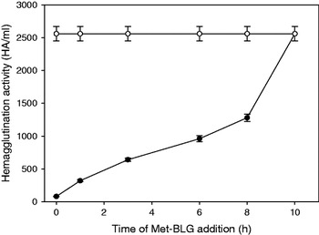
Influence of the time of addition of 50 μg/ml Met-BLG (•) on its antiviral activity against H1N1 infecting MDCK cells (1 MOI) as incubated for 24 h. (○): control without addition of Met-BLG. The results are the averages of three replicates with the standard deviation.

Compared with viral control, the number of intact viral particles in virus treated for 1 h at room temperature with 25 μg/ml Met-BLG was reduced by over 65%. This reduction was not accompanied by an important increase in amount of virus fragments but by significant alterations of the remaining viral particles. Some images showed the aggregation of the deformed viral particles and viral remnants, what might be caused by the penetration of used sodium tungstate into the interior of the viral particles as a result of altered viral coat or through its fixation at the surface of the deformed envelopes.
The reduction by about 65% of the amount of intact virus in the presence of Met-BLG is apparently due to the direct effect of esterified protein on the viral particles. Consequently, Met-BLG may be able to act directly on the viral coat proteins, contributing to viral deformation and consequent aggregation leading to reduction of activity.
Influence of the time of Met-BLG addition
Met-BLG (50 μg/ml) was added to H1N1-infected MDCK cells after different intervals from infection. The results showed a gradual decrease of the anti-hemagglutination effect of Met-BLG when its addition after infection was delayed (Fig. 6). Moreover, the anti-hemagglutination effect disappeared when Met-BLG was added 10 h after infection. This may indicate that after one cycle of virus replication, the virus concentration becomes too high to be inhibited by the applied Met-BLG concentration.
Influence of multiple additions of Met BLG
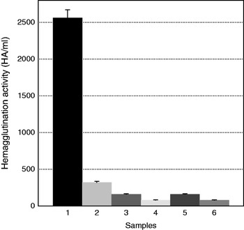
The antiviral activity of 50 μg/ml Met-BLG was tested after multiple additions of the protein (Fig. 7). The higher the number of additions, the higher the antiviral effect. Treatments # 4 and 6 were of similar antiviral magnitude (addition of 100 μg/ml Met-BLG), albeit they differ since treatment # 4 consisted in 2 supplementary additions (50 μg/ml at t 24 h and t 32 h) and treatment # 6 consisted in 1 addition of 100 μg/ml at t 32 h. Consequently, Met-BLG/virus ratio is the most important factor determining the extent of antiviral activity. There was no difference between treatments # 3 and 5, which consisted of the additions of 50 μg/ml after 24 h and 32 h, respectively. This difference in time of addition was useless on viral activity as compared to the effect of concentration of added Met-BLG.
Hemagglutination activity of H1N1 virus inoculated into MDCK cell lines at 1 MOI after 48 h of incubation as affected by different additions of Met-BLG. (1): no substance addition (viral control); (2): addition of 50 μg/ml Met-BLG at the 0 time; (3): addition of 50 μg/ml Met-BLG at 0 time and 50 μg/ml after 24 h; (4): addition of 50 μg/ml Met-BLG at 0 time, 50 μg/ml after 24 h and 50 μg/ml after 32 h; (5): addition of 50 μg/ml Met-BLG at 0 time and 50 μg/ml after 32 h; (6): addition of 100 μg/ml Met-BLG at 0 time and 50 μg/ml after 32 h.

Mahmoud Sitohy would like to thank for funding from ‘Imhotep’ Programme Hubert Curien and from Mission des Relations Internationales of INRA. The authors express their gratitude to Dr Cédric Gaillard (INRA, BIA) for his help in analysis of the electron microscopy pictures.