Percutaneous closure of patent ductus arteriosus has become the treatment of choice in many centres. There is a wide variety of devices to perform the technique with good results and minimal complication rate.Reference Porstman, Wierny and Warnke 1 – Reference Brunetti, Ringel and Owada 3 In patients with large ductus arteriosus and pulmonary hypertension, transcatheter closure has been achieved with success using the Amplatzer Duct Occluder or even the Amplatzer Muscular Ventricular Septal Defect Occluder.Reference Thanopoulos, Tsaousis, Djukic, Al Hakim, Eleftherakis and Simeunovic 4 – Reference Zabal, García-Montes and Buendía-Hernández 6 In some patients, the anatomic characteristics of the ducts, with very large diameter or very short length, make them difficult or even impossible to close with such devices. There have been some case reports where the Amplatzer Septal Occluder, intended for atrial septal defect closure, has been used to close this type of ducts with success.Reference Spies, Ujivari and Schräder 7 – Reference Yang, Zhous and Hu 11 We present a series of patients with large and hypertensive patent ductus arteriosus treated using the Amplatzer Septal Occluder.
Materials and methods
Patients
From April, 2006 to October, 2011, we treated 178 patients with large (⩾5 mm) and/or hypertensive (pulmonary artery systolic pressure ⩾40 mmHg) patent ductus arteriosus. In this retrospective and descriptive study, we analyse a group of 17 cases (9.5%) who received an Amplatzer Septal Occluder as final treatment. The group had 11 female patients (64.7%), and the mean age was 18.6±12.1 years (range 4–44) and weight was 39±15.9 kg (range 16–57). We had eight patients younger than 18 years and five of them were <20 kg.
Catheterisation procedure
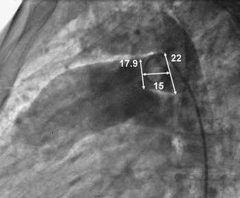
Informed, written consent was obtained and complete cardiac catheterisation was performed with venous and arterial femoral access, under general anaesthesia for children and conscious sedation for adults. In cases with pulmonary artery systolic pressure >50 mmHg, oxygen and pharmacologic tests, and pulmonary wedge angiography were performed to assess reversibility of pulmonary hypertension. In cases where pulmonary pressure did not decrease at least 20% breathing 100% oxygen, we performed a test occlusion with a compliant balloon for 10–15 minutes and repeated measurements. If response to 100% oxygen and occlusion was non-conclusive, pharmacological challenge with adenosine or inhaled nitric oxide was done. Reversibility of pulmonary hypertension was established when at least one of the following parameters was achieved: more than 20% decrease in pulmonary artery systolic pressure, more than 20% increase in pulmonary to systemic flow ratio (Qp/Qs), or a normal arterial morphology with more than five monopedial arteries in the pulmonary wedge angiography.Reference Zabal, García-Montes and Buendía-Hernández 6 Aortic angiographies were performed in straight lateral and right anterior oblique 45–60° projections to define anatomic characteristics of the ducts and to perform measurements (Fig 1). Once the device was in place, and before release, angiograms were made in the aorta and pulmonary artery to exclude device obstructions. Angiographic immediate residual shunt was classified as severe (contrast jet that reaches the pulmonary valve), moderate (contrast jet that reaches the pulmonary trunk but not the valve), mild (contrast that paints just the pulmonary branches), and trivial (“foaming” of contrast through the device). No heparin was used during the procedures unless embolisation of the device occurred and/or the procedure was prolonged for more than 90 minutes. If used, a dose of 100 U/kg (maximal dose of 5000 U) was administered with a target-activated coagulation time between 200 and 250 seconds.
Sample figure of measurements performed on a type A patent ductus arteriosus. Pulmonary end 17.9 mm, aortic end 22 mm, and length 15 mm. In this case, a 24 mm ventricular septal defect occluder was initially delivered but it embolised. A 28 mm Amplatzer Septal Occluder was used finally.

Follow-up
Patients were discharged the following morning on prophylactic antibiotics and 100 mg aspirin. Follow-up visits were scheduled 1 and 6 months after the procedure, and yearly thereafter with a clinical exam, x-ray, and transthoracic echocardiogram. Cardiac catheterisation was performed in two selected cases.
Statistical analysis
Data were compiled in an Excel worksheet and processed with a Prisma graphic software. Categorical variables are expressed as number and percentage, and continuous variables as average or median, standard deviation and limits.
Results
All patients had pulmonary artery systolic pressure ⩾40 mmHg (71.3±31.8, range 40–164), which was 73±22% (range 31–100) of the systemic pressure. Oxygen saturations were the same at the left ventricle and femoral artery in all cases but one, which had 90% and 81%, respectively (pulmonary artery systolic pressure=118 mmHg and aortic systolic pressure=120 mmHg). This patient decreased her pulmonary artery systolic pressure to 60 mmHg after 100% oxygen and ductal occlusion without modification on the systemic pressure. The group mean pulmonary artery pressure was 50.1±22.7 mmHg, Qp/Qs was 3.14±1.36, the smallest ductal diameter at the pulmonary end was 12.5±3.8 mm (range 7.9–18.8), and at the aortic end was 20.2±7.7 mm (range 12–28); 14 cases (82.3%) had ductus arteriosus type A, two (11.8%) type C, and one (5.9%) type D.Reference Krichenko, Benson, Burrows, Möes, McLaughlin and Freedom 12 One of the cases, a 25-year-old woman with a 33-week pregnancy, was urgently admitted with pulmonary oedema.
With regard to the anatomy of the type A ducts, they had a pulmonary end diameter of 12.4±3.34 mm and an aortic ampulla diameter of 21.63±8.42 mm, and thus the ampulla was 1.77±0.32 times larger than the pulmonary end.
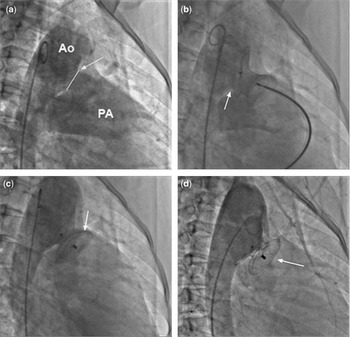
In 11 patients, we began the procedure using a different type of device – six with Amplatzer Duct Occluder and five with Amplatzer Muscular Ventricular Septal Defect Occluder – and in six patients we selected an Amplatzer Septal Occluder as the first option. In the 11 patients where we began the procedure with a different device, it was changed because of device embolisation in six (35.3%), three immediately in the catheterisation lab and another three within 24 hours of the procedure. All these devices were percutaneously retrieved and changed. In the other five, the device was changed because of easy passage through the duct or significant protrusion into the left pulmonary artery (Fig 2).
Aortic angiographies in right anterior oblique 45° of one of the cases. ( a ) A large and short ductus arteriosus (arrow) can be observed with a measurement of 17.4 mm at the pulmonary end and 20.8 at the aortic end. ( b ) After balloon occlusion test with an expanded diameter of 24 mm, we intended occlusion with a 24 mm Amplatzer Muscular Ventricular Septal Defect Occluder. The distal disc dislodges easily from the inferior margin of the aortic ampulla (arrow). ( c ) Final angiography after delivering and releasing a 30 mm Amplatzer Septal Occluder. Note that the ductal ampulla is completely covered by the distal disc and the proximal disc (arrow) is inside the pulmonary artery with very little protrusion to the left pulmonary artery. A mild residual shunt is present. ( d ) Angiographic control 16 months after occlusion. Note the good device position (arrow) with no residual shunt. Ao=Aorta; PA=pulmonary artery.

After the experience with these cases, we began to use the Amplatzer Septal Occluder as our initial device, and the device waist diameter was selected according to the duct diameter at the aortic end (around 1:1) with a mean of 20.8±6.7 mm (range 12–34). The device to patent ductus arteriosus diameters final analysis showed that device waist was 64% larger than the pulmonary end diameter and only 3% larger than the aortic end. All Amplatzer Septal Occluders were delivered successfully. There was no gradient across the aorta, and only in one case (5.9%) we found an 8 mmHg gradient between the pulmonary artery and the left pulmonary branch. Immediate residual shunt was moderate in six patients (35.3%), mild in eight (47%), trivial in two (11.8%), and no shunt in one (5.9%). Pulmonary artery systolic pressure decreased from 71.3±31.8 to 48.9±10.8 mmHg after occlusion (p=0.0015) and the pulmonary artery mean pressure from 50.1±22.7 to 37±9.7 mmHg (p=0.002). Fluoroscopy time was 21.5±18.4 minutes (range 3.6–70) and procedure time was 95.9±33.4 minutes (range 30–180). Despite 82.3% of mild and moderate immediate residual shunt, only one case presented haemolysis with a haemoglobin decrease of 1.24 mol/L and a maximal bilirubin increment of 29 mmol/L, which ceased in 2 days. All patients were discharged from the hospital within 48 hours of the final procedure with 100 mg aspirin daily for 6 months and antibiotic prophylaxis.
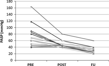
Follow-up was possible in 15 patients (88.2%) for 28.4±14.4 months (range 6.3–53.5); all but one, with a trivial residual shunt, showed complete closure. Echocardiograms suitable for pulmonary pressure measurements (using the tricuspid regurgitation jet velocity) were obtained in 14 patients with a mean pulmonary artery systolic pressure of 31.4±10.5 (range 20–60) (Fig 3). There were two patients with pre-closure pulmonary artery systolic pressure of 90 and 85 mmHg who agreed to be recatherised at 8 and 16 months after patent ductus arteriosus closure, with complete closure of the duct and pulmonary artery systolic pressure of 30 and 35, respectively. The patient with the highest pre- and post-closure pulmonary artery systolic pressure (164 and 80) showed continuous improvement with an echo measured pulmonary artery systolic pressure at 19 months of 60 mmHg. He is the only patient under sildenafil treatment. No complications at follow-up have been reported.
Graphic showing evolution of the pulmonary artery systolic pressures (PASPs) before (PRE), immediately after closure (POST), and at follow-up (FU) in the 14 patients with suitable measurements.

Discussion
Percutaneous closure of patent ductus arteriosus is an excellent alternative to surgical treatment and, in recent years, it has become the best treatment option, even in cases with pulmonary hypertension, where Amplatzer Duct Occluder and Amplatzer Muscular Ventricular Septal Defect Occluder have been used.Reference Ghasemi, Pandya and Reddy 2 – Reference Zabal, García-Montes and Buendía-Hernández 6 In some countries, there are devices similar to the Amplatzer Duct Occluder in larger sizes (up to 26 mm), and thus patent ductus arteriosus larger than 12 mm can be closed with these devices.Reference Fatema 10 There have been case reports of large ductus arteriosus with pulmonary hypertension that were closed with an Amplatzer Septal Occluder with good results.Reference Spies, Ujivari and Schräder 7 – Reference Yang, Zhous and Hu 11 We present here a series of 17 patients, including children and adults, with large ductus arteriosus and pulmonary hypertension who live at high altitude (2240 m). In most patients, we made a first attempt to close the patent ductus arteriosus with an Amplatzer Duct Occluder or an Amplatzer Muscular Ventricular Septal Defect Occluder with no success. In these cases, we changed the devices for an Amplatzer Septal Occluder considering the size of the previous device, the position of the first device after release, and the diameter of the aortic end of the duct, to decide the size of the Amplatzer Septal Occluder to be used. With these parameters, we selected an Amplatzer Septal Occluder with a distal disc large enough to completely cover the aortic end of the ductus. After doing an analysis of the device size to patent ductus arteriosus diameters in these cases, we found that the device waist was 64% larger than the pulmonary end diameter and only 3% larger than the aortic end. We selected the Amplatzer Septal Occluder as the first choice device when the aortic ampulla was 1.5 times larger than the pulmonary end, and when the duct was very short (type C). We chose a device size similar to the aortic end of the duct. We have found in such cases better apposition of the device to the ductal walls and less protrusion of the proximal disc into the left pulmonary artery. Finally, all cases were successfully closed with the Amplatzer Septal Occluder. Regarding pulmonary hypertension, all cases had systolic pressures ⩾40 mmHg (65% ⩾60 mmHg) but, with a thorough evaluation, all cases decreased their pulmonary pressures significantly after closure. We have found patent ductus arteriosus balloon occlusion test very useful to determine reversibility in these cases. Pulmonary pressure continues decreasing in the follow-up and only one patient is under sildenafil treatment. No complications have been reported at follow-up and, with echocardiographic study, all but one case are completely closed and no obstruction of the aorta or left pulmonary artery has been detected. We suggest the use of an Amplatzer Septal Occluder as the first choice device in cases of large (⩾8 mm) type C ducts, and in cases with type A ducts where the aortic ampulla diameter is more than 1.5 times the pulmonary end diameter.
Limitations
This device is not specifically designed for patent ductus arteriosus closure, but it performed very well in these cases. Although the series is small, it demonstrates that the device can be used in such circumstances.
Conclusion
The Amplatzer Septal Occluder is a good alternative to percutaneously treat large and hypertensive patent ductus arteriosus, although we have available devices specifically designed for this condition.
Acknowledgements
None.
Financial Support
This research received no specific grant from any funding agency, commercial or not-for-profit sectors.
Conflicts of Interest
Dr. Carlos Zabal is proctor and consultant for St. Jude Medical.