INTRODUCTION
Excavating sponges are some of the most dominant bioeroders on coral reefs, and some species are particularly strong competitors for space (e.g. Vicente, Reference Vicente1978; McKenna, Reference McKenna, Lessios and MacIntyre1997; López-Victoria et al., Reference López-Victoria, Zea and Weil2006; Chaves-Fonnegra & Zea, Reference Chaves-Fonnegra, Zea, Custódio, Hajdu, Lôbo-Hadju and Muricy2007). Bioerosion by excavating sponges can account for over 90% of carbonate loss from coral skeletons – specifically by macroborers (e.g. MacGeachy & Stearn, Reference MacGeachy and Stearn1976; Mallela & Perry, Reference Mallela and Perry2007), and remove up to 30 kg of CaCO3 m−2 yr−1 of reef matrix (e.g. Schönberg, Reference Schönberg2002; Calcinai et al., Reference Calcinai, Azzini, Bavestrello, Gaggero, Cerrano, Custódio, Hajdu, Lôbo-Hadju and Muricy2007) contributing to the balance between accretion and bioerosion (Glynn, Reference Glynn and Birkeland1997). However, in excess these sponges can drive reefs into erosional states (e.g. Acker & Risk, Reference Acker and Risk1985; Nava & Carballo, Reference Nava and Carballo2008; Perry et al., Reference Perry, Spencer and Kench2008), restructuring the reef and possibly causing collapse in extreme cases (Rose & Risk, Reference Rose and Risk1985; Carballo et al., Reference Carballo, Bautista-Guerrero and Leyte-Morales2008; Kennedy et al., Reference Kennedy, Perry, Halloran, Fine, Carricart-Ganivet, Iglesias-Prieto, Form, Wisshak, Schönberg and Mumby2013).
At least 35 species of Caribbean reef sponges are known bioeroders, and 19 are from the genus Cliona (van Soest et al., Reference Van Soest, Boury-Esnault, Hooper, Rützler, de Voogd, Alvarez de Glasby, Hajdu, Pisera, Manconi, Schönberg, Janussen, Tabachnick, Klautau, Picton, Kelly, Vacelet, Dohrmann, Díaz and Cárdenas2014). Some coral-excavating sponges can overgrow and kill entire coral colonies up to several metres in diameter using chemical (enzymes or allelopathic metabolites) and physical (etching cells) mechanisms to excavate and invade substrates of other organisms (Pomponi, Reference Pomponi1979; Sullivan et al., Reference Sullivan, Faulkner and Webb1983; Sullivan & Faulkner, Reference Sullivan, Faulkner and Rützler1990; Schönberg & Wilkinson, Reference Schönberg and Wilkinson2001; Zundelevich et al., Reference Zundelevich, Lazar and Ilan2007; Chaves-Fonnegra et al., Reference Chaves-Fonnegra, Castellanos, Zea, Duque, Rodríguez and Jiménez2008; Schönberg, Reference Schönberg, Wisshak and Tapanila2008).
Decline in coral cover across the Caribbean Sea and Gulf of Mexico region is widely reported and has been attributed to a variety of natural and anthropogenic stressors (e.g. Hughes, Reference Hughes1994; Gardner et al., Reference Gardner, Côté, Gill, Grant and Watkinson2003; Kennedy et al., Reference Kennedy, Perry, Halloran, Fine, Carricart-Ganivet, Iglesias-Prieto, Form, Wisshak, Schönberg and Mumby2013), including excavating sponges, which have grown significantly in these areas (Rützler, Reference Rützler2002; López-Victoria & Zea, Reference López-Victoria and Zea2005; Ward-Paige et al., Reference Ward-Paige, Risk, Sherwood and Jaap2005). Increases in density and cover of bioeroding sponges can be directly and indirectly enhanced by various factors that are damaging to stony corals such as thermal events and eutrophication (Holmes, Reference Holmes, Lessios and MacIntyre1997, Reference Holmes2000; Rützler, Reference Rützler2002; Schönberg & Suwa, Reference Schönberg, Suwa, Custódio, Lôbo-Hajdu, Hajdu and Muricy2007; Schönberg et al., Reference Schönberg, Suwa and Hidaka2008; Schönberg & Ortiz, Reference Schönberg, Ortiz and Riegl2009; Carballo et al., Reference Carballo, Bautista, Nava, Cruz-Barraza and Chavez2013). As the various coral reef stressors continue, understanding the impact of excavating sponges on coral growth is important. Accordingly, these sponges are included in monitoring and reef restoration projects throughout the Caribbean Sea and Gulf of Mexico Region (e.g. Lang, Reference Lang2003; Gilliam et al., Reference Gilliam, Brinkhuis, Ruzicka and Walton2013), giving special attention to Cliona delitrix Pang, 1973. This species is destructive to live corals (Chaves-Fonnegra & Zea, Reference Chaves-Fonnegra, Zea, Custódio, Hajdu, Lôbo-Hadju and Muricy2007, Reference Chaves-Fonnegra and Zea2011) and acutely responds to eutrophication (Rose & Risk, Reference Rose and Risk1985; Ward-Paige et al., Reference Ward-Paige, Risk, Sherwood and Jaap2005; Chaves-Fonnegra et al., Reference Chaves-Fonnegra, Zea and Gómez2007). Cliona delitrix is abundant on offshore reefs of south-east Florida where it commonly invades 11 different local coral species and affects 4% of stony coral colonies (Chiappone et al., Reference Chiappone, Rutten, Miller, Swanson, Custódio, Hajdu, Lôbo-Hadju and Muricy2007; Halperin, Reference Halperin2014). However, in this area corals with excavating sponges are not moved and reattached during projects of impact minimization, mitigation or restoration (Ken Banks, Broward County Natural Resources Planning and Management Division, personal communication), and coral cover from these colonies is lost.
When excavating sponges colonize stony corals, they invade the skeleton that is created and defended by the coral, its ‘host’. The ecological interaction between these organisms was first considered as epizoism (Antonius & Ballesteros, Reference Antonius and Ballesteros1998) or infestation (Glynn, Reference Glynn and Birkeland1997), and parasitism is a possibility. However, it may be facultative parasitism, as the sponge is able to grow on calcium carbonate terraces, and also on corals, and could complete its life cycle without being metabolically dependent on the host (see definitions of parasitism in Lincoln et al., Reference Lincoln, Boxshall and Clark1998). Recent studies consider these organisms are in asymmetric competition: where both the sponge and coral are ‘fighting’ for the same resource (skeleton), but the sponge tends to gradually displace and overgrow entire coral colonies (Rützler, Reference Rützler2002; López-Victoria et al., Reference López-Victoria, Zea and Weil2006; Chaves-Fonnegra & Zea, Reference Chaves-Fonnegra and Zea2011). In this study we will refer to the sponge-coral interaction as asymmetric competition.
Considering that Cliona delitrix is clearly an important bioeroder that has become abundant (Ward-Paige et al., Reference Ward-Paige, Risk, Sherwood and Jaap2005), and thus detrimental to live corals, restoration techniques are suggested to improve the chances of survival for adult corals colonized by this sponge. But to date, no studies have experimentally tested any technique. Previous studies on competitive interactions in the marine environment have shown that removing one competitor can allow recovery of the other (Tanner, Reference Tanner1995; Jompa & McCook, Reference Jompa and McCook2002). Thus, the aim of this study was to explore the direct effect of C. delitrix on Montastraea cavernosa Linnaeus, 1767 tissue loss, and its possible recovery through a restoration method that eliminates C. delitrix and alters the competitive nature of the coral-sponge interaction. We also evaluate the efficacy of the technique to prevent C. delitrix regrowth.
MATERIALS AND METHODS
Sponge removal and cavity filling
To determine the effect of Cliona delitrix removal on stony coral tissue loss or growth, 33 Montastraea cavernosa corals colonized by small sponges (up to 10 cm in diameter) were utilized, as they excavate shallower cavities (usually <5 cm) and are easier to remove from coral colonies. In July 2012, sponges were manually eliminated from 22 of the affected coral colonies. Most of the sponge tissue and affected coral skeleton was extracted using a hammer and chisel, taking care not to leave resulting fragments on the reef. The remaining sponge tissue in the resulting cavities was cleaned out using a steel wire brush.
The cavities in the coral skeletons were then filled to promote coral tissue overgrowth. A pH balanced cement mixture was applied to 11 of the coral colonies (Reef Innovations Inc., St. Cloud, Florida), and 11 were filled with two-part epoxy (ALL FIX©, Cir-Cut Corp., Philadelphia, PA). Cavities were filled by manually applying the fill material to the same level as the surrounding colony surface and smoothing the edges against the adjacent live coral tissue. These two fill materials were selected because they have been used in previous restoration efforts and corals were reported to effectively overgrow them (Collier et al., Reference Collier, Dodge, Gilliam, Gracie, Gregg, Jaap, Mastry and Poulos2007; Young et al., Reference Young, Schopmeyer and Lirman2012). The 11 coral-sponge colonies in which sponges were left untouched were monitored as controls.
Coral tissue loss monitoring and analysis
Images of each coral colony – for both controls and treatments – were taken using a mounted digital camera at the beginning of the experiment, 6 and 12 months after sponge removal. Presence of visible C. delitrix tissue was also noted on the coral colony surface or around the refilled cavity, along with the presence of other bioeroders (i.e. other sponges, polychaetes, barnacles).
In order to determine coral tissue loss or growth between monitoring periods 0–6 months, and 0–12 months, images were compared digitally. First, all images were standardized in ArcGIS 10.1© software (ESRI©, Redlands, CA). Using the ‘georeferencing’ tool, we matched features such as unique coral polyps or worm tubes between images to align the colony images to the same viewing angle and size.
CPCe 4.1© software (National Coral Reef Institute, Dania Beach, FL) was used for tracing and surface area calculations. For each control colony, the live coral tissue border surrounding the sponge was traced, and for each removal colony, the live coral tissue boundary around the filled cavity was traced (Figure 1). In either case, the traced area is referred to as the ‘dead area’. Images from all three time periods (0, 6 and 12 months) were traced three times each, and the three surface area measurements per time period were averaged to obtain a mean surface area value.
Examples of typical coral colonies and traced respective ‘dead areas’ at t = 6 months. (A) Colony from the removal treatment showing the light fill material and surrounding darker live coral tissue with minor partial mortality. (B) Control colony containing intact C. delitrix sponge surrounded by exposed coral skeleton. (C) Traced ‘dead area’ for the above colony containing fill material and exposed coral skeleton. (D) Traced ‘dead area’ for the above colony containing sponge individual and exposed coral skeleton.

For analysis of both the 6 and 12 month monitoring periods, the per cent change in ‘dead area’ (DA) was calculated using the following formula:
 $$\%\,{\rm change\,in}\,{\rm DA}\, = \,(({\rm DA}_f -{\rm DA}_i )/{\rm DA}_i )\, \times {\rm 1}00,$$
where DAf is the final ‘dead area’ and DAi represents the initial ‘dead area’. A positive percentage change represents coral tissue loss (final ‘dead area’ > initial ‘dead area’), and a negative percentage change represents coral tissue growth over the ‘dead area’. To test for differences in coral tissue mortality (change in ‘dead area’) between treatment groups and fill materials, Wilcoxon rank sum tests (e.g. Sokal & Rohlf, Reference Sokal and Rohlf1995) were conducted for each monitoring period.
To determine if C. delitrix reappeared on the coral colonies in the removal treatment, the filled removal cavities and the exposed coral skeleton surrounding these cavities were scraped during monitoring to eliminate sediment, algae and tunicates. Control colonies were also scraped clean during each monitoring event so the sponge-coral boundary was clearly visible in images.
To test for differences in C. delitrix reappearance between fill materials, a chi-squared test was conducted for the 12-month monitoring period. Additionally, to determine whether initial C. delitrix size played a role in sponge reappearance, a t-test was conducted between the groups in which the sponge remained absent or was visibly present within the 12-month monitoring period.
RESULTS
Per cent change in ‘dead area’ between treatments
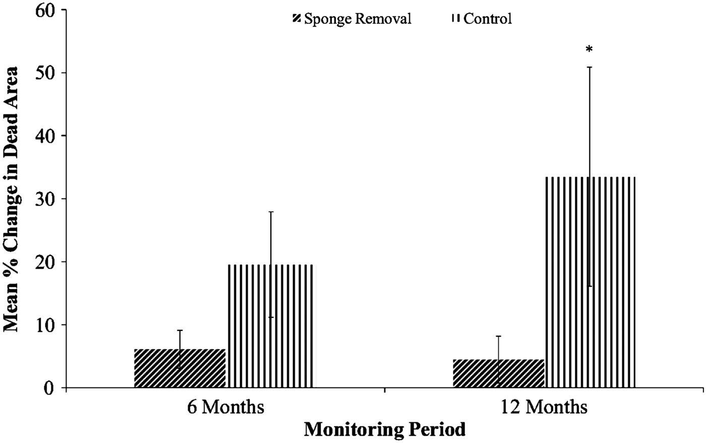
The change in size of the ‘dead area’ over time varied between treatment groups and between monitoring periods. The control group, i.e. corals in which the sponge remained in place, showed the greatest increase in the ‘dead area’, i.e. the sponge grew over time (Figure 2). Coral colonies in the sponge removal treatment (cement and epoxy pooled) displayed a much smaller increase in the ‘dead area’, representing a lesser amount of coral tissue loss. Although in the controls (sponge remained) coral tissue loss was about three times greater than in the pooled removal group after 6 months, effects of sponge removal proved to be statistically insignificant (Wilcoxon rank sum test with mean ranks of control and removal treatments of 20.55 and 14.14 respectively; S = 185, Z = 1.76, P = 0.08). After 12 months however, the change in ‘dead area’ was significantly larger in the control group than in the sponge removal group (mean ranks of control and removal treatments were 23.00 and 13.55 respectively; S = 230, Z = 2.62, P < 0.01) (Figure 2).
Mean per cent change in ‘dead area’ ± SE per treatment for both 6 and 12 months after C. delitrix removal. The asterisk represents significant statistical differences comparing both treatments at 12 months (P < 0.05).
Per cent change in ‘dead area’ compared between fill materials
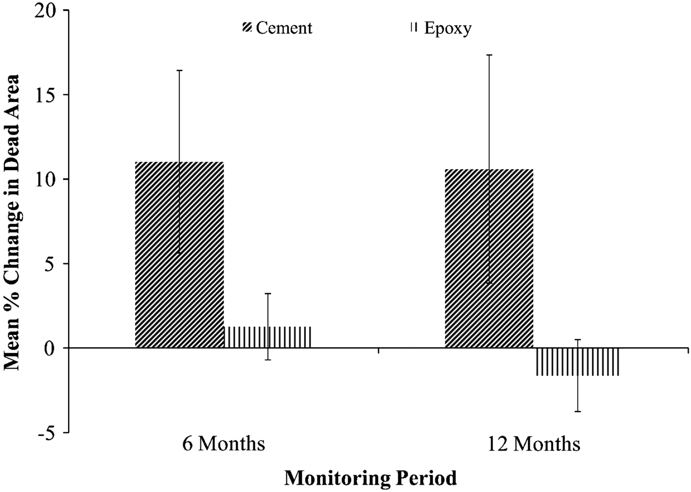
The coral colonies with cement patches showed a greater increase in the ‘dead area’ than those repaired with marine epoxy, both at 6 and 12 months after sponge removal (Figure 3). In the cement group the increase in ‘dead area’ was consistent across both monitoring periods, while colonies filled with epoxy actually showed a decrease in the ‘dead area’ after 12 months representing coral tissue overgrowth. Despite the latter observation, fill materials were determined to be statistically similar at both 6 and 12 months, and did not significantly vary over time (Wilcoxon rank sum test, 6 months – mean ranks of cement and epoxy were 13.82 and 9.18 respectively; S = 101, Z = −1.64, P = 0.10; 12 months – mean ranks of cement and epoxy were 14.09 and 8.91 respectively; S = 98, Z = −1.84, P = 0.07).
Mean per cent change in ‘dead area’ ± SE per fill material at both 6 and 12 months after C. delitrix removal.
Presence of Cliona delitrix after removal
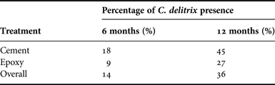
After sponge removal and cavity filling, experimental colonies were left without any visible portions of C. delitrix tissue on the surface of the fill material or surrounding dead coral skeleton. After 6 months, however, 14% of the coral colonies from the removal treatments showed presence of C. delitrix, and this percentage increased to 36% after 12 months (Table 1). Visible sponge tissue was approximately 0.5 cm in diameter. Reappearance of the sponge on the surface was observed on exposed coral skeleton located adjacent to the removal cavity, in old dead areas and in previously live areas of coral tissue where the sponge was not initially present.
Frequency of M. cavernosa colonies showing C. delitrix tissue on colony surface 6 and 12 months after sponge removal.
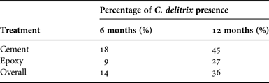
Cliona delitrix recurrence after 12 months post-removal was more pronounced in coral colonies of the cement group for both monitoring periods. Differences between fill materials were not statistically significant (X 2 = 0.79, P = 0.37, N = 22). We additionally examined if the initial sponge size had an influence on the re-appearance, and no statistical effect was detected, (t = 1.10, P = 0.29). In one coral colony the sponge tissue re-appeared after 6 months, but disappeared at 12 months, indicating that the previously visible sponge had died.
DISCUSSION
Removal of Cliona delitrix temporarily reduced coral tissue loss in Montastraea cavernosa on an individual coral colony basis. Differences in coral tissue loss between control and removal treatments were statistically significant after 12 months, suggesting that coral colonies needed a recovery period beyond 6 months to start overgrowing the fill materials after sponge removal. This also shows that it takes a year for the sponge to significantly reduce coral tissue (control colonies).
Coral overgrowth of the removal cavities, as determined through image analysis, was only witnessed in a portion of experimental colonies, mostly in the epoxy treatment. This may be due to the tendency of M. cavernosa to grow upwards and form a dome over dead areas before growing laterally across them (e.g. López-Victoria et al., Reference López-Victoria, Zea and Weil2006), which was not examined in this study. In addition, our efficiency in filling the removal cavities could have affected coral overgrowth of dead areas. Particularly for the cement-filled colonies, areas of bare dead skeleton were present between the infilled cavity and the live coral tissue. Not covered by the fill material, these dead areas provided suitable substrate for colonization of other fouling organisms between monitoring and ‘cleaning’ events that can negatively affect coral growth (López-Victoria et al., Reference López-Victoria, Zea and Weil2006; Chaves-Fonnegra & Zea, Reference Chaves-Fonnegra and Zea2011).
Both fill materials showed an average reduction in the amount of coral tissue lost compared with the controls. Coral overgrowth of the removal cavities only occurred on average in the epoxy treatment, while in the cement group, live coral tissue was lost from colonies. This result contrasts with a previous study of Acropora palmata that found similar coral growth rates over both fill materials (Forrester et al., Reference Forrester, O'Connell-Rodwell, Baily, Forrester, Giovannini, Harmon, Karis, Krumholz, Rodwell and Jarecki2011), but the two species overgrow dead areas differently and may have affected this result. Differences in coral growth between treatments can be attributed to a greater cover of algae, sediment and tunicates on the cement than on the epoxy fills. Cement contained a portion of calcium carbonate not present in the epoxy that may have created a more ‘natural’ substrate preferred by fouling organisms. Alternatively, epoxy appeared to attach better to the newly established cavity, as well as to surrounding dead coral skeleton. Due to its smoothness, epoxy may have also ‘caught’ fewer recruits than the cement. Both fill materials were ‘fresh’ surfaces when applied and thus lacked the appropriate microbial biofilm necessary for attachment of fouling marine invertebrates, which could have produced a lag period for colonization compared with dead coral skeleton (e.g. Zardus et al., Reference Zardus, Nedved, Huang, Tran and Hadfield2008).
The sponge removal technique maintained an absence of C. delitrix from over 60% of the experimental corals in this study, indicating an apparent relief of the coral from the sponge's excavating activities during this period. Two possible explanations for C. delitrix reappearance in the remaining colonies include: (1) regrowth and resurfacing of remaining sponge tissue left behind after removal, and (2) new colonization of the dead coral substrate by sponge recruits. If the observed sponge tissue was indeed resurfacing of tissue remnants from within the colony, this shows amazing regrowth potential of the species, as most of the sponge individual had been eliminated, completely covered with fill material and restricted from open access to seawater for nutrition or waste removal. Alternatively, it is possible that visible sponges were new recruits, as C. delitrix has an extended reproductive cycle in Florida with multiple (three to five) spawning events throughout the year and a preference for recently dead coral substrate clean of other organisms (Chaves-Fonnegra, Reference Chaves-Fonnegra2014). To confirm which explanation applies to this scenario, opening of the filled cavity to follow sponge filaments inside the coral and genetic testing would be necessary. The periodic scraping of the filled cavities in both treatments during monitoring events could have also aided sponge reappearance by eliminating other competitors (i.e. macroalgae, tunicates, etc.) that would have smothered any remaining sponge tissue attempting to make its way to the colony surface, or covered any exposed skeleton, preventing new C. delitrix colonization.
This experiment showed that eliminating the sponge allows for the potential recovery of the coral, further demonstrating that excavating sponges have a detrimental effect on coral growth, as found in previous studies (Schönberg & Wilkinson, Reference Schönberg and Wilkinson2001; Rützler, Reference Rützler2002; López-Victoria et al., Reference López-Victoria, Zea and Weil2006; Chaves-Fonnegra & Zea, Reference Chaves-Fonnegra and Zea2011). Montastraea cavernosa is a slow-growing species, thus monitoring experimental colonies beyond 12 months would provide further insights into the effect of sponge removal on coral individuals and the extent of sponge or attachment of new Cliona delitrix recruits. Considering that competitive interaction with macroalgae reduced the reproductive output of M. annularis (Foster et al., Reference Foster, Box and Mumby2008), future studies are encouraged to test if the competitive interaction with C. delitrix has the same effect on M. cavernosa.
Management and restoration considerations
During impact minimization, mitigation or restoration projects involving stony coral in south-east Florida, reattachment or relocation of corals with excavating sponge colonization is discouraged (Ken Banks, personal communication). Resource managers’ rationale is that the time, money, and effort required to relocate and reattach these affected colonies are wasted resources because the coral will inevitably die. Our findings show that given the availability of additional resources common to any coral relocation or reattachment project (divers, hammers, chisels, epoxy), coral mortality associated with C. delitrix can be somewhat reduced to preserve the remaining live coral tissue using epoxy as the fill material. However, considering that C. delitrix reappeared in 36% of the coral colonies after a year of study, our method is not suitable to control the sponge colonization. Also, this technique introduces a large amount of foreign material in the form of cement and epoxy that is not justifiable given the results of our study. This practice is also expensive to carry out due to the amount of manufactured fill materials and diver salaries that are necessary. Because this method is also fairly labour intensive, it would be difficult to utilize at reef depths where dive time is restricted, and it would not make an impact on populations of C. delitrix in such habitats. Left untouched, remaining sponge populations at these depths would reproduce and reseed shallower reef areas where all sponges had been removed previously, counteracting previous efforts. For all of the above reasons, it is highly unlikely that this technique could be applied on the large reef-scale to make a considerable impact on local C. delitrix populations.
Cliona delitrix and other excavating sponges are natural components of Caribbean coral reefs, and contribute to the balance between construction and destruction of coral reefs (Glynn, Reference Glynn and Birkeland1997). But due to current climate changes and anthropogenic influences, the impact of this group of sponges on stony corals has been exacerbated (López-Victoria & Zea, Reference López-Victoria and Zea2005; Ward-Paige et al., Reference Ward-Paige, Risk, Sherwood and Jaap2005; Bell et al., Reference Bell, Davy, Jones, Taylor and Webster2013; Carballo et al., Reference Carballo, Bautista, Nava, Cruz-Barraza and Chavez2013; Wisshak et al., Reference Wisshak, Schönberg, Form and Friewald2014). Instead of attempting to control excavating sponges by direct human intervention, we suggest other alternatives to minimize the growth and proliferation of C. delitrix such as improvements in water quality. For example, lowering the amount of nitrogen and ammonium in sewage may reduce the abundance and cover of excavating sponges, such as C. delitrix (see Ward-Paige et al., Reference Ward-Paige, Risk, Sherwood and Jaap2005). Also, the reduction of fossil fuel emissions (i.e. CO2) is necessary to avoid the loss of corals and slow processes that may negatively affect them (Ateweberhan et al., Reference Ateweberhan, Feary, Keshavmurthy, Chen, Schleyer and Sheppard2013; Kennedy et al., Reference Kennedy, Perry, Halloran, Fine, Carricart-Ganivet, Iglesias-Prieto, Form, Wisshak, Schönberg and Mumby2013). These actions would in turn play a larger role in preserving present stony corals and reefs for future generations. Indeed, left to themselves, some corals are capable of escaping excavating sponges by growing upwards and forming domes (López-Victoria et al., Reference López-Victoria, Zea and Weil2006), which under less polluted conditions may allow them to survive into the future.
ACKNOWLEDGEMENTS
Thank you to the various members of the CRRAM lab at Nova Southeastern University who helped with fieldwork including C. Walton, M. Lopez-Padierna, L. Larson, C. Bliss, J. Mellein, S. Bush, Z. Ostroff, P. Espitia, N. D'Antonio, L. Kabay, K. Correia and K. Cucinotta. A special thanks to D. Fahy for his help in developing the research idea, and the same for B. Walker who assisted with image standardization techniques. This manuscript was submitted as partial fulfilment of the MSc degree to A.A. Halperin at the Nova Southeastern University Oceanographic Center, Dania Beach, Florida. Sponge removal was conducted in accordance with Special Activity License Number: SAL-11-1358-SRP, granted by the Florida Fish and Wildlife Conservation Commission.
FINANCIAL SUPPORT
This work was supported by joint effort of the Coral Reef Restoration, Assessment and Monitoring Laboratory at the Nova Southeastern University Oceanographic Center.