Implications
The empirical selection of phenotypic traits of economic interest carried out by the bovine breeders will be assisted gradually by the exploitation of knowledge concerning the bovine genome. However, such a revolution in the methods of selection henceforth presents several difficulties. To speed up the identification of genes controlling the musculoskeletal functions in cattle, we have set up a chemical mutagenesis programme based on a sensitised screen using the Gdf8 knockout mouse as genetic background to assess skeletal muscle hypertrophy. The increased understanding of gene's function in the mouse is undeniably bringing a wealth of information that, when combined with comparative genomics, can accelerate our knowledge on the bovine genome. Our strategy led to the identification of new mouse mutants displaying defects in the muscle and skeleton. Detailed characterisation of these selected mutants might unravel new pathways or genes involved in physiological functions. We might provide the molecular means for a next generation of marker-assisted or gene-assisted selection in livestock populations to improve product quality and competitivity.
Introduction
Comparing genomes provides a tremendous insight into the biology of the organisms and constitutes a major resource to reveal functionally conserved regions. With the annotation of the human and mouse genome sequences and the availability of the sequence of the bovine genome, cattle genomics research is greatly accelerating. It allows to carry out phenotypic and functional analyses of orthologous genes in the mouse, the major model organism for the study of mammalian genomes. Thus, the causal mutations for double-muscling in cattle were identified based on mouse model in which Gdf8 was knocked out, producing in mouse a similar phenotype as the one observed in several cattle breeds (McPherron and Lee, Reference McPherron and Lee1997; Grobet et al., Reference Grobet, Martin, Poncelet, Pirottin, Brouwers, Riquet, Schoeberlein, Dunner, Menissier, Massabanda, Fries, Hanset and Georges1997, Reference Grobet, Poncelet, Royo, Brouwers, Pirottin, Michaux, Menissier, Zanotti, Dunner and Georges1998 and Reference Grobet, Pirottin, Farnir, Poncelet, Royo, Brouwers, Christians, Desmecht, Coignoul, Kahn and Georges2003). For several genetic disorders in cattle, the existence of a mouse mutant associated with similar phenotypes in human were important keys to discover the relevant gene, as in Chediak–Higashi syndrome (Kunieda et al., Reference Kunieda, Ide, Nakagiri, Yoneda, Konfortov and Ogawa2000), chondrodysplastic dwarfism (Takeda et al., Reference Takeda, Takami, Oguni, Tsuji, Yoneda, Sato, Ihara, Itoh, Kata, Mishina, Womack, Moritomo, Sugimoto and Kunieda2002), anhidrotic ectodermal dysplasia (Drögemüller et al., Reference Drögemüller, Distl and Leeb2001) and more recently syndactyly (Duchesne et al., Reference Duchesne, Gautier, Chadi, Grohs, Floriot, Gallard, Caste, Ducos and Eggen2006). Similarly, a mouse knockout for Dgat1 gene was the starting point for the discovery of a mutation in the bovine gene and its effect as quantitative trait loci (QTL) for milk fat content. Large-scale mutagenesis strategies produce comprehensive collections of mouse mutations allowing the identification of effector genes in a systematic, efficient and high-throughput manner. In particular, phenotype-driven mutagenesis programmes using chemical N-ethylnitrosourea (ENU) have led in the past years to a rich worldwide collection of mutant lines, essential for systematic and comprehensive gene function studies of the mouse genome (Soewarto et al., Reference Soewarto, Klaften and Rubio-Aliaga2009). The primary premise behind a phenotype-driven approach such as ENU mutagenesis is that it allows an unbiased investigation of the genetic bases of a heritable trait. Both the identification of the biological consequences of mutations in uncharacterised genes and the assignment of novel roles to previously studied genes have been highlighted. In many cases, the connection between cloned genes and mutant phenotypes is unexpected, supporting the use of an analytical strategy that is not biased by presumptions with regard to gene functions. The ENU mutagenesis efficiently creates point mutations in spermatogonial stem cells (Guenet, Reference Guenet2004) that usually have effects on single genes. The identification of a mutated gene is only dependent on the screening process set up for a given variable trait. Moreover, ENU mutagenesis has the potential to create an exclusive collection of loss- and gain-of-function mutations, providing a range of phenotypes.
Taking advantage of the power of comparative genomics to speed up the identification of genomic regions or genes controlling the musculoskeletal functions in farm animal, especially cattle, we set up an ENU-sensitised programme to isolate new mutations in mouse leading to muscle or bone phenotypes. As it has been successfully shown in lower organisms such as Drosophila melanogaster, Caenorhabditis elegans and Saccharomyces cerevisiae (Loo et al., Reference Loo, Laurenson, Foss, Dillin and Rine1995; Raftery et al., Reference Raftery, Twombly, Wharton and Gelbart1995; Bach et al., Reference Bach, Vincent, Zeidler and Perrimon2003), modifier chemical mutagenesis screens are powerful genetic tools to define the relative contribution of signalling interactions to physiological processes. Recently, such a sensitised genetic system has been developed in mouse allowing a similar dissection of mammalian signalling pathways (Satterthwaite et al., Reference Satterthwaite, Willis, Kanchanastit, Fruman, Cantley, Helgason, Humphries, Lowell, Simon, Leitges, Tarakhovsky, Tedder, Lesche, Wu and Witte2000; Carpinelli et al., Reference Carpinelli, Hilton, Metcalf, Antonchuk, Hyland, Mifsud, Di Rago, Hilton, Willson, Roberts, Ramsay, Nicola and Alexander2004; Rubio-Aliaga et al., Reference Rubio-Aliaga, Soewarto, Wagner, Klaften, Fuchs, Kalaydjiev, Busch, Klempt, Rathkolb, Wolf, Abe, Zeiser, Przemeck, Beckers and de Angelis2007; Buac et al., Reference Buac, Watkins-Chow, Loftus, Larson, Incao, Gibney and Pavan2008). In a commonly used type of genetic screen, organisms carrying a defined mutation affecting the process of interest are further mutagenised and screened for second-site mutations, or modifiers, that enhance or suppress the severity of the parental phenotype (Simon et al., Reference Simon, Bowtell, Dodson, Laverty and Rubin1991; Karim et al., Reference Karim, Chang, Therrien, Wassarman, Laverty and Rubin1996). The initial mutation often renders the system responsive to subtle changes in modifiers that would not be penetrant on a wild type background. This sensitivity often permits a dominant screen, decreasing the time and resources required to identify new mutations.
In this study, we designed a sensitised ENU modifier screen using the mutant mouse model with defects in the myostatin/Gdf8 gene, which is critical for the normal muscle development (Grobet et al., Reference Grobet, Pirottin, Farnir, Poncelet, Royo, Brouwers, Christians, Desmecht, Coignoul, Kahn and Georges2003). Mice lacking the myostatin gene have a widespread increase in skeletal muscle mass resulting from a combination of muscle fibre hypertrophy and hyperplasia associated with alterations of haematological and biochemical blood parameters. This phenotype presents significant similarities to the double-muscling seen in cattle (Arthur, Reference Arthur1995; Bellinge et al., Reference Bellinge, Liberles, Iaschi, O'brien and Tay2005). Mutants also show a decrease in the subcutaneous fat and a generalised increase in bone density and strength (Elkasrawy and Hamrick, Reference Elkasrawy and Hamrick2010). Unlike the majority of sensitised screens in mouse in which direct mutagenesis of knockout males that are deficient in a key gene are used, we choose to ENU-treat wild type C57BL/6J males. The rationale behind our design is based on the hypermuscular phenotype of the Gdf8 −/− mice, which may interfere with the efficiency of the in vivo mutagenesis. Furthermore, generating F1 animals heterozygous at the Gdf8 locus may allow us to highlight low changes not revealed on a wild type background.
We screened about 3000 individuals in the F1 progeny that carry heterozygous mutations induced by ENU, and identified seven independent lines of high interest. Two lines exhibit a hypermuscular phenotype, the other five show various skeletomuscular phenotypes. Functional analyses are currently being undertaken. As far as we can tell, most of the mutagenesis programmes have focused up to now on the location of mutations linked to human diseases (Clark et al., Reference Clark, Goldowitz, Takahashi, Vitaterna, Siepka, Peters, Frankel, Carlson, Rossant, Nadeau and Justice2004; Cordes, Reference Cordes2005) and none has been oriented towards the improvement of traits for domestic animals. These new mouse mutant lines will provide significant information about modifiers of the Gdf8 signalling pathway.
Material and methods
Sensitised ENU mutagenesis
Male C57BL/6J mice were purchased from Charles River (Germany). Gdf8 knockout mice were a generous gift from L. Grobet (University of Liège, Belgium). They were generated on a mixed background of 129/Sv and CD1 mice and subsequently backcrossed 17 times to FVB strain (Grobet et al., Reference Grobet, Pirottin, Farnir, Poncelet, Royo, Brouwers, Christians, Desmecht, Coignoul, Kahn and Georges2003). All the mice were bred and housed under specific pathogen free conditions in The Limoges University animal facility according to international ethical guidelines. The housing room was maintained on a 12 h light/dark cycle. C57BL/6J males (10 to 12 weeks old) were injected intraperitoneally with three weekly doses of 90 mg/kg ENU (Serva, Heidelberg, Germany) as previously described (Soewarto et al., Reference Soewarto, Blanquet and de Angelis Hrabe2003).
Efficiency of the in vivo mutagenesis is scored by the period of sterility that follows the ENU treatment. Only F0 ENU-treated males displaying a period of sterility (period between the last ENU injection and the calculated time of conception) of more than 65 days have been used to breed with Gdf8 −/− homozygous females to obtain the F1 progeny.
Phenotypic screening
The F1 offspring, transheterozygote animals carrying heterozygous mutations induced by ENU and a mutated allele for Gdf8, were screened through a series of morphological and physiological parameters. After weaning, in parallel to a morphological study derived from the SHIRPA protocol (Rogers et al., Reference Rogers, Fisher, Brown, Peters, Hunter and Martin1997; Brown et al., Reference Brown, Chambon and de Angelis2005), weight and body size measurements are recorded every 3 days the first month, then every week till 10 weeks. These tests allow us to estimate both overall adiposity and the set of musculature, especially the development of hind- and forelimb muscle mass. Blood count test and clinical chemistry analyses were performed at 6 and 8 weeks, respectively. Haematology parameters were evaluated using an automated haematological analyser (ABC Vet, Scil, Altorf/Strasbourg, France). The blood (50 μl) was collected in EDTA-coated tubes. Blood biochemical explorations including 26 parameters (uric acid, albumin, alkaline phosphatase, alanine aminotransferase, amylase, aspartate aminotransferase, total bilirubin, calcium, cholinesterase, cholesterol, creatine kinase, creatine kinase-MB, creatinine, gamma-glutamyl transpeptidase, glucose, iron, HDL/LDL-cholesterol, lactate, lactate dehydrogenase, magnesium, phosphorus, transferrin, triglycerides, urea and total proteins) were measured. Mice were fasted overnight (16 h) for blood collection on the following morning. Blood samples (500 μl) were collected in lithium heparin tubes under isofluorane anaesthesia by retro-orbital puncture following recommended ethical guidelines. After centrifugation, plasmatic analyses were performed using a KONELAB 30 automat (Thermo scientific, Cergy Pontoise, France) on the day of collection. To identify phenotypic variants, we collected normative data from C57BL/6J control mice (n = 40), FVB control mice (n = 40) and C57BL/6J × FVB mixed background mice (n = 40). Mice with at least one parameter ⩾2.5 s.d. above or ⩾2.5 s.d. below the mean of the control mice were defined as phenotypic variants, and considered as putative candidates for further examination. Statistics were calculated with a Student's t-test. If a variant was identified in the haematological or clinical chemistry screening, measurements were repeated 2 weeks after the first analysis to confirm the phenotype.
Confirmation cross
Any phenovariant showing parameters above or below 2.5 s.d. of the mean is considered as interesting for further study. The Mendelian inheritance of the trait needs first to be confirmed by mating mutant animals with Gdf8 −/− mice. These G2 animals, checked for the mutated phenotype are used to both map the genomic region and to refine the phenotypic analyses.
Mapping
A panel of 60 MIT polymorphic markers between FVB and C57BL/6J was used for a genome-wide linkage analysis as previously described by Besson et al. (Reference Besson, Nalesso, Herpin, Bizot, Messaddeq, Romand, Puech, Blanquet and Herault2005). PCR amplifications of microsatellite markers were carried out on DNA from G2 or G3 affected animals, leading to the map of a chromosomal region to an approximately 30 cM interval. Further fine mapping was performed using more closely spaced markers.
Results
ENU mutagenesis screen for modifiers of the Gdf8 phenotype
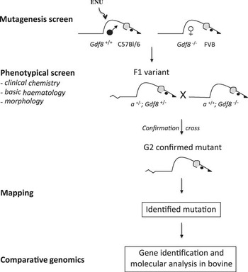
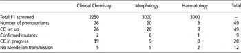
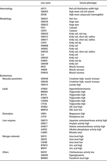
The strategy of our ENU-sensitised screen is shown in Figure 1. Since the beginning of our project, a total of 136 C57BL/6J mice were injected with 90 mg/kg ENU in three weekly doses. Thirty ENU males (22%) were excluded because they were sterile or the sterility period was <65 days, and therefore it would not ensure that the F1 offspring would derive from ENU-exposed spermatogonial stem cells. By breeding the ENU-injected males with Gdf8 −/− female mice, we generated about 3000 F1 mice that underwent the phenotyping workflow. We identified 48 abnormal phenotypes that have been successively put in confirmation cross. Nine confirm the Mendelian inheritance of the phenotype, 28 are still under investigation, and 11 do not transmit the phenotype or died before transmission (Table 1). At present, we focus our study on seven lines. Incorporation of two different inbred strains in our screen allows efficient mapping of the C57BL/6J mutagenised allele possible by subsequent crosses to FVB.
Design of the sensitised genetic screen. To identify modifier mutations that interact with the Gdf8 signalling pathway, we used myostatin knockout mutant mice (denoted by Gdf8 −/− allele). The ENU-treated C57BL/6J wild type males were bred with Gdf8 −/− females (FVB genetic background). The F1 progeny was subjected to a defined phenotypic screen. Interesting F1 variants were tested for inheritance of the phenotype by backcrossing to Gdf8 −/− animals. G2 animals were then phenotyped. ENU mutation is marked as ‘a’ locus; ENU = N-ethylnitrosourea.

Inheritance testing of F1 animals with abnormal phenotypes CC

CC = confirmation cross.
Mutant lines generated in Gdf8 modifier dominant screen

Gdf8 mutant phenotype
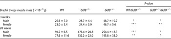
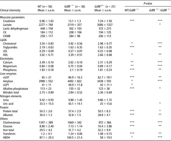
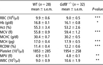
We characterised the phenotype of adult heterozygous and homozygous Gdf8 knockout mice in detail to be able to identify ENU-modified phenotypes from the original Gdf8 −/− ones. As described by McPherron et al. (Reference McPherron, Lawler and Lee1997), Gdf8 −/− mice show a large and widespread increase in skeletal muscle mass, resulting from a combination of muscle cell hyperplasia and hypertrophy. This phenotype is more obvious in males than in females. In heterozygous Gdf8 +/− mice, we observed an age-related hypermuscular phenotype. Until 5 months old, Gdf8 +/− animals are not distinguishable from the wild type, and subsequently they have a greater muscle development, although less pronounced than in homozygous Gdf8 −/− mice (Table 2). Both genotypes were analysed for clinical chemistry and basic haematology parameters. Out of the 26 clinical chemical parameters, 17 were significantly different in Gdf8 −/− animals compared to wild type littermates, including 10 with a P-value <0.001 (Table 3). It is known that creatinine and glucose levels in blood rise under certain circumstances, such as muscle mass increase. As expected, higher blood levels of creatinine and glucose were observed in Gdf8 −/− mice. Plasma alkaline phosphatase activity and phosphorus content increased in homozygous animals compared to their wild type littermates. Triglyceride concentration significantly decreased in both Gdf8 −/− and Gdf8 +/− mice. All these differences were taken into account when screening for phenovariants in our sensitised screen. In addition, a significant lower number of red blood cells were found in Gdf8 −/− mice compared to wild type. This change is associated with higher mean corpuscular values (Table 4).
Muscle development in Gdf8 knockout mice

WT = wild type.
Triceps muscles from 3- or 20-week-old mice were collected and weighed. Each value (n = 5) represents the mean ± s.e.m.
P-value from two-sample t-test: ***P < 0.001; **P < 0.01; *P < 0.05.
Comparison of clinical chemistry parameters between heterozygous Gdf8 +/− homozygous Gdf8 −/− and WT mouse littermates

WT = wild type; CK = creatine kinase; CKMB = creatine kinase MB; LDL = low density lipoprotein; HDL = high density lipoprotein; sGOT = serum glutamooxaloacetate transferase; sGPT = serum glutamopyruvate transferase; HBDH = hydroxybutyrate deshydrogenase.
P-value from two-sample t-test: ***P < 0.001; **P < 0.01; *P < 0.05.
Comparison of haematological parameters between homozygous Gdf8 −/− mice and WT littermates

RBC = red blood cells; Hb = haemoglobin; Hct = haematocrit; MCV = mean corpuscular volume; MCHC = mean corpuscular haemoglobin concentration; MCH = mean corpuscular haemoglobin; RCDW = red cell distribution width; MPV = mean platelet volume; WBC = white blood cells.
P-value from two-sample t-test: ***P < 0.001; **P < 0.01; *P < 0.05.
Skeletomuscular phenotypes
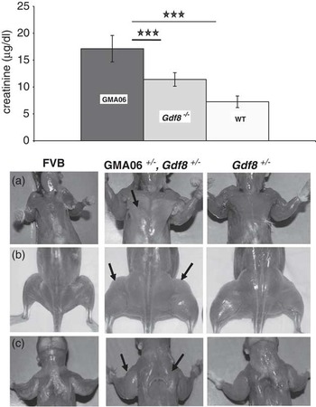
The seven mutant lines of major interest showed either altered creatinine levels (GMA06 and GMA45) or skeletal defects (Cut, GMA11, GMA12, GMA24 and GMA35). All the other mutant lines were cryopreserved. In the GMA06 mutant line, animals present high levels of plasma creatinine associated with a hypermuscular phenotype (Figure 2). GMA06 males weigh approximately 43% more than control FVB or FVB × C57BL/6J wild type mice and 21% more than FVB Gdf8 −/− mice. This weight difference is less pronounced in GMA06 females compared to FVB Gdf8 −/− mice (∼15%). We carried out a preliminary rough mapping of the mutation in GMA06. The identified mutation shows a linkage to the chromosome 13 in an interval of 8 cM, in between D13Mit288 and D13Mit76 markers. The GMA45 mutant line is still under investigation. A more detailed phenotypic and molecular analysis may reveal parallelisms with the former line.
Characterisation of the GMA06 phenotype. Top: plasmatic creatinine levels from homozygous Gdf8 GMA06 mutant, wild type and homozygous Gdf8 −/− mice. Bottom: increased skeletal muscle mass in GMA06 mutant compared to wild type and homozygous Gdf8 −/− mice. (a) pectoral muscles (b) lower limb (c) upper limb and neck of skinned animals.

The four lines Cut, GMA11, GMA12 and GMA35 display different malformations of the skeleton such as fusion of sacral or caudal vertebrae and absence of several lumbar vertebrae. All mice exhibit variable expressivity in tail phenotypes, ranging from kinked tails of various degrees, to short tails and even no tails. An interesting observation in this study was provided by genotype–phenotype correlation in GMA11 and GMA12 mice. Their phenotype is always more severe when mice are homozygous at the Gdf8 locus. Haematological and clinical chemistry parameters did not vary in these four lines when compared to controls.
The GMA24 mutant mice display reduced phosphorus levels in blood by 38% in females and 53% in males. No other change was observed in this mutant line.
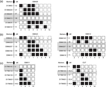
We started to map the mutations and identify a region of 18 cM and 29 cM on proximal chromosome 17 for the Cut and GMA11 lines, respectively. We detected linkage to regions on chromosome 9 for GMA12 and GMA24. Subsequent genotyping of mutant animals in these regions localised GMA12 to a 29 cM interval flanked by D9Mit129 and D9Mit192 markers and to a 18 cM region from centromere to D9Mit205 for GMA24 (Figure 3).
Mapping. For each identified line, mutant original C57BL/6J × FVB F1 mutant mice (carrying heterozygous mutations induced by N-ethylnitrosourea and a mutated allele for Gdf8) were backcrossed to Gdf8 −/− FVB mice. Genomic DNA was isolated from the backcross progeny for genetic analyses. Genome-wide scanning with 60 microsatellite markers showed linkages between (a) GMA06 mutation and chromosome 13, (b) GMA24, GMA12 mutations and chromosome 9, (c) GMA11, CUT mutations and chromosome 17. No other regions of the genome provided evidence of linkage. Markers are shown with their respective positions (cM) on the chromosomes. The black boxes represent FVB alleles and the white ones correspond to C57BL/6J × FVB alleles. The number of progeny that inherited each haplotype is given below the boxes. The greyed box indicates the chromosomal interval bearing the mutation.

Discussion
The current investigation focused on the development of an alternative approach to QTL mapping and its contribution to identify regions or genes controlling skeletomuscular development in cattle. QTL strategy is incontestably very powerful (Boichard et al., Reference Boichard, Grohs, Bourgeois, Cerqueira, Faugeras, Neau, Rupp, Amigues, Boscher and Leveziel2003; Abe et al., Reference Abe, Saburi, Hasebe, Nakagawa, Kawamura, Saito, Nade, Misumi, Okumura, Kuchida, Hayashi, Nakane, Mitsuhasi, Nirasawa, Sugimoto and Kobayashi2008) but extremely cumbersome and costly when dealing with large animals such as ruminants. Although recent developments in livestock genomics have revolutionised the mapping and the identification of QTL, the fine mapping remains a real challenge. The limiting step is not the number of genetic markers on a map, nor the knowledge of the DNA sequence of the detected region, but the functional properties of a given DNA segment. Thus, our goal is to get genomic and functional information on a specific DNA segment in other species, and especially in mouse, helping to dissect the exact molecular mechanism of the QTL. Our modifier screen is based on the identification of mutations directly or indirectly interfering with the Gdf8 pathway altered in the cattle ‘double-muscling’ phenotype. Gdf8 plays a crucial role during myogenesis but also in bone development and morphogenesis, as it has recently been revealed (Hamrick et al., Reference Hamrick, Shi, Zhang, Pennington, Thakore, Haque, Kang, Isales, Fulzele and Wenger2007; Kellum et al., Reference Kellum, Starr, Arounleut, Immel, Fulzele, Wenger and Hamrick2009; Elkasrawy and Hamrick, Reference Elkasrawy and Hamrick2010). In this genetic context, we identify a greater number of mutations affecting the skeletomuscular system than in a wild type setting (www.helmholtz-muenchen.de/en/ieg/group-functional-genetics/enu-screen/mutants-generated/index.html; www.mousegenome.bcm.tmc.edu/ENU/ENUMutantSources.asp). The subtle differences that we have found show the power of our strategy. The myostatin gene is located on chromosome 1. None of our identified mutations lies on this chromosome, showing that we have isolated dominant mutations acting with the Gdf8 locus. Fine mapping is still in progress, and does not allow us to discriminate between a dominant mutation affecting an already known molecular partner of Gdf8 or an unknown gene directly or indirectly interacting with Gdf8. The GMA06 and GMA45 lines are of high interest. Although these lines are currently under investigation, our preliminary phenotypic characterisation show that the hypertrophy in GMA06 mutants is more pronounced than in Gdf8 knockout mice, and even more marked if the GMA06 mice are on a Gdf8 −/− genetic background. Further analyses are required to dissect the underlying mechanisms of these differences. Many studies have detected bovine QTL for both dairy and beef traits. Although some QTL for growth and carcass traits have been identified on chromosome 20, the ortholog of mouse chromosome 13, these QTL only show either a suggestive level of linkage or a large interval of linkage (http://www.genome.iastate.edu/cgi-bin/QTLdb/index; http://genomes.sapac.edu.au/bovineqtl/). Fine mapping of these regions on BTA20 is still needed. However, once the role of a highlighted gene will be validated in our mouse model, the corresponding orthologous region in cattle will be defined based on the bovine sequence. It is also interesting to note that we found in our screen a large number of mutants with skeletal defects. These morphological abnormalities are more severe in homozygous Gdf8 −/− mice, consistent with the role of Gdf8 in bone development (Hamrick et al., Reference Hamrick, Shi, Zhang, Pennington, Thakore, Haque, Kang, Isales, Fulzele and Wenger2007; Kellum et al., Reference Kellum, Starr, Arounleut, Immel, Fulzele, Wenger and Hamrick2009). The mechanisms by which myostatin regulates bone formation are not well understood, but it is clear that myostatin has direct effects on the proliferation and differentiation of mesenchymal stem cells (Rebbapragada et al., Reference Rebbapragada, Benchabane, Wrana, Celeste and Attisano2003; Artaza et al., Reference Artaza, Bhasin, Magee, Reisz-Porszasz, Shen, Groome, Meerasahib and Gonzalez-Cadavid2005; Guo et al., Reference Guo, Flanagan, Jasuja, Kirkland, Jiang and Bhasin2008). Our new mouse models might help to unravel the exact role of myostatin in regulating osteogenesis. The improvement of the livestock is a basic issue, by itself vital. It will ensure a safer and healthier food supply. It is undeniable that the increased understanding of gene's function through mutagenesis programmes in the mouse is bringing a wealth of information that, when combined with comparative genomics, can accelerate our knowledge of the bovine genome. Finally, our approach might give clues to the origin of the diversity of the double-muscling phenotype. On the basis of the similar phenotype induced by the Gdf8 knockout in the mouse, our sensitised screen will identify modifiers of the muscular phenotype in a pure genetic background.
Acknowledgements
We are indebted to Professor L. Grobet for kindly providing the Gdf8 mice. We thank K. Pasquier, S. Desforges for help with animal manipulation. We are grateful to A. Germot and D. Petit for critical reading of the manuscript. We sincerely thank Professor R. Julien for his continuous support and interest in our study. This study was supported by the French National Institute for Agricultural Research and by the Limousin Regional Council.