Introduction
Osteomas of the temporal bone are benign tumours that usually involve the external auditory canal. Middle-ear osteomas are rare, and generally solitary and small. The male-to-female ratio is 2:1, with 70 per cent of affected patients aged under 40 years. Middle-ear osteomas can be asymptomatic, but a conductive hearing loss can be observed in the majority of cases.
This paper presents an extremely rare case of malleoincudal osteoma that led to conductive hearing loss despite an unusually normal otomicroscopic appearance, and it highlights the usefulness of costal cartilage for ossicular chain reconstruction after tumour removal. The authors assert that all procedures contributing to this work complied with the ethical standards of the relevant national and institutional guidelines on human experimentation (Comitato Etico Valpadana).
Case report
A 37-year-old woman presented with a 2-year history of progressive, right-sided hearing loss. The patient denied previous otitis media, trauma, otological surgery or a family history of otosclerosis.
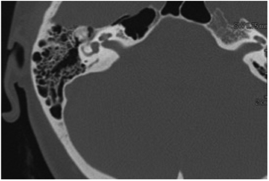
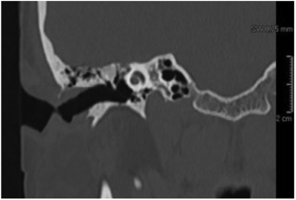
Physical examination revealed a normal tympanic membrane. Pure tone audiometry showed a right-sided conductive hearing loss. The pre-operative air–bone gap was 45 dB HL. Unenhanced high-resolution computed tomography (CT) of the temporal bone revealed a right-sided epitympanic mass arising from the malleus head and contiguous with the incus (Figures 1 and 2).
Axial computed tomography scan showing the right-sided malleo-incudal osteoma.

Coronal computed tomography scan showing the osteoma arising from the malleus head.

The patient underwent a closed mastoido-epitympanectomy under general anaesthesia (23 April 2020). The middle ear was inspected; this revealed that the malleus and incus were fixed. The incudostapedial joint was divided, demonstrating a mobile stapes. A mastoido-epitympanectomy was performed, revealing a bony mass arising from the malleus, extending to the incus, and fixing the malleus and incus to the tegmen.
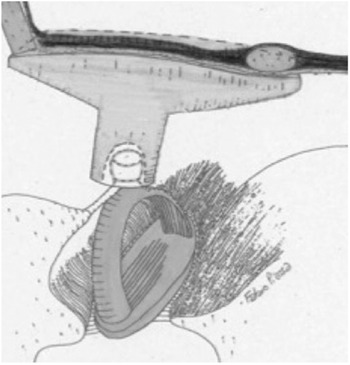
After dividing the neck of the malleus, the malleus head and the incus with associated malleoincudal osteoma were removed. A small area of the tegmen was drilled to ensure total removal of the lesion. The tegmen was repaired with bone pâté, and ossiculoplasty was performed using allogenic costal cartilage. A T-shaped cartilage partial ossicular replacement prosthesis (PORP) was sculpted. At the end of the shaft, an indentation was created with a diamond burr in order to accommodate the capitulum of the stapes. The head of the PORP was placed in contact with the tympanic membrane and the malleus handle (Figure 3).
Cartilage partial ossicular replacement prosthesis: an indentation is created in order to accommodate the capitulum of the stapes.

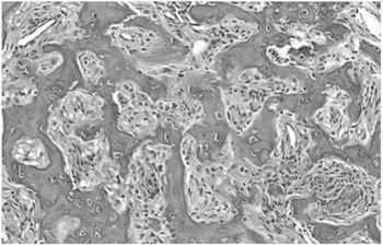
Inspection of the gross specimen revealed a firm mass arising from the malleus head. Histological evaluation of the specimen showed findings consistent with a diagnosis of malleoincudal osteoma (Figure 4). The post-operative course was uneventful. At one-month follow up, the air–bone gap was 15 dB HL.
Histopathological findings showing trabeculae of lamellar bone and intertrabecular fibrovascular tissue. (H&E; ×10)

Discussion
Middle-ear osteomas are very rare entities. Usually, they are solitary, but multiple middle-ear osteomas have been reported in the literature.
Osteomas of the temporal bone often arise along the tympanomastoid or tympanosquamous suture line. Yoon et al.Reference Yoon, Yoon and Lee1 reviewed 34 reported middle-ear osteomas and added 2 cases: 5 osteomas arose from the incus and 3 from the malleus. Other sites of involvement were: the pyramidal process, facial nerve canal, aditus, epitympanum, promontory, hypotympanum, lateral semicircular canal, posterior-inferior middle ear, annulus and cochleariform process. In 1989, Milroy et al.Reference Milroy, Phelps, Michaels and Grant2 reported a middle-ear osteoma that was inseparable from the incus. In 1994, RamadanReference Ramadan3 reported an osteoma arising from the malleus: the osteoma could be separated from the malleus before excision.
Histologically, osteomas show abundant fibrovascular channels and trabeculae of lamellar bone. On high-resolution CT, osteomas usually appear as a circumscribed bony mass. Less frequently, the signal density can be soft tissue.
Usually, osteomas are slow-growing tumours. Arensburg et al.Reference Arensburg, Belkin and Wolf4 observed that middle-ear osteomas tend to be small, and size tends to remain stable. Therefore, we believe that the wait-and-see policy can be adopted for small asymptomatic middle-ear osteomas; however, surgical treatment is preferred in cases of significant conductive hearing loss.
• Middle-ear osteomas can be asymptomatic, but conductive hearing loss is observed in most cases
• Otoscopic examination often reveals a white mass behind the tympanic membrane
• Middle-ear osteoma should be considered in unilateral and progressive conductive hearing loss patients with normal otoscopy and no relevant history
• The wait-and-see policy can be adopted for small, asymptomatic middle-ear osteomas; surgical treatment is preferred in significant conductive hearing loss cases
• Allogenic costal cartilage prostheses are excellent for ossicular chain reconstruction, as costal cartilage is stiffer than tragal or conchal cartilage
Ossicular chain reconstruction can be performed at the time of tumour removal. When autologous ossicles are not available, cartilage can be used for ossicular chain reconstruction.Reference Yung5 Our preference is allogenic costal cartilage, which is stiffer than tragal or conchal cartilage, and more suitable for ossiculoplasty.Reference Quaranta, Fernandez-Vega Feijoo, Piazza and Zini6 Chole and KimReference Chole and Kim7 reported no extrusions in a large series of ossiculoplasties performed with pre-sculpted banked cartilage. Moreover, unlike ossicles, cartilage does not stick to the scutum, the facial nerve or the promontory.Reference Farrior and Nichols8 The safety of homografts has been debated for a long time.Reference Glasscock, Jackson and Know9 More recently, Lubbe and FaganReference Lubbe and Fagan10 revisited the risk involved in using homograft ossicles in otological surgery.
Conclusion
This unique case report emphasises consideration of a middle-ear osteoma in cases of unilateral and progressive conductive hearing loss with a normal otomicroscopic appearance in patients with no history of ear infection, trauma or prior surgery, and with no family history of hearing loss.
Based on 30 years of experience with allogenic cartilage prostheses, the authors believe that a partial cartilage ossicular replacement prosthesis (PORP) is preferable when autologous ossicles are not available because they are involved by the tumour. Unlike synthetic materials, the extrusion rate of cartilage is low. Moreover, unlike ossicles, cartilage does not fix to the bone. Cartilage prostheses can be beneficial for patients suffering from conductive hearing loss associated with a malleoincudal osteoma, as they are associated with good and stable post-operative hearing and a low incidence of failures.
Acknowledgement
The authors wish to express their deep gratitude and sincere thanks to Antonella Piazza (an English-speaking teacher) for her substantial contribution to this work.
Competing interests
None declared