Introduction
Brazil has been the worldwide leader in the in vitro production (IVP) of bovine embryos since 2013, as approximately 70% of these embryos are obtained from Brazil (IETS, 2015). The use of IVP in Brazil is unique, with strong impetus for replacing multiple ovulation and embryo transfer (MOET) by ovum pick up (OPU)/IVP (Pontes et al., Reference Pontes, Melo-Sterza, Basso, Ferreira, Sanches, Rubin and Seneda2010). One of the main reasons for this situation is that Bos indicus (zebu) cattle, the largest herd in the country, have more ovarian follicles and more oocytes recovered by OPU than Bos taurus cattle (Pontes et al., Reference Pontes, Nonato-Junior, Sanches, Ereno-Junior, Uvo, Barreiros, Oliveira, Hasler and Seneda2009). In this system, one of the greatest challenges is to overcome geographical distance between oocyte donor farms and IVP laboratories because the conditions during oocyte transport influence both the rate and quality of blastocysts at the end of the process (Marinho et al., Reference Marinho, Untura, Morotti, Moino, Rigo, Sanches, Pontes and Seneda2012). In this sense, the use of a system, partially or fully portable, for in vitro maturation (IVM), fertilization (IVF) and culture (IVC) would be highly beneficial to the IVP bovine embryo industry.
Although the IVP of bovine embryos has developed considerably in recent decades, embryos are still susceptible to a wide range of stressful situations, including inadequate culture medium and culture conditions, such as pH (Lane, Reference Lane2001), temperature (Sugiyama et al., Reference Sugiyama, McGowan, Phillips, Kafi and Young2007) and oxygen tension (Corrêa et al., Reference Corrêa, Rumpf, Mundim, Franco and Dode2008). The oxygen tension most frequently used in IVP systems is often higher (20% O2) than that found in the female reproductive tract (3–9% O2) and it is believed that this high tension induces excessive generation of reactive oxygen species (ROS), such as hydrogen peroxide (H2O2), hydroxyl radical and peroxyl radical, which can cause deleterious effects on the embryo development (Thompson et al., Reference Thompson, Simpson, Pugh, Donnelly and Tervit1990; Martín-Romero et al., Reference Martín-Romero, Miguel-Lasobras, Domínguez-Arroyo, González-Carrera and Álvarez2008). Additionally, beneficial effects of low oxygen concentration on in vitro developmental competence of oocytes have been reported in cattle (Yuan et al., Reference Yuan, Van Soom, Coopman, Mintiens, Boerjan, Van Zeveren, de Kruif and Peelman2003), mouse (Rinaudo et al., Reference Rinaudo, Giritharan, Talbi, Dobson and Schultz2006) and humans (Ciray et al., Reference Ciray, Aksoy, Yaramanci, Karayaka and Bahceci2009).
Therefore, the aim of this study was to evaluate the quantitative–qualitative production of IVP bovine embryos under three incubation systems: conventional bench (high O2 tension), mini bench (low O2 tension) and portable (low O2 tension). In addition, for each incubation system, ROS levels were evaluated at the different stages: post-IVM oocytes, cleaved embryos (day 2) and blastocysts (day 7).
Materials and methods
Chemicals
All chemicals used in this study were purchased from Sigma-Aldrich Chemicals Co. (St. Louis, MO, USA) unless otherwise indicated.
Experimental design
Bovine oocytes/embryos were subjected to IVM, IVF and IVC in three experimental groups or incubators: conventional bench – CONV (Thermo-Forma, Thermo Fischer, Waltham, MA, USA) with high oxygen tension (5% CO2, 20% O2 in air), mini bench – MINI (Eve, WTA, Cravinhos, Brazil) with low oxygen tension (5% CO2, 5% O2, 90% N2) and portable – PORT (LabMix, WTA) also with low oxygen tension.
Oocyte collection and IVM
Ovaries obtained at a local slaughterhouse were transported to the laboratory in a thermos box containing saline solution (NaCl 0.9% w/v) at 34ºC. Ovaries were washed in pre-warmed fresh saline and cumulus–oocyte complexes (COCs) from follicles of 2–8 mm were aspirated with an 18-gauge needle connected to a syringe. COCs were selected under a stereomicroscope (SMZ800, Nikon Co., Tokyo, Japan) and only COCs enclosed in three compact layers of cumulus cells and homogeneous cytoplasm were selected. For IVM, COCs were washed in HEPES-buffered TCM-199 (H-TCM) medium and distributed into groups of 50 COCs in wells containing 500 μl of maturation medium which consisted of TCM-199 supplemented with 10% (v/v) fetal bovine serum (FBS) (Gibco, Grand Island, NY, USA), 20 μg/ml follicle stimulating hormone (FSH) and luteinizing hormone (LH) (Pluset; Hertape-Calier, Spain), 0.2 mM sodium pyruvate, 0.1 mM cysteamine, 1× antibiotic antimycotic, 10 ng/ml epidermal growth factor (EGF), 1 μg/ml estradiol and 1 mM l-glutamine under mineral oil. COCs were incubated at 38.5°C for 22 h in each experimental group (incubator).
Nuclear maturation
For nuclear maturation analysis, oocytes of each experimental group were denuded, washed in phosphate-buffered saline (PBS) supplemented with 0.4% bovine serum albumin (BSA) and stained with 10 μM Hoechst 33342 reagent in PBS/BSA for 15 min at room temperature. The stained oocytes were washed in PBS/BSA and mounted with glycerol between a slide and coverslip and observed under an optical microscope (Eclipse E400, Nikon) equipped with UV-2E/C filter (330–380 nm). Oocytes were classified for nuclear configuration in germinal vesicles, and metaphase I and metaphase II oocytes to obtain the maturation rate (MII oocytes) for each experimental group.
IVF and IVC
Frozen semen was thawed in a water bath at 37°C for 25 s. Sperm was then centrifuged three times (700 g for 25 min, 500 g for 10 min or 400 g for 10 min) in Brackett & Oliphant (BO) medium (Brackett & Oliphant, Reference Brackett and Oliphant1975) supplemented with 5 mM caffeine and 20 IU/ml heparin (Calbiochem, San Diego, CA, USA). Sperm was diluted with BO medium containing 20 mg/ml BSA to achieve a concentration of 20 × 106 spermatozoa/ml. Matured COCs were washed with H-TCM and incubated in 100 μl droplets of sperm suspension covered with mineral oil at 38.5°C in each incubator. After 6 h of incubation, presumptive zygotes were denuded by vortexing, washed in H-TCM and transferred to wells containing 500 μl synthetic oviduct fluid (SOF) covered with mineral oil. Embryos were cultured for 8 days at 38.5°C in each incubator. On days 2 (D2), 7 (D7) and 8 (D8) after IVF, the percentages of cleaved, blastocyst and hatching, were evaluated, respectively. Similar to that performed for nuclear maturation, expanded blastocysts were stained (Hoechst 33342) and the total number of cells counted.
Assessment of ROS levels
From each experimental group, samples of post-IVM oocytes and embryos (D2 and D7) were evaluated for ROS concentration using the fluorescent probe 6-carboxy-2′,7′-dichlorodihydrofluorescein diacetate (H2DCFDA). Oocytes/embryos were incubated for 30 min in 10 μM H2DCFDA dissolved in DMSO in 50 μl droplet of PBS supplemented with 0.1% (w/v) polyvinyl alcohol (PBS/PVA) at 38.5°C in a dark, humidified and 5% CO2 atmosphere (Yang et al., Reference Yang, Hwang, Kwon, Kim, Choi and Oh1998; Wang et al., Reference Wang, Tian, Zhang, He, Ji, Li, Tan and Liu2014). After incubation, structures were washed and placed into four-well cell culture plates containing 50 μl PBS/PVA. Fluorescence emitted was captured using GFP filter (450–490 nm) 2 s after exposure to UV light fluorescence using an inverted microscope (Eclipse TE2000, Nikon). The fluorescence intensities expressed were analyzed in arbitrary fluorescence units (AFUs) using the Infinity Analyze software (Lumenera Co., Ottawa, Canada).
Statistical analyses
Before analysis, data were tested for normality using the Shapiro–Wilk test. Rates of matured oocytes, cleavage, expanded blastocysts, hatching and total cells numbers were evaluated by one-way analysis of variance (ANOVA). ROS levels were analyzed using the Kruskal–Wallis test. Analyses were performed using the GraphPad InStat 7.0 software, at the significance level of 5%.
Results
After IVM for 22 h, 201 oocytes were assessed for nuclear maturation. Oocytes in metaphase I were observed in 12.3% (10/81), 14.8% (9/61) and 22.0% (13/59) for CONV, MINI and PORT groups, respectively. The majority of oocytes reached MII stage in all groups, with 70.4% (CONV), 50.8% (MINI) and 57.6% (PORT). There were no differences among treatments (P>0.05).
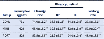
Concerning embryo production, the results obtained from 19 replicates are summarized in Table 1. After IVF, 1889 presumptive zygotes were cultured in the different incubation systems. Concerning the cleavage rate, no significant difference was observed between CONV and MINI groups (74.0 vs. 65.0%, P>0.05). However, the CONV group was superior to PORT (74.0 vs. 59.5%, P<0.05). Regarding the blastocyst rate, both on D7 and D8, the same pattern was observed. Finally, regarding the hatching rate, MINI and PORT groups had the highest values (50.0 vs. 46.0, P>0.05). The number of cells per expanded blastocyst was verified and no significant difference (P>0.05) was observed among the three experimental groups: 122.2±29.1 (CONV), 145.9±57.1 (MINI) and 99.0±49.5 (PORT).
Effect of incubation system (conventional – CONV, mini bench – MINI and portable – PORT) on development of in vitro produced bovine embryos

Different superscript letters within same column denote statistical difference (P<0.05) between groups. Data are presented as mean percentage±standard deviation (SD). Cleavage and blastocyst rates were calculated from the total presumptive zygotes. Hatching rate was calculated from the number of blastocysts at D8.
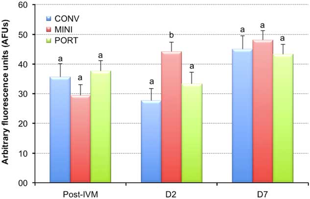
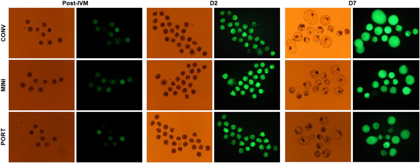
Randomly selected from each experimental group, 130 post-IVM oocytes, 148 cleaved embryos (D2) and 128 blastocysts (D7) were stained and the fluorescence emitted was verified and expressed in AFUs. The main effects of the incubation system on the intracellular ROS content are demonstrated in Figs 1 and 2. Regarding the post-IVM oocytes, no significant difference (P>0.05) was observed among the experimental groups, which showed values varying between 29.4 and 37.6 AFUs. Concerning the ROS levels in D2 embryos, a significant difference was observed (P<0.05) between MINI group (44.2±3.1) in comparison with CONV (27.7±3.7) and PORT (33.3±3.2) groups. Finally, for blastocysts, no statistical difference (P>0.05) was observed, but at this embryonic development stage the highest mean values of ROS levels were verified, varying between 43.3 to 48.1 AFUs (Fig. 1). This finding is also subjectively confirmed when observing the blastocysts fluorescence images captured at D7 of culture (Fig. 2).
ROS levels [mean±standard error of the mean (SEM)] in post-IVM oocytes and embryos (at D2 and D7) cultured in different incubation systems (conventional – CONV, mini bench – MINI and portable – PORT) and stained with H2DCFDA. Different letters denote statistical difference (P<0.05).

Representative images (×100 magnification) of post-IVM oocytes and embryos (at D2 and D7) cultured in different incubation systems (conventional – CONV, mini bench – MINI and portable – PORT) and stained with H2DCFDA. Bright field (left column) and fluorescence (right column) are shown for each developmental stage. The green fluorescence indicates arbitrary fluorescence units (AFUs) of ROS levels.

Discussion
This study sought to find solutions to two specific needs of the IVP bovine embryos industry. Firstly, due to great distances between oocyte donor farms and laboratories, to propose a totally portable incubation system to carry out the several steps of IVP (IVM, IVF and IVC). Secondly, to verify the efficiency of these different steps when the structures (oocytes/embryos) are subjected to incubation systems with low or high oxygen tension. The supraphysiological concentration (high oxygen tension) has been associated with increased concentrations of ROS, which alters the culture environment, therefore hindering the embryo development (Fatehi et al., Reference Fatehi, Roelen, Colenbrander, Schoevers, Gadella, Beverst and van den Hurk2005). ROS levels were verified during the several steps and for each incubation system.
In embryo culture, the high volume conventional culture incubators were adapted and equipped with humidification to minimize both evaporative changes and osmolarity of culture medium. Conversely, there is currently a worldwide trend in human-assisted reproduction laboratories to use mini-incubators in which the final CO2 tension is not regulated by the incubator but premixed in the gas supply company cylinder.
In these pieces of equipment, the small chamber volume allows a rapid recovery of the initial culture conditions and, consequently, an economy in the gas mixture (Lee et al., Reference Lee, Grazi and Seifer2013).
The results obtained in the present study indicated that the nuclear oocytes maturation was not affected by the incubation system. In our study, the maturation rate varied between 50.8 and 70.4%, which was lower than the rates verified in other studies with bovine oocytes, which describe rates between 80 and 90% (Diógenes et al., Reference Diógenes, Guimarães, Leme, Maurício and Dode2017). However, our results are consistent with previous studies in which it was observed that there was no difference in IVM rate of bovine oocytes produced under 20% and 5% of oxygen tension (Pereira et al., Reference Pereira, Machado, Costa, Serapiao, Viana and Camargo2010; Mingoti et al., Reference Mingoti, Castro, Méo, Barretto and Garcia2011).
Oocyte transportation in incubators with an uncontrolled gaseous atmospheric tension can lead to alterations in the pH of the transport medium and increased oxidases activity, therefore culminating in increased ROS production (Guemra et al., Reference Guemra, Santo, Zanin, Monzani, Sovernigo, Ohashi, Verde Leal and Adona2014). Apparently, the portable incubation system used in our study (with a controlled gaseous atmospheric tension) was efficient in avoiding these deleterious effects, because the oocyte maturation rate and ROS levels were similar to the other groups. Ambrogi et al (Reference Ambrogi, Dall’Acqua, Rocha-Frigoni, Leão and Mingoti2017) also avoided these negative effects when the transport-maturation medium was supplemented with an antioxidant mixture.
Except for the hatching rate, all other parameters of embryo production, i.e., cleavage (D2) and blastocyst (D7-D8) rates, were lower in the PORT group. These results suggest that the action of PORT system had on oocyte maturation was not enough for a longer culture period (seven to eight days). Conversely, the CONV and MINI systems presented the same efficiency for several parameters. The effects of in vitro oxygen tension on embryonic development are still controversial. Although oxygen concentration in the mammals’ oviduct varies between 2% and 9% (Fischer & Bavister, Reference Fischer and Bavister1993), several studies find success culturing at 20% (Thompson et al., Reference Thompson, Simpson, Pugh, Donnelly and Tervit1990; Oliveira et al., Reference Oliveira, Saraiva, Cruz, Mazeti, Oliveira, Lopes and Garcia2013). Studies find increased development when embryos are cultured in high oxygen (20%) tension (Mingoti et al., Reference Mingoti, Castro, Méo, Barretto and Garcia2011). In addition, the combination of oxygen tension and FBS supplementation plays a major role in the lipid profile of bovine embryos (Ferreira et al., Reference Ferreira, Saraiva, Catharino, Garcia, Gozzo, Sanvido, Santos, Lo Turco, Pontes, Basso, Bertolla, Sartori, Guardieiro, Perecin, Meirelles, Sangalli and Eberlin2010).
In our study, the ROS levels were assessed in post-IVM oocytes, cleaved embryos and blastocysts. Only for the cleaved embryos an effect was observed of the incubation system. At this stage, the embryos cultured in the MINI system had the ROS highest levels. Studies have implicated ROS in many aspects of cell growth and development, not only in cell death/senescence and are found in equilibrium in cells under normal physiological conditions. Low ROS concentrations are capable of mediating cellular functions through intracellular signalling steps activated by various stimuli (Khazaei & Aghaz, Reference Khazaei and Aghaz2017). The cells redox balance appears to be an important part of the development, such that non-toxic ROS concentrations, via alterations in redox balance, are considered to be a mechanism that regulates several cell functions, particularly in the preimplantation embryo (Harvey et al., Reference Harvey, Kind and Thompson2002). Under special conditions, such as embryos’ IVP, ROS production increases and results in decreased in vitro embryos’ development (Guérin et al., Reference Guérin, El Mouatassim and Menezo2001). However, the highest levels of ROS observed in D2 embryos cultured in the MINI system did not cause negative effects on the later development of these embryos until the blastocyst stage.
Finally, portable or semi-portable systems have some advantages in comparison with expensive incubators and gas mixtures for IVM, IVF and IVC. Under field conditions and as a method for gamete transportation under controlled gas atmospheres these systems may therefore be a valuable and low-cost alternative to conventional systems (Avery et al., Reference Avery, Melsted and Greve2000).
In conclusion, under our experimental condition, it was possible to conclude that: (i) the fully portable incubation system (PORT) was able to obtain IVP embryos, but not as efficient as the CONV system; and (ii) although it produced a high ROS level on D2 of culture, the intermediate system (MINI) was as efficient as the CONV system of incubation for blastocyst production. This option may be more economical for the in vitro produced bovine embryo industry.
Ethics statements
The authors assert that all procedures contributing to this work comply with the ethical standards of the guidelines on the care and use of experimental animals. This study was approved by the Ethics Committee on Animal Use of the State University of Ceará (CEUA, no. 4024452/2017).
Financial support
This work was supported by Conselho Nacional de Desenvolvimento Científico e Tecnológico (CNPq, Brasília, Brazil) and Fundação Cearense de Apoio ao Desenvolvimento Científico e Tecnológico (FUNCAP, Fortaleza, Brazil).
Conflict of interest
The authors hereby declare that there are no conflicting interests.