Introduction
Techniques have changed enormously since Robert Hungate (Reference Hungate1966) wrote his seminal monograph ‘Rumen microbiology’. Throughout his career, Hungate and his contemporaries advanced their science via painstaking observation alongside techniques that, although revolutionary in their time, now sometimes appear weak and even misleading. They relied heavily on manual dexterity and investigated a relatively small number of cultivated microbes. The power of molecular methods in analysing a microbial ecosystem is staggering in comparison. Present technology relies mostly on sequences within or close to small subunit ribosomal RNA (rRNA) genes. However, as functional genomics and microarrays gather momentum, it seems that other quantum leaps may be possible. The aim of this review is to assess briefly our present understanding of microbial diversity in two gut ecosystems, namely the human intestine and the rumen, in the context of examples that illustrate how certain microorganisms, despite the wide genetic diversity, can uniquely occupy highly specific ecological/metabolic niches within the ecosystem, carrying out key reactions for the well-being of the host animal.
Microbial diversity in the human intestine and the rumen
It is salutary to reflect upon how little we understood about the microbial ecology of the human intestine until recently. The importance of gut fermentation in human health was established in the 1970s and 1980s (Hill, Reference Hill1983; Schiffman, Reference Schiffman1986), a perception that continues to gather pace. Yet, the bacterial species thought to predominate in the ecosystem were mainly Gram-negative, mostly belonging to the Bacteroides group, because, generally speaking, they were the ones that were recovered using the general-purpose media commonly in use (Savage, Reference Savage2001).
The fact that the true composition of the human intestinal flora was very different from being dominated by Bacteroides, Bifidobacterium and Lactobacillus spp. became apparent in the 1990s, when methods based on the analysis of 16S rRNA gene sequences began to become routine (Weisburg et al., Reference Weisburg, Barns, Pelletier and Lane1991). All bacteria, archaea and higher organisms synthesise protein in ribosomes, which are complexes of RNA and protein. The smaller subunit in bacteria is a particle that sediments at 16 Svedberg units in the ultracentrifuge – hence the rRNA associated with the small ribosomal subunit is known as 16S rRNA – and as protein synthesis is an activity that is essential for life throughout evolution, so the sequences tend to be conserved. Over evolutionary time, although the function has been conserved, the sequences have gradually become divergent, particularly in variable regions that may be less important to ribosome function. The degree of divergence can provide quantitative indications of how the far the organisms harbouring the different sequences have diverged from each other during evolution.
Suau et al. (Reference Suau, Bonnet, Sutren, Godon, Gibson, Collins and Dore1999) sequenced and characterised 284 clones of 16S rRNA genes from a single human faecal sample, finding 82 species, many (76%) of which had previously been unknown. Hayashi et al. (Reference Hayashi, Sakamoto and Benno2002) analysed 744 clones from three subjects and came to similar conclusions, namely that there were about 130 species of which 75% were unknown. More recently, the true diversity of the bacterial flora of the human intestine has become much clearer, the result of a much larger cloning and sequencing study on three human volunteers (Eckburg et al., Reference Eckburg, Bik, Bernstein, Purdom, Dethlefsen, Sargent, Gill, Nelson and Relman2005). Mucosal tissue was taken from six healthy mucosal sites during colonoscopy and from faeces collected 1 month later. In all, 13 355 gene sequences were analysed, by far the largest to date from the human intestine. The study, although a landmark, is imperfect, because of the small number of individuals involved and also because the samples were either of faeces or of mucosal samples taken during colonoscopy. Thus, the same in-depth information on the luminal population in the upper parts of the intestine is missing. Nevertheless, the results represent the best global view of the human intestinal microbiota available to date.
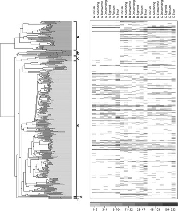
The sequences ordered themselves across the 16S rRNA phylogenetic tree in a manner that illustrated very clearly the numerical importance of the Firmicutes phylum compared with others (Figure 1). The Firmicutes includes genera such as Roseburia, Clostridium and Faecalibacter, which are the truly most predominant species of the human intestine, not the more easily cultivated Bacteroidetes, although the Bacteroidetes still represent a significant proportion of the total flora. Escherichia coli, the archetypal model intestinal bacterium beloved of biochemists and geneticists for generations, is a γ-Proteobacterium, a phylum that is almost insignificant in the overall scheme of bacterial diversity in this study.
Frequency and phylogenetic relationships between 16S rRNA clones obtained from three human volunteers. The Y-axis is a neighbour-joining phylogenetic tree containing one of each of the 395 phylotypes obtained from these individuals. a – Bacteroidetes; b – non-Alphaproteobacteria; c – Actinobacteria; d – Firmicutes; e – Fusobacteria; f – Alphaproteobacteria. Each column is labelled according to subject (A, B, C) and anatomical site. The abundance of the different sequences is indicated by the intensity of the gray-scale. From Eckburg et al. (2005) Science 308, 1635–1638. Reprinted with permission from AAAS.

The study by Eckburg et al. (Reference Eckburg, Bik, Bernstein, Purdom, Dethlefsen, Sargent, Gill, Nelson and Relman2005) illustrates some very important principles applying to gut microbiology. The numbers of different sequences are indeed very high. However, Eckburg et al. (Reference Eckburg, Bik, Bernstein, Purdom, Dethlefsen, Sargent, Gill, Nelson and Relman2005) estimated that there were 395 different phylotypes present in the three individuals – a large number, but by no means an infinite one. The word ‘phylotype’ is used because there is no real definition of species in bacteria that corresponds with that in higher life forms. Most authors make an arbitrary definition of what constitutes a bacterial species based entirely on sequence identity – usually 97% identity is used. However, there are arguments that a combination of sequence similarity and phenotypic properties should be used to distinguish the biotypes (Stackebrandt and Ebers, Reference Stackebrandt and Ebers2006). A second principle to be noted is that 80% of the sequences were from species that have not been cultivated. This is sometimes interpreted incorrectly to mean that the species cannot be cultivated. While this may be true, it is much safer to assume that they have not yet been cultivated, whether due to an inappropriate medium being used or some other factor. Given time and patience, one might expect many of these bacteria eventually to be cultivated, see below.
A third important general principle was established that the bacterial communities were more similar within individuals than between individuals. Indeed, many species were present in all samples taken from one individual but they were not present in others. Individual A had a small number of Bacteroidetes throughout the tract, in contrast to the others, who possessed a much larger number. Figure 1 illustrates the persistence within individuals of the Bacteroidetes. Other studies had demonstrated the principle that different individuals had very different flora composition (Zoetendal et al., Reference Zoetendal, Akkermans and De Vos1998; Suau et al., Reference Suau, Bonnet, Sutren, Godon, Gibson, Collins and Dore1999; Tannock et al., Reference Tannock, Munro, Harmsen, Welling, Smart and Gopal2000; Hayashi et al., Reference Hayashi, Sakamoto and Benno2002). There is, as some of the studies illustrate, a temporal persistence as well. Electrophoretic profiling of the rRNA sequences present indicate that differences between individuals persist for many weeks (Zoetendal et al., Reference Zoetendal, Akkermans and De Vos1998; Tannock et al., Reference Tannock, Munro, Harmsen, Welling, Smart and Gopal2000). Host factors are clearly of major importance in determining the flora present in the gut, although the precise mechanisms that cause the host to exert this effect are not really known (Hooper et al., Reference Hooper, Wong, Thelin, Hansson, Falk and Gordon2001).
The molecular ecology studies of the human intestinal flora have been complemented by similar studies carried out in the rumen (Whitford et al., Reference Whitford, Forster, Beard, Gong and Teather1998; Tajima et al., Reference Tajima, Aminov, Nagamine, Ogata, Nakamura, Matsui and Benno1999 and Reference Tajima, Arai, Ogata, Nagamine, Matsui, Nakamura, Aminov and Benno2000). These studies illustrated similar principles. Edwards et al. (Reference Edwards, McEwan, Travis and Wallace2004) drew all the sequence information then present in public-access databases, formulated the sequences together in a single phylogenetic tree and concluded that the number of phylotypes in the rumen was rather similar to the number determined by Eckburg et al. (Reference Eckburg, Bik, Bernstein, Purdom, Dethlefsen, Sargent, Gill, Nelson and Relman2005) in humans. A total of 341 phylotypes was identified, once again with the majority being Firmicutes at 54% of the total. The Bacteroidetes were relatively more abundant than in the human gut, at 40% of the total. The rumen sequences indicated that 89% of the sequences were most similar to species that had not been cultivated.
One might imagine that this wonderfully diverse type of ecosystem would have many different organisms performing the same function, and that there would be much duplication of activities in competing species. Generally speaking, this is true. Most species ferment a range of sugars, for example, competing in different ways to capture different oligosaccharides and sugars. However, there are certain metabolic niches that are occupied by a very small number of bacterial species. It might perhaps be expected that these metabolic activities would be relatively obscure, and not have a great impact on the fermentation or the host. However, there are a few examples where, spectacularly, this is not the case. The functions are essential for the well-being either of the entire microbial community or of the host animal.
Rumen cellulolytic bacteria – so important, so few species
Cellulose breakdown has been the force driving the evolution of ruminants. Their ability to use grasses and other fibre-containing plants efficiently sets them apart from even those animals which, via a hindgut fermentation, can effect a digestion that enables them to graze similar forages. The abundance of cellulose in the diet of most ruminants means that all the diverse species mentioned above have the opportunity to obtain energy from the polymer. It is remarkable how few possess the ability to digest the most recalcitrant, crystalline form of cellulose that forms the backbone of most forage fibres. Ruminal ciliate protozoa (Coleman, Reference Coleman1985 and Reference Coleman1986; Devillard et al., Reference Devillard, Bera-Maillet, Flint, Scott, Newbold, Wallace, Jouany and Forano2003) and anaerobic fungi (Akin and Borneman, Reference Akin and Borneman1990; Gordon and Phillips, Reference Gordon and Phillips1993) contribute to the process, but the bulk of the activity is due to only two bacterial genera, Fibrobacter and Ruminococcus (Chesson and Forsberg, Reference Chesson and Forsberg1997). Fibrobacter succinogenes is the main species from that genus found in the rumen (Stewart et al., Reference Stewart, Flint and Bryant1997). It is a high GC Gram-positive species, atypical of the most numerous Firmicutes and Bacteroidetes. Ruminococcus albus and Ruminococcus flavefaciens are the two most common cellulolytic ruminococci. They are phylogenetically much more similar to the majority of ruminal bacteria, being central in the Firmicutes phylum (Edwards et al., Reference Edwards, McEwan, Travis and Wallace2004).
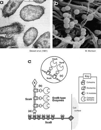
Genomic sequence information is now becoming available for several species of ruminal bacteria. In R. flavefaciens, 36% of the genome encodes glycosyl hydrolase genes (M. Rincon, personal communication), indicating that the release of sugars from oligo- and polysaccharides is of central importance to this species. However, the genome of the non-cellulolytic species, Clostridium proteoclasticum, contains more than 100 glycoside hydrolases (G. T. Attwood, personal communication), and around 80 have been identified in Prevotella ruminicola (M. Morrison, personal communication), which also is not cellulolytic. One key difference between the truly cellulolytic species and the others appears to be the spatial organisation of the fibre-digesting enzymic machinery. Remarkably, F. succinogenes and the Ruminococcus spp. appear to have evolved completely different strategies in this regard. Elegant enzymological and genetic investigations have revealed a cell surface-associated anchor-like structure that enables hydrolases to bind in close proximity to each other in a branched scaffoldin-based complex (Figure 2; Rincon et al., Reference Rincon, Ding, McCrae, Martin, Aurilia, Lamed, Shoham, Bayer and Flint2003). Presumably it is the close proximity of hydrolases of different specificity, held in place by dockerins, that enables R. flavefaciens to digest fibre efficiently. The genome sequence of F. succinogenes contains no dockerins, however (M. Morrison, personal communication). The way F. succinogenes appears to have evolved to cope with the digestion of a complex, insoluble substrate is they have an extraordinarily pleomorphic cell envelope, in contrast to the rigid cell envelope of R. flavefaciens (Costerton et al., Reference Costerton, Damgaard and Cheng1974; Latham et al., Reference Latham, Brooker, Pethipher and Harris1978). The hydrolases of F. succinogenes do not appear to associate with each other, but several of them anchor directly to the cell envelope (McGavin et al., Reference McGavin, Lam and Forsberg1990). The flexibility of the cell envelope then presumably affords them the ability to overcome the steric problems presented by the cellulose fibre.
Morphology of the ruminal cellulolytic bacteria (a) Fibrobacter succinogenes and (b) Ruminococcus flavefaciens. (c) Cellulosome structure in R. flavefaciens. Reproduced from Rincon et al. (Reference Rincon, Ding, McCrae, Martin, Aurilia, Lamed, Shoham, Bayer and Flint2003) with permission.

Biohydrogenation in the rumen
Unsaturated fatty acids, particularly linolenic acid (LNA; cis-9,cis-12,cis-15-18:3) and linoleic acid (LA; cis-9,cis-12-18:2), occur abundantly in forages and other ruminant feedstuffs, yet are present at low concentrations in meat and milk. Tissue lipids of ruminants have been known for a long time to be more saturated than those of non-ruminants (Banks and Hilditch, Reference Banks and Hilditch1931). As the consumption of saturated fats, dairy products and ruminant meats is often associated with an increased incidence of coronary heart disease in humans (Menotti et al., Reference Menotti, Kromhout, Blackburn, Fidanza, Buzina and Nissinen1999), the transformation of unsaturated fatty acids to saturated fatty acids, or biohydrogenation, in ruminants represents a major human health issue in societies that consume substantial amounts of ruminant products.
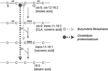
The biohydrogenation process has been known for a long time to occur in the rumen as the result of microbial metabolic activity (Shorland et al., Reference Shorland, Weenink and Johns1955). If ruminal biohydrogenation of unsaturated fatty acids can be controlled, it may be possible to improve the healthiness of ruminant meats and milk by increasing their unsaturated fatty acids composition in general and the n-3 fatty acids in particular (Scollan et al., Reference Scollan, Choi, Kurt, Fisher, Enser and Wood2001). Intermediates in the biohydrogenation of linoleic acid include conjugated linoleic acids (CLA, mainly cis-9,trans-11-18:2) (Figure 3). Dietary CLA are believed to confer potential health benefits in humans, including cancer prevention, decreased atherosclerosis, improved immune response and altered protein/energy metabolism (Kritchevsky, Reference Kritchevsky2000; Whigham et al., Reference Whigham, Cook and Atkinson2000). CLA are present at higher concentrations in ruminant products than in corresponding meats from non-ruminants or in vegetable oils (Chin et al., Reference Chin, Storkson, Ha and Pariza1992). Thus, we would wish to increase the flow of CLA from the rumen in order to enhance the CLA content of ruminant products. However, not only CLA flow is important: recent experiments suggest that dietary vaccenic acid (VA, trans-11-C18:1), the biohydrogenation product of the main isomer of CLA formed in the rumen, cis-9,trans-11-C18:2, may be of benefit in the prevention of cancer through endogenous conversion to cis-9,trans-11-C18:2 in mammalian tissues (Miller et al., Reference Miller, McGrath, Stanton and Devery2003; Mensink, Reference Mensink2005).
Main bacterial species responsible for biohydrogenation of linoleic acid in the rumen.

One might perhaps expect that many species of ruminal microorganism would be involved in biohydrogenation, because unsaturated fatty acids are so abundantly available. Early work by Polan et al. (Reference Polan, McNeill and Tove1964) suggested that, among the bacteria, Butyrivibrio fibrisolvens possessed greatest biohydrogenation activity. Later studies demonstrated that many species could metabolise linoleic acid to a limited extent, producing a variety of products (Harfoot and Hazlewood, Reference Harfoot and Hazlewood1997). However, the predominance and extraordinarily high activity of B. fibrisolvens means that it is usually much more important than other species in this activity (Maia et al., Reference Maia, Chaudhary, Figueres and Wallace2007). B. fibrisolvens does not form stearate (18:0) from VA, however. Before recent studies, the only bacteria carrying out stearate formation were identified as Fusocillus spp. (Kemp et al., Reference Kemp, White and Lander1975). The genus description, like the cultures themselves, has not survived. Cultures deposited in culture collections have proved to be non-viable, thus the bacteria had not been subjected to modern molecular phylogenetic analysis. Clearly the ‘Fusocillus’ was the key in controlling the flow of CLA and VA from the rumen. Recently, van de Vossenberg and Joblin (Reference van de Vossenberg and Joblin2003) isolated from a grazing cow a bacterium which could also form stearate from linoleate. It was phenotypically similar to ‘Fusocillus’ and their analysis indicated that it was phylogenetically close to Butyrivibrio hungatei. Studies of a larger group of isolates and of more 16S rRNA sequences (Paillard et al., Reference Paillard, McKain, Chaudhary, Walker, Pizette, Koppova, McEwan, Kopecny, Vercoe, Louis and Wallace2006) have shown that the C18:0-forming isolates previously identified as ‘Fusocillus’ spp. or B. hungatei were found to occupy a specific branch of the Butyrivibrio tree, which included Clostridium proteoclasticum. The assignment of these bacteria to the Clostridium genus (Attwood et al., Reference Attwood, Reilly and Patel1996) was therefore incorrect. Biohydrogenation was necessary in these species to enable the bacteria to detoxify the unsaturated fatty acids. Toxicity was greatest with the C. proteoclasticum isolates, hence the need to hydrogenate the monounsaturated VA, which is not toxic to B. fibrisolvens (Maia et al., Reference Maia, Chaudhary, Figueres and Wallace2007).
Biohydrogenation is associated with ruminal protozoa, but it now seems that this activity is entirely due to ingested bacteria (Devillard et al., Reference Devillard, McIntosh, Newbold and Wallace2006). Likewise, the anaerobic ruminal fungi carry out biohydrogenation, producing CLA, but the activity is so low compared to B. fibrisolvens as to be negligible (Maia et al., Reference Maia, Chaudhary, Figueres and Wallace2007). Thus, once again, a very important activity seems to reside in very few of the many biotypes of microorganism, in this case a small branch of bacteria related to B. fibrisolvens, in the ruminal ecosystem.
Oxalate utilisers
Oxalic acid (ethanedioic acid) is ingested in many foods consumed by humans, including coffee, chocolate and beer, and is highest in spinach and rhubarb (Stewart et al., Reference Stewart, Duncan and Cave2004). Oxalic acid is also formed endogenously in the liver (Holmes and Assimos, Reference Holmes and Assimos1998). High oxalate consumption can lead to kidney disease, the result of the formation of stones composed of calcium oxalate (Neuhaus et al., Reference Neuhaus, Belzer, Blau, Hoppe, Sidhu and Leumann2000). Dietary oxalate is not usually a health problem, however, because it is metabolised in the body. Remarkably, it is microbial activity rather than host enzyme activity that detoxifies the oxalate. All 8- to 10-year olds appear to possess this activity (Sidhu et al., Reference Sidhu, Allison and Peck1997). In adults, there seems to be a propensity to lose the capability to metabolise oxalate, mostly the consequence of antibiotic therapy, in which cases the problem of calcium oxalate kidney stones is exacerbated (Sidhu et al., Reference Sidhu, Hoppe, Hesse, Tenbrock, Bromme, Rietschel and Peck1998). In accord with the theme of this review, it seems that the presence or absence of a single bacterium, quite atypical of the other intestinal species, can have a dramatic effect on metabolism by and the health of the host animal. Oxalobacter formigenes was isolated first from the rumen (Dawson et al., Reference Dawson, Allison and Hartman1980) and is present in the gut of other herbivores (Allison et al., Reference Allison, Dawson, Mayberry and Foss1985) but it appears to be the key oxalate degrader in humans. It is a member of the β-Proteobacteria, one of the least numerous phyla in the human colon, and relies on oxalate for its energy – no sugars are fermented (Allison et al., Reference Allison, Dawson, Mayberry and Foss1985). O. formigenes is sensitive to many of the most commonly used antibiotics in humans, which is why oxalate metabolism can be compromised by antibiotic therapy. Cystic fibrosis patients have an exceptionally high incidence of kidney stone formation, some 20-fold higher than in the general population, coupled with a low incidence of O. formigenes colonisation (Sidhu et al., Reference Sidhu, Hoppe, Hesse, Tenbrock, Bromme, Rietschel and Peck1998).
When the link between human faecal oxalate degradation and O. formigenes colonisation was incontrovertibly made using in vitro continuous cultures and human volunteers (Duncan et al., Reference Duncan, Richardson, Kaul, Holmes, Allison and Stewart2002), a therapeutic potential became obvious. Inoculating oxalate non-metabolising individuals with O. formigenes could prevent calcium oxalate kidney stones. Strictly speaking, whether this re-inoculation with a normal member of the human flora can be called ‘probiosis’ rather depends on the strict definition of what is a probiotic. Nevertheless, it is a striking example of a single organism taking an extremely specialised, irreplaceable metabolic role in the presence of hundreds of other species.
The Leucaena story
‘The Leucaena story’ is the most celebrated example in ruminal microbiology of how a single species of bacteria can transform the activity of the entire gut ecosystem and thereby enhance the health of the host animal. Leucaena leucocephala is a leguminous plant that was investigated for its potential as a forage for ruminants in Australia during the 1960s (Shelton and Brewbaker, Reference Shelton and Brewbaker1994). Its nutritional potential was very high, except for problems of toxicity. Leucaena caused toxicity symptoms, including excessive salivation, poor live-weight gain, ulceration of the oesophagus and rumen and poor breeding performance, most of which was thought to stem from a severe goitrogenic effect of a phytochemical present in the plant. The cause was established to be mimosine, a non-proteic amino acid. Mimosine itself was not toxic, but mimosine was converted in the rumen to 4-hydroxy-4(H)-pyridone (DHP), a potent goitrogen (Hegarty et al., Reference Hegarty, Court, Christie and Lee1976 and Reference Hegarty, Lee, Christie, Court and Haydock1979).
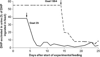
The sequence of events that seems to have transpired was that personal observations of ruminants grazing Leucaena in Hawaii and Indonesia prompted the investigators to speculate that the absence of toxicity in some locations might be caused by the further metabolism of DHP by ruminal bacteria not present in the Australian animals. A simple, elegant experiment was carried out in Indonesia using Australian goats that had been exposed to Leucaena and flown to Indonesia (Jones and Lowry, Reference Jones and Lowry1984). Two of the four animals were infused with ruminal digesta from a native Indonesian goat. The two untreated goats continued to excrete DHP, while DHP excretion in the two infused animals rapidly fell to zero (Figure 4). It was subsequently demonstrated that cultures of ruminal bacteria from a goat fed Leucaena, without ill effect, in Hawaii could prevent DHP excretion and toxicity in Australian cattle receiving Leucaena (Jones and Megarrity, Reference Jones and Megarrity1986). There was only one species responsible, a Gram-negative anaerobe now named Synergistes jonesii, a unique anaerobe not closely related to any other ruminal bacteria (Allison et al., Reference Allison, Mayberry, McSweeney and Stahl1992). Its geographical distribution appears to be limited, being absent in Africa and much of Asia as well as Australia (Jones, Reference Jones1994).
Excretion of the goitrogen, DHP, following inoculation of Australian goats with ruminal digesta from Indonesian goats. Inoculation is indicated by arrows. The Australian goats were previously unable to degrade DHP to non-toxic products, making Leucaena leucocephala toxic to these animals. The Indonesian goats showed no toxic effects of Leucaena, as they were colonised with bacteria able to degrade DHP. Inoculation of the Australian goats enabled the DHP-degrading bacteria to be transferred and detoxification to take place. From Jones and Lowry (1984).

Conclusions
The progress of the Leucaena project and the successful inoculation of humans with oxalate-degrading bacteria illustrates how the theme of this paper, namely that important functions can be carried out by unique species even in an ecosystem as complex as the rumen or human gut, is not only intrinsically interesting but can lead to benefits to animal and human health.
Acknowledgements
The Rowett Research Institute receives core funding from the Scottish Executive Environment and Rural Affairs Department. Genomic data on ruminal bacteria were obtained partly from USDA-funded projects carried out by the North American Consortium for Genomics of Fibrolytic Ruminal Bacteria.