Introduction
Insertion of the electrode during cochlear implantation inevitably causes mechanical trauma and the already damaged inner ear is affected by the additional trauma.Reference Ahmad, Choudhury, De Mason, Adunka, Finley and Fitzpatrick 1 Insertion of the electrode into the scala tympani triggers an apoptotic process. In addition to cochlear structures, spiral ganglion cells are also damaged. Therefore, atraumatic techniques are of primary importance in cochlear implant surgery. Ongoing research attempts to develop techniques to prevent or reverse such damage.Reference Leake, Stakhovskaya, Hetherington, Rebscher and Bonham 2
Local dexamethasone application has been shown to be effective in preventing electrode trauma in guinea pigs.Reference Eastwood, Chang, Kel, Sly, Richardson and O'Leary 3
Insulin-like growth factor 1 is a peptide of which serum levels are determined by growth hormone. It is mainly secreted by the liver. Moreover, it can be synthesised by recombinant DNA technology. Its use in chronic liver disease, cystic fibrosis, wound healing, acquired immune deficiency syndrome muscle wasting, burns, osteoporosis, Crohn's disease, anorexia nervosa, immunodeficiency syndromes and neurological disorders has been considered.Reference Rosenbloom 4 Recombinant insulin-like growth factor 1 has been shown to reverse glucocorticoid-resistant sudden hearing loss.Reference Nakagawa, Sakamoto, Hiraumi, Kikkawa, Yamamoto and Hamaguchi 5
We aimed to examine the effect of insulin-like growth factor 1 in the prevention of inner-ear damage caused by mechanical trauma in a rat model. Specifically, a dummy electrode was inserted through the round window and the effect of local, slow-release insulin-like growth factor 1 was examined.
Materials and methods
Animals
The study was conducted on 21 healthy female Wistar albino rats. All animals had normal otoscopic examination findings. Rats were kept in a 12-hour dark/bright light environment, at a temperature of 21–22 °C, with free access to food and water. The rats’ weight ranged between 250 g and 330 g (mean weight (± standard deviation) was 273.3 g ± 16.22 g). The background noise level was below 50 dB.
Groups
The rats were divided into three groups. In group 1 (sham group), an intracochlear dummy electrode was inserted through the round window and left in place. In group 2 (control group), after the same insertion procedure, saline-soaked porcine skin gelatine was placed on the round window. In group 3 (study group), after the same procedure, insulin-like growth factor 1 soaked porcine skin gelatine was placed on the round window membrane.
Anaesthesia
General anaesthesia was achieved via intraperitoneally injected 45 mg/kg ketamine hydrochloride plus 5 mg/kg xylazine. Additional anaesthesia was achieved using 25 mg/kg ketamine hydrochloride plus 5 mg/kg xylazine.
Surgical procedure
In all groups, the right ears of rats were chosen for the surgical procedure. After appropriate disinfection and 1 ml of lidocaine infiltration, a posteroinferior retroauricular incision was made and muscles were bluntly dissected. Under microscope, the bulla was opened with a number 11 surgical blade and the middle ear was entered. The round window membrane was punctured centrally with a needle.
Dummy electrode
A fluorocarbon line with a 0.28 mm diameter and a 0.5 cm length was chosen as a dummy electrode.
Interventions
In group 1 rats, the right ear round window was perforated with a needle. A perforation with a diameter of 1 mm was created. The dummy electrode with a diameter of 0.28 mm was pushed about 5 mm into the scala tympani, and the round window opening was sealed with muscle tissue. The incision was sutured with size 4-0 rapidly absorbable suture (Vicryl Rapide; Ethicon, New Brunswick, New Jersey, USA).
The group 2 rats were treated identically to those in group 1. In addition, 3 mg of normal saline-embedded porcine skin gelatine (Sigma, St Louis, Missouri, USA) was placed on the round window before it was sealed with muscle tissue.
The group 3 rats were treated in an identical manner to those in group 1. Additionally, 500 µg recombinant human insulin-like growth factor 1 (Increlex; Ipsen Pharma, Ettlingen, Germany) embedded in 3 mg porcine skin gelatine (Sigma) was placed on the round window before it was sealed with muscle tissue.
Audiological assessment
The GSI Audera system (Grason Stadler, Madison, Wisconsin, USA) was used for auditory brainstem response (ABR) measurements. The ABR measurements were obtained under anaesthesia using Viasys disposable electroencephalograph subdermal needle electrodes. Animals of which both ears were normal were included.
The ABR thresholds were obtained with clicks at 2–4 kHz, and tone bursts at 6 kHz and 8 kHz. Hearing thresholds were recorded pre-operatively, immediately after the surgical procedure and 14 days post-operatively. Post-operative hearing thresholds were compared to those obtained immediately after the surgical procedure. A decrease in ABR thresholds of 10 dB or more was considered an improvement and an increase of 10 dB or more was considered deterioration.
Following post-operative audiological assessment, all animals were sacrificed, after application of intracardiac formaldehyde. The right temporal bones were dissected and transferred to formaldehyde solution for histopathological examination.
Histopathological examination
Mucosa around the round window was dissected. Samples were fixed in 10 per cent formaldehyde solution for 24–48 hours, after which they were decalcified in ethylenediaminetetraacetic acid solution for 3 weeks. Sections of 5 µm thickness, prepared according to the routine paraffin tissue follow-up protocol, were stained with haematoxylin and eosin and examined with light microscopy.
Organ of Corti, basilar membrane, stria vascularis, spiral ganglion, spiral limbus, ganglion cell and satellite cell oedema, inflammation, congestion, and vacuolisation parameters were examined. The same histologist graded the damage as mild (1 point), medium (2 points) or severe (3 points).
Ethical considerations
The study was conducted in accordance with the Helsinki convention (1986) and the national animal protection law (law number 5199), and carried out with the approval of the local ethics committee (approval number 2012-094).
Results
Hearing thresholds
At 2 kHz and 4 kHz, one rat had deterioration, one rat showed some improvement and the rest had stable ABR thresholds after intervention in the study group. At 6 kHz, four rats showed improvement, and thresholds in the rest remained stable. At 8 kHz, four rats showed improvement, one had deterioration and two remained stable after intervention. The sham and control group rats showed no improvement at any frequency; about half of the rats had deterioration of hearing loss and the rest remained stable. Hearing thresholds as measured by ABR audiometry are shown in Table I.
Auditory brainstem response thresholds

Values represent means ± standard deviations. ABR = auditory brainstem response; pre-op = pre-operative; post-op = post-operative; IGF = insulin-like growth factor
The mean post-operative 2–4 kHz ABR threshold was significantly higher than the mean pre-operative threshold in group 1. The mean 2–4 kHz ABR threshold was significantly higher immediately after the intervention than pre-operatively, and the mean post-operative 2–4 kHz ABR threshold was significantly higher than the mean pre-operative threshold in group 3.
The mean 6 kHz and 8 kHz ABR thresholds were significantly higher immediately after the intervention than pre-operatively in all groups. The mean post-operative 6 kHz and 8 kHz ABR thresholds were significantly higher than the pre-operative thresholds in all groups. The mean 6 kHz ABR threshold was significantly lower post-operatively than immediately after the intervention in group 3.
Mean differences between pre- and post-operative ABR thresholds at 6 kHz, and between those immediately after the intervention and post-operatively, were lower in group 3 than in group 1. The mean difference between post-operative and pre-operative ABR thresholds at 6 kHz in group 3 was lower than the same mean difference in group 2.
Histopathological grading
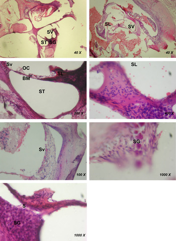
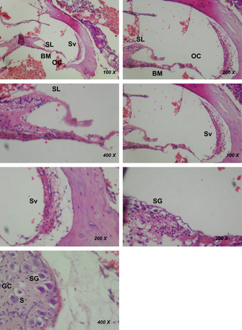
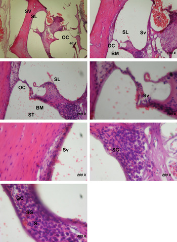
Group 3 had statistically significantly higher histopathological scores compared to both group 1 and group 2 (p < 0.05) (Table II). Histopathological examples of each group can be seen in Figures 1–3.
An example of inner-ear histology (H&E) for a rat in the electrode group (group 1), showing disruption of normal histological structure, increase of inflammatory cells and vascularisation, oedema in the spiral ganglion, decrease in neurons, and disordered satellite cells due to electrode trauma, as observed under a light microscope. SV = scala vestibuli; ST = scala tympani; SG = spiral ganglion; SL = spiral limbus; Sv = stria vascularis; OC = organ of Corti; BM = basilar membrane; S = satellite cell

An example of inner-ear histology (H&E) for a rat in the electrode plus normal saline group (group 2), showing disruption of the structure of the organ of Corti located on the basilar membrane, congestion in the stria vascularis, oedema, vacuolisation, chromatolysis in the spiral ganglion cells and disordered satellite cells, as observed under a light microscope. SL = spiral limbus; Sv = stria vascularis; BM = basilar membrane; OC = organ of Corti; SG = spiral ganglion; GC = ganglion cell (neuron); S = satellite cell

An example of inner-ear histology (H&E) for a rat in the electrode plus insulin-like growth factor group (group 3), showing preservation of the structure of the organ of Corti located on the basilar membrane, decreased congestion in the stria vascularis, decreased oedema, vacuolisation and nearly normal spiral ganglia cells, as observed under a light microscope. SV = scala vestibuli; SL = spiral limbus; OC = organ of Corti; Sv = stria vascularis; BM = basilar membrane; ST = scala tympani; SG = spiral ganglion; GC = ganglion cell (neuron); S = satellite cell

inner-ear histological damage

IGF = insulin-like growth factor
Discussion
In cochlear implant patients, residual hearing can sometimes be lost in the immediate or late post-operative period. This can be due to the loss of partially functional hairy cells and the apoptotic process triggered by electrode insertion into the scala tympani. Apoptosis-preventing agents and atraumatic techniques are therefore important for preserving residual hearing.Reference Carlson, Driscoll, Gifford, Service, Tombers and Hughes-Borst 6 The most common locally or systemically used substance is dexamethasone.Reference Eastwood, Chang, Kel, Sly, Richardson and O'Leary 3 Local hypothermia could also prevent damage in the cochlea.Reference Smith, Eshraghi, Whitley, van de Water and Balkany 7
Although a great deal of effort is made to carry out cochlear implantation in an atraumatic fashion, mechanical trauma and resultant damage to the spiral ganglion cells is inevitable. Therefore, routine use of otoprotective agents during cochlear implantation is advised.
Intra-operative cytoprotective agents have short-term effects. Furthermore, as the presence of a foreign body in the scala tympani persists, the apoptotic process continues, and tissue damage can occur in the long- or mid-term. Corticosteroid-emitting electrodes have been developed to address this issue.Reference Farahmand, Mirzadeh, Imani, Jolly and Farhadi 8 , Reference Niedermeier, Braun, Fauser, Kiefer, Straubinger and Stark 9 In addition to dexamethasone, erythropoietin, adenosine, ganglion-derived neurotrophic factor and brain-derived neurotrophic factor have been used in in vivo studies.Reference Quesnel, Nguyen, Campo, Hermine, Ribeil and Elmaleh 10
Lee et al. used porcine skin gelatine to deliver insulin-like growth factor 1 in a rat model for sudden deafness due to noise.Reference Lee, Nakagawa, Okano, Hori, Ono and Tabata 11 Following successful results, this substance has been used in glucocorticoid-resistant cases of sudden deafness.Reference Nakagawa, Sakamoto, Hiraumi, Kikkawa, Yamamoto and Hamaguchi 5
Insulin-like growth factor 1 is a peptide and does not cross the cell membrane. It works by binding to the receptors in the membrane. It is effective in almost all organs and plays an important role in embryonic development. Its nanomolar concentration continues into adult life. In adulthood, insulin-like growth factor plays a role in functions such as normal cellular metabolism, proliferation and protection against apoptotic stimuli.Reference Butt, Firth and Baxter 12
In their studies conducted in insulin-like growth factor deficient mice, Camarero et al. showed that developmental abnormalities appear in the cochlea and spiral ganglion cells, and apoptosis is increased related to caspase-3 secretion in spiral ganglion cells.Reference Camarero, Avendano, Fernandez-Moreno, Villar, Contreras and de Pablo 13 Additionally, organ of Corti cells had abnormal innervation.Reference Camarero, Avendano, Fernandez-Moreno, Villar, Contreras and de Pablo 13 Frago et al. studied the insulin-like growth factor effect on chicken embryo otic vesicles, and showed that inner-ear cell growth and survival increases, and neural growth factor caused apoptosis, may be prevented with insulin-like growth factor 1.Reference Frago, Cañón, de la Rosa, León and Varela-Nieto 14
In our study, hearing loss and cochlear damage were induced through mechanical trauma to the scala tympani through the round window, associated with the insertion of a dummy electrode. Hearing loss (determined using ABR audiometry) and histopathological effects were evaluated.
The ABR threshold changes indicated that electrode trauma caused similar hearing loss in all study groups. At the two-week follow up, the local application of insulin-like growth factor resulted in ABR threshold improvement at 6 kHz. The control and sham groups showed no improvement. The difference between pre- and post-operative hearing ABR thresholds was statistically significantly lower in the study group compared to the control and sham groups. This suggests that local insulin-like growth factor 1 application is effective at preserving inner-ear functionality.
Histopathological data were consistent with audiological findings. Histopathological data showed that the groups with no insulin-like growth factor 1 administration had disruption in the organ of Corti structure, congestion in the stria vascularis, oedema and vacuolisation, chromatolysis in the spiral ganglion cells, and disruption in satellite cell organisation. In the insulin-like growth factor 1 applied group, less stria vascularis congestion, oedema and vacuolisation were present, and the structure of the organ of Corti and spiral ganglion cells was near-normal. The effect of an insulin-like growth factor 1 eluting electrode in a guinea pig model has been examined before in one study.Reference Kikkawa, Nakagawa, Ying and Tabata 15 That study showed better audiological and histopathological outcomes. Our results are similar, and a better outcome was obtained with topical insulin-like growth factor 1 application compared to the control group.
-
• Inner-ear damage due to mechanical trauma causes histopathological changes and hearing loss
-
• In a rat model, local insulin-like growth factor 1 application caused better hearing and histopathological scores compared to a control group
-
• Local insulin-like growth factor 1 application may be effective at preventing or reversing inner-ear damage
Data obtained from our study suggest that local insulin-like growth factor 1 application after cochlear trauma had beneficial effects on inflammatory, degenerative and apoptotic processes, and this was reflected on audiological findings.
Conclusion
A relatively non-invasive method using porcine insulin-like growth factor 1 embedded skin gelatine application to the round window may be valuable for potential inner-ear damage associated with procedures that cause mechanical trauma, such as cochlear implantation and stapes surgery.