INTRODUCTION
A new genus and species of small centric diatom was described by Gerloff (Reference Gerloff1968) under the name Potamodiscus kalbei. A few years later, Gaarder et al. (Reference Gaarder, Fryxell and Hasle1976) showed that these entities were not diatoms, but instead were the siliceous plate-scales surrounding the cell of a then unidentified protist. Soon after, researchers in England (Belcher & Swale, Reference Belcher and Swale1978) and in Norway (Thomsen, Reference Thomsen1978) simultaneously published their apparently independent conclusion that loose scales of the type species of Pinaciophora (P. fluviatilis (Greeff, 1873) Penard, Reference Penard1904) were identical to P. kalbei, thus rendering Potamodiscus invalid.
Pinaciophora is one of a few genera of benthic heterotrophic protists of the order Rotosphaerida Rainer, Reference Rainer and Dahl1968 (= Cristidiscoidida Page, 1987). Rotosphaerids are silica-scale-bearing organisms, all with filopodia that facilitate phagotrophic nutrition and a slowly rolling or creeping form of motility. All rotosphaerids lack the centroplast (= axoplast, or central granule) of centrohelid heliozoa, but otherwise are ‘heliozoan' in appearance in a light microscope (LM). In recent compilations of protistan classification systems, the rotosphaerids have been either included among other protists of uncertain taxonomic position (Patterson et al., Reference Patterson, Simpson, Rogerson, Lee, Leedale and Bradbury2002) or have been totally omitted (Corliss, Reference Corliss1994; Adl et al., Reference Adl, Simpson, Farmer, Andersen, Anderson, Barta, Bowser, Brugerolle, Fensome, Fredericq, James, Karpov, Kugrens, Krug, Lane, Lewis, Lodge, Lynn, Mann, McCourt, Mendoza, Moestrup, Mozley-Standridge, Nerad, Shearer, Smirnov, Spiegel and Taylor2005), perhaps a testament to their general unfamiliarity.
Rainer (Reference Rainer and Dahl1968) assigned five genera to his sub-order Rotosphaeridia: Rabdiophrys, Pompholyxophrys, Pinaciophora, Pinaciocystis and Lithocolla. Thomsen (Reference Thomsen1978, Reference Thomsen1979), Manton & Sutherland (Reference Manton and Sutherland1979), and Croome (Reference Croome1987a, Reference Croomeb) greatly expanded the scope of the genus Pinaciophora to include many more species with plate-scales similar to those of P. fluviatilis but including species having spine-scales of various morphologies. Roijackers & Siemensma (Reference Roijackers and Siemensma1988) split up the total complement of 14 Pinaciophora species known at that time into two genera, by transferring all but P. fluviatilis and P. rubicunda (Hertwig & Lesser, 1874) emend. Roijackers & Siemensma, Reference Roijackers and Siemensma1988 to the related genus Rabdiophrys Rainer, Reference Rainer and Dahl1968, thus isolating all spine-scale bearing species from those two species (P. fluviatilis and P. rubicunda) possessing plate-scales only.
Mikrjukov (Reference Mikrjukov1999) further altered the taxonomy of this group by excluding Pinaciocystis and Lithocolla and including the new genus Rabdiaster Mikrjukov, Reference Mikrjukov1999, thus restricting the order Rotosphaerida to these four genera: Pinaciophora, Rabdiophrys, Rabdiaster and Pompholyxophrys. One result of Mikrjukov's (1999) revision was the reunification of all ‘pinaciophorids', with and without spine-scales, within the genus Pinaciophora (with the exception of those with a single large hole in the centre of their plate-scales, which were assigned to Rabdiophrys). Mikrjukov (Reference Mikrjukov1999) also separated out those ‘pinaciophorids' lacking holes in their plate-scales and placed them in Rabdiaster. While these latter two components of Mikrjukov's revision relating to Rabdiophrys and Rabdiaster are logical and justifiable, the arguments supporting the reunification of the remaining Pinaciophora species irrespective of the presence of spine-scales is not justifiable, in my opinion. The reasons Mikrjukov (Reference Mikrjukov1999) provided for the Pinaciophora reunification were subjective and based in large part on the presumption that spineless species of Pinaciophora might be a ‘natural' occurrence. There is, however, no published evidence for the presence of spine-scales in the type species, P. fluviatilis. Neither is there evidence for the loss (under unfavourable environmental conditions) of one of the scale types in other rotosphaerid genera known to possess both plate-scales and spine-scales.
Pinaciophora fluviatilis is strictly a freshwater taxon with distinctive plate-scale morphology. Several earlier reports of loose scales identified as P. fluviatilis from marine habitats are erroneous. The ‘plate-scale only' character typifying P. fluviatilis has been strengthened by the findings reported here for the marine species P. rubicunda and P. marina sp. nov., both of which lack spine-scales. Pinaciophora marina may account for several of the erroneous reports of P. fluviatilis referred to above. The purpose of this paper is to propose a solution to the ‘Pinaciophora problem' by introducing the new genus Thomseniophora gen. nov. to accommodate those ‘Pinaciophora' species known to possess both plate-scales and spine-scales. In addition, 13 marine rotosphaerid taxa are described from Canadian marine habitats, eight of which are new to science.
MATERIALS AND METHODS
Methods used in sampling near-shore benthic Atlantic and Pacific Oceans habitats, and laboratory methods relating to light microscopy and electron microscopy were described in detail in Nicholls (Reference Nicholls2009a, Reference Nichollsb). Specific locations are described where appropriate in the type species habitat descriptions given in the Results section, below. Unless stated otherwise, all taxa described here were found in a near-shore sediment/seawater sample collected at low tide on 8 December, 2008 from the Pacific Ocean on Canada's west coast (Haida Gwaii—Queen Charlotte Islands, British Columbia; 53.241°N 131.898°W); salinity = 32 ppt.
RESULTS
SYSTEMATICS
Phylum: CERCOZOA Cavalier-Smith, 1998
Class: IMBRICATEA Cavalier-Smith & Chao, 2003
Order: ROTOSPHAERIDA Rainer, Reference Rainer and Dahl1968
Family: POMPHOLYXOPHRYIDAE Page, 1987
NEW GENUS
Thomseniophora gen. nov.
[=Pinaciophora (partum)]
DIAGNOSIS
Single-celled, free-living rotosphaerids with a silica-scaled periplast comprised both plate-scales and spine-scales. Plate-scales double walled with multiple holes in the distal surface; spine-scales tangential with an elongated shaft and a swollen basal structure. Distinguished from Rabdiophrys Rainer, Reference Rainer and Dahl1968 which has a single, central large hole in the plate-scales; distinguished from Rabdiaster Mikrjukov, Reference Mikrjukov1999, which lacks holes in the plate-scales; distinguished from Pinaciophora (sensu Penard, Reference Penard1904) which lacks spine-scales.
ETYMOLOGY
The genus name is in honour of Helge Thomsen, noted Danish protistologist, who discovered and described most of the Pinaciophora species now assigned to Thomseniophora.
TYPE SPECIES (LECTOTYPE)
Thomseniophora denticulata (Thomsen, Reference Thomsen1978) comb. nov.; figure 16, p. 365 in Thomsen (Reference Thomsen1978; Protistologica 14, 359–373).
TYPE LOCALITY
Dybsø Fjord, Denmark.
New combinations
Thomseniophora denticulata (Thomsen, Reference Thomsen1978) comb. nov.
[=Pinaciophora denticulata Thomsen, 1978]
[=Rabdiophrys denticulata (Thomsen, Reference Thomsen1978) Roijackers & Siemensma, 1988]
Thomseniophora thomseni (Thomsen, Reference Thomsen1979) comb. nov.
[=Rabdiophrys thomseni Roijackers & Siemensma, 1988]
(including figure 15 in Thomsen (Reference Thomsen1978)
Thomseniophora triangulata (Thomsen, Reference Thomsen1978) comb. nov.
[=Pinaciophora triangulata Thomsen, 1978]
[=Rabdiophrys triangulata (Thomsen, Reference Thomsen1978) Roijackers & Siemensma, 1988]
Thomseniophora tasmanica (Croome, 1987) comb. nov.
[=Pinaciophora tasmanica Croome, 1987]
[=Rabdiophrys tasmanica (Croome, 1987) Roijackers & Siemensma, 1988]
Thomseniophora ovalis (Croome, 1987) comb. nov.
[=Pinaciophora ovalis Croome, 1987]
Thomseniophora tridentata (Thomsen, Reference Thomsen1978) comb. nov.
[=Pinaciophora tridentata Thomsen, 1978]
[=Rabdiophrys tridentata (Thomsen, Reference Thomsen1978) Roijackers & Siemensma, 1988]
Thomseniophora bifurcata (Thomsen, Reference Thomsen1978) comb. nov.
[=Pinaciophora bifurcata Thomsen, 1978]
[=Rabdiophrys bifurcata (Thomsen, Reference Thomsen1978) Roijackers & Siemensma, 1988]
Thomseniophora candelabrum (Thomsen, Reference Thomsen1978) comb. nov.
[=Pinaciophora candelabrum Thomsen, 1978]
[=Rabdiophrys candelabrum (Thomsen, Reference Thomsen1978) Roijackers & Siemensma, 1988]
Thomseniophora paucipora (Thomsen, Reference Thomsen1978) comb. nov.
[=Pinaciophora paucipora Thomsen, 1978]
[=Rabdiophrys paucipora (Thomsen, Reference Thomsen1978) Roijackers & Siemensma, 1988]
Thomseniophora spiculata (Manton & Sutherland, Reference Manton and Sutherland1979) comb. nov.
[=Pinaciophora spiculata Manton & Sutherland, Reference Manton and Sutherland1979]
[=Pinaciophora spiculata Manton (in Thomsen (Reference Thomsen1978)]
[=Rabdiophrys spiculata (Manton & Sutherland, Reference Manton and Sutherland1979) Roijackers & Siemensma, 1988]
Thomseniophora turisfenestrata (Wujek & O'Kelly, 1991) comb. nov.
[=Rabdiophrys turisfenestrata Wujek & O'Kelly, 1991]
[=Pinaciophora turisfenestrata (Wujek & O'Kelly, 1991) Mikrjukov, 1999]
NEW AND LITTLE-KNOWN TAXA
GENUS THOMSENIOPHORA
Thomseniophora denticulata (Thomsen, Reference Thomsen1978) comb. nov.
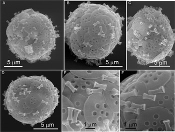
(Figure 1A–F)
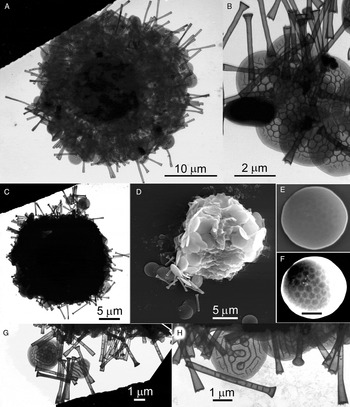
This species was relatively common in samples collected 8 December 2008 from the Pacific Ocean (Haida Gwaii) location. The Canadian specimens of T. denticulata agree well with Thomsen's (1978) description of this taxon from Denmark. Cells 11–13 µm in diameter (6 whole dried cells, scanning electron microscopy (SEM); Figure 1 A–D), covered in circular plate-scales 3.0–3.7 µm in diameter and in short tubular spine-scales 1.1–1.6 µm long, with slightly flared apices and swollen bases (Figure 1E, F). Plate-scales had convex distal surfaces and narrow (0.09 µm wide), but well-developed marginal rims. Plate-scales with usually 5–9 holes (0.32–0.42 µm in diameter) that were usually arranged in a circular pattern of 5–7 holes at a distance of about 1/3 of the scale diameter from the scale rim, with an additional 0–2 holes near the centre of the scale. The spine-scales were narrowest (0.28–0.39 µm) at about the mid-point of their length. The flared apices (up to 0.75 µm diameter) had scalloped rims resulting in a series of 4–8 marginal ‘teeth' of variable size and shape. The swollen bases of the spine scales had a series of 5–8 holes 0.10 µm in diameter that led to the interior of the hollow scale. This structure was fused to a circular base-plate, the proximal surface of which was slightly concave, 0.5–0.7 µm in diameter and 0.1 µm thick.
Thomseniophora denticulate: (A–D) scanning electron microscopy images of whole cells with their investitures of plate-scales ornamented with large holes and short tubular spine-scales; (E, F) details of scale morphology.

Thomseniophora candelabrum (Thomsen, Reference Thomsen1978) comb. nov.
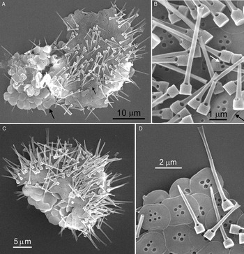
(Figure 2A–D)
This species is characterized by very elaborate spine-scales with a complex basal structure described as ‘basket-like' by Thomsen (Reference Thomsen1978), with which three intact whole specimens from Canada's west coast (Haida Gwaii, Pacific Ocean) agree very well. In the Canadian material, plate-scales were double walled with a smooth, unornamented proximal surface (Figure 2A, large arrow) and a distal surface (Figure 2A, small arrow) with 3–8 usually circular holes ranging in size from 0.13 to 0.35 µm in diameter. Very large and very small holes were sometimes found together on a single scale (Figure 2D). Plate-scales were subcircular, 2.2–3.5 µm in diameter/length with the longer more elliptically-shaped scales characterized by slight marginal concavities about mid-way along both edges of the longest dimension of the scale.
Thomseniophora candelabrum: (A, C) scanning electron microscopy images of whole (collapsed) cells with their investitures of scales showing the distal surfaces of plate-scales patterned with centrally-located holes (small arrow) and the plane unornamented proximal surfaces of other plate-scales (large arrow); the elongated spine-scales have swollen basal structures and flattened, flared apices; (B, D) details of scale morphology; black arrow shows the rim on a plate-scale; white arrow shows one of usually four holes on the base of a spine-scale.

Spine-scale shafts were hollow and 4-sided through most of their length of 2–7 µm, and tapered to a flattened but flared distal end (Figure 2D). The base of the spine shaft widened abruptly into a hollow cylinder about 0.6–0.8 µm in diameter with 5–6 rows of small pores encircling the diameter of this structure. In the intermediate zone between the base of the spine shaft and the porous cylinder there were four large (about 0.2 µm in diameter) holes, each in the same plane as each of the four walls of the spine shaft (Figure 2B). Occasionally, an ‘extra' hole located between a pair of the more regularly positioned holes was observed (i.e. not aligned with the flat surface of the spine shaft (Figure 2B, white arrow)).
The plate-scales of the Canadian population of T. candelabrum resemble more those from the Danish Sound than those from the type locality of this species (Dybsø Fjord, Denmark: Thomsen, Reference Thomsen1978), but the spine-scales of the Canadian material resemble more those from Dybsø Fjord than those from the Danish Sound. The differences in plate-scales related to the deviation from circular shape to a more elliptical shape with indented margins along the opposite long edges; the differences in spine-scales relate to the presence or absence of a ‘banister-like' structure surrounding the intermediary zone between the base of the spine shaft and the enlarged cylindrical spine base. The single spine-scale of P. candelabrum illustrated by Takahashi (Reference Takahashi1981) closely resembles those in the Canadian population, as do the scales reported for Arctic Canada by Vørs (Reference Vørs1993). It would appear that there may be some minor geographical variation in scale structure of this species. Scales structure is, however, highly distinctive in P. candelabrum and cannot be confused with that of any other known Thomseniophora species.
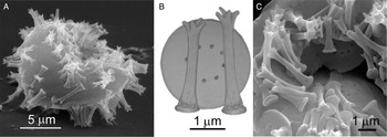
Thomseniophora emarginata sp. nov.
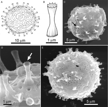
(Figure 3A–E)
Thomseniophora emarginata sp. nov.: (A, B) diagrammatic representation of the investiture of plate-scales and spine scales covering a cell (A) and of a single spine-scale (B); (C, E) scanning electron microscopy images of whole cells with their scaled periplasts; an atypical spine-scale apex with its internal septum (black arrow); (D) distal end of a typical spine-scale with its denticulate margin (white arrow).

DIAGNOSIS
Cells spherical, 20–25 µm in diameter, covered with both plate- and spine-scales (visible in high magnification, phase contrast light microscopy). Plate-scales are circular–elliptic (3–5 µm) but with one or two (usually) strongly rounded indentations occupying about one-quarter of the margin of the scale; plate-scales with a well developed marginal rim (about 0.13 µm wide) and a convex distal surface perforated with 6–12 circular holes, about 0.2–0.4 µm in diameter. Spine-scale, a short hollow tube (1.5–3.3 µm long) with a swollen base about 1.1–1.2 µm in diameter, and a slightly flared distal end about 0.8 µm wide with 6–8 marginal small teeth. Occasional spine-scales have a greater flare at the distal end with an internal septum dividing the opening leading to the hollow shaft of the scale into two parts. Spine base is reinforced with a series of thickened longitudinal ribs.
ETYMOLOGY
The specific epithet (emarginata) refers to the rounded indentations in the circumference of the otherwise circular scales of this species.
TYPE SPECIMEN
TYPE LOCALITY
Pacific Ocean beach near Gillatt (Grassy) Island, Haida Gwaii (Queen Charlotte Islands, British Columbia, Canada (53.241°N 131.898°W); salinity = 32 ppt; sample collected 8 December 2008.
MATERIAL FROM TYPE LOCALITY
Retained by the author (aqueous dilute formalin solution) as preserved sample No. V-2111.
REMARKS
The circular shape of the plate-scales is ‘deformed' by the large rounded indentations that occupy about one-quarter of the circumference of the scale (Figure 3A, E). This shape is somewhat similar to, but more strongly developed than that found in the plate scales of the a2/b2 variant of T. candelabrum (Thomsen, Reference Thomsen1978); other details of scale structure, especially the spine-scales, clearly indicate, however, that T. emarginata is distinct from T. candelabrum. The image of the single plate-scale provided by H.A. Thomsen to Gaarder et al. (Reference Gaarder, Fryxell and Hasle1976; their figure 13) may well have been from a Norwegian (Hordaland) specimen of T. emarginata. Similarly, occasional aberrant scales with marginal indentations resembling those found consistently in T. emarginata have been reported for other Thomseniophora species (e.g. Manton & Sutherland, (Reference Manton and Sutherland1979); their figure 4), but without whole intact specimens from these collections, it is not possible to establish their identity with certainty.
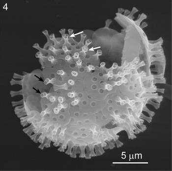
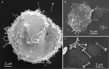
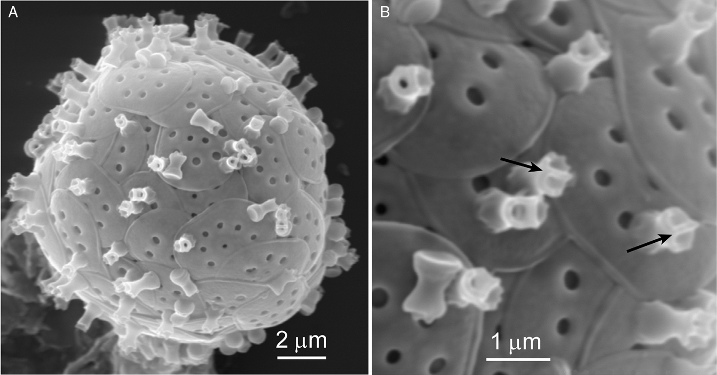
An additional specimen found along with T. emarginata from the same British Columbia collection site (Figure 4) shows some resemblance to T. emarginata in the general shape and size of plate-scales and spine-scales, but with two notable exceptions: (1) the holes in the plate-scales are larger; and (2) all of the spine-scales appear to have septa that create two or three separate holes leading to the interior of the spine shaft (compare Figure 4 with Figure 3E). Only the single specimen shown in Figure 4 was found, so it is not known if its morphology is consistently different enough to represent a separate species. The occurrence of terminal septa in spine-scales was found only rarely in several specimens of T. emarginata, so its more abundant presence in the specimen shown in Figure 4 may have little or no taxonomic relevance given the other similarities in scale morphology (but for the larger holes in the plate-scales).
Thomseniophora emarginata ? variant ?: scanning electron microscopy image of a partial periplast cell covering of scales with spine-scales having internal septa creating two access holes (black arrows) and three access holes (white arrows) to the distal part of the scale.

The spine-scales of T. emarginata are unique, but there is some resemblance to those of the freshwater Rabdiaster apora (Croome, Reference Croome1987a) and to those of Thomseniophora thomseni (see especially the drawings in Roijackers & Siemensma (Reference Roijackers and Siemensma1988); their figure 37). Spine-scales of R. apora are longer and relatively narrower with a differently structured base; its plate-scales are circular and lack holes. Plate-scales of T. thomseni lack the strong lateral indentations of T. emarginata and its spine-scales are about twice as long and have a much more elaborately structured septum at the spine terminus.
Thomseniophora biparta sp. nov.
(Figure 5A–G)
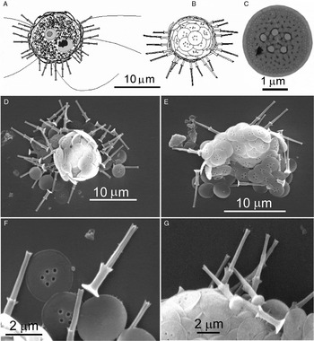
Thomseniophora biparta sp. nov.: (A, B) diagrammatic representations of a cell showing a food vacuole, nucleus and filopodia (A) and of the investiture of plate-scales and spine-scales covering the cell (B); (C) a single plate-scale (transmission electon microscopy); (D, E) scale-covered periplast of collapsed whole cells; (F, G) details of scale structure; the distal surfaces of plate-scales have holes, the proximal surfaces do not.

DIAGNOSIS
Cells spherical, 8–12 µm in diameter, covered with both plate- and spine-scales (visible in high magnification, phase contrast light microscopy). Movement is a very slow tumbling action mediated by several fine filopodia about 2 times the cell diameter in length. Plate scales circular to occasionally slightly elliptical, 1.7–3.5 µm in diameter (most commonly 2.5–3 µm in diameter) with a slightly thickened rim about 0.8 µm wide. Proximal surface of plate scales concave, lacking ornamentation; distal surface convex with 3–7 circular holes 0.15–0.25 µm in diameter. Spine-scales in two parts; the basal portion, about 2/5 of the total scale length, consists of a circular base-plate 1.0–1.2 µm in diameter from which arises a sub-cylindrical (weakly hour-glass-shaped) tube of about 0.7 µm diameter in the slightly constricted mid-region. This structure terminates in a flared apex with 3–6 marginal ‘teeth'. The longer apical portion consists of a sub-cylindrical shaft (slightly constricted in the mid-region) with a mid-section diameter of about 0.3 µm and with 2–3 longitudinal ribs that terminate at the apex in 2–3 small tooth-like projections. Total spine-scale length, 3.0–6.5 µm (most commonly, 4–5 µm).
ETYMOLOGY
The specific epithet (biparta) refers to the two morphologically distinct components of the spine-scales of this taxon.
TYPE SPECIMEN
TYPE LOCALITY
Benthic near-shore sand, north shore, Sable Island, (Atlantic Ocean, Nova Scotia, 43°56′27″N 59°58′0″W); salinity = 33 ppt; collected 20 August 2007.
MATERIAL FROM TYPE LOCALITY
Retained by the author (aqueous dilute formalin solution) as preserved sample No. V-2081.
REMARKS
Because T. biparta was relatively abundant and the only rotosphaerid found in the Sable Island sample, it was possible to link LM observations of it to the scale-covered specimens examined with SEM. Plate-scales of T. biparta bear some resemblance to those of Pinaciophora rubicunda (Hertwig & Lesser, 1874) Roijackers & Siemensma, Reference Roijackers and Siemensma1988, Thomseniophora paucipora and P. tasmanica, Croome (Croome, Reference Croome1987a). Pinaciophora rubicunda, however, has larger and predominantly elliptical scales with much larger and more plentiful centrally located pores on the distal surface. The plate-scales of T. biparta are larger with larger and differently arranged central pores than those of either T. paucipora or T. tasmanica. The highly characteristic bipartite spine-scales of T. biparta are very unlike those of any previously described rotosphaerid species.
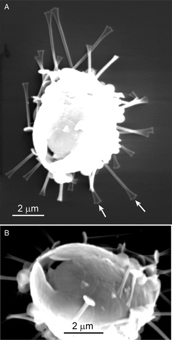
Thomseniophora brevispina sp. nov.
Figure (6A, B)
Thomseniophora brevispina sp. nov.: (A) scanning electron microscopy image of a whole cell and its investiture of plate-scales and spine-scales; (B) details of scale structure; arrows show the septum dividing the distal opening to the spine shaft into two parts.
DIAGNOSIS
Cells about 10–15 µm in diameter. Plate-scales oval in outline with parallel sides and rounded ends, about 3 µm wide and 4 µm long. Distal surface of the plate-scales with a series of 7–9 holes 0.2–0.3 µm in diameter forming a ring located about 1/4 of the scale width from the margin of the scale; 1–2 additional smaller holes, 0.15–0.2 µm in diameter, are located near the centre of the scale. Spine-scales are short (0.9–1.3 µm), hollow tubes with a marked mid-length constriction where spine shaft diameter is about 0.04 µm. The flared circular base of the spine-scale has a concave outer surface 0.06–0.08 µm in diameter, above which are a series of depressions around the spine base giving a scalloped edge appearance to the base in a polar view of the scale. The flared distal ends of the spine-scales are characterized by a septum that divides the opening to the internal column of the spine shaft into two separate cavities with entrance holes of about 0.15 µm in diameter.
ETYMOLOGY
The specific epithet (brevispina) denotes the shortness of the spine-scales in this species.
TYPE SPECIMEN
TYPE LOCALITY
Pacific Ocean beach near Gillatt (Grassy) Island, Haida Gwaii (Queen Charlotte Islands, British Columbia, Canada (53.241°N 131.898°W); salinity = 32 ppt; sample collected 8 December 2008.
MATERIAL FROM TYPE LOCALITY
Retained by the author (aqueous dilute formalin solution) as preserved sample No. V-2111.
REMARKS
Thomseniophora brevispina joins a small group of Thomseniophora species with spine-scales having septate, flared distal ends; the others being T. thomseni, T. minima and T. emarginata. The spine-scales of T. thomseni are 2–6 times larger than those of T. brevispina and have much more elaborately structured terminal septa. Thomseniophora emarginata plate-scales have fewer and larger holes on the distal surfaces and margins with marked lateral concavities; spine-scales are 2–3 times larger with differently structured spine ends. For differences with T. minima, see below.
Thomseniophora minima sp. nov.
(Figure 7A, B)
Thomseniophora minima sp. nov.: (A) scanning electron microscopy image of a whole cell and its investiture of plate-scales and spine-scales; (B) details of scale structure; arrow shows a septum dividing the distal opening to the spine shaft into two parts.

DIAGNOSIS
Cells about 15 µm in diameter. Plate-scales circular/elliptical, 2.0–2.3 × 3.3–3.6 µm; distal surface ornamented with 5–10 large holes (0.35–0.45 µm in diameter). Spine-scales, 0.6–0.9 µm long with a floral-like terminus consisting of a funnel-like opening with a scalloped rim (6–14 well-developed rounded denticles) and divided by 0–2 internal septa creating 1–3 separate cavities leading to the hollow centre of the spine shaft. Base of the spine swollen with 6–8 circular depressions at the bottom of which may be a small (0.05 µm) hole through to the hollow centre of the spine.
ETYMOLOGY
The specific epithet (minima) denotes the shortness of the spine-scales in this taxon.
TYPE SPECIMEN
TYPE LOCALITY
Pacific Ocean beach near Gillatt (Grassy) Island, Haida Gwaii (Queen Charlotte Islands, British Columbia, Canada (53.241°N 131.898°W): salinity = 32 ppt; sample collected 8 December 2008.
MATERIAL FROM TYPE LOCALITY
Retained by the author (aqueous dilute formalin solution) as preserved sample No. V-2111.
REMARKS
The four taxa T. thomseni, T. emarginata, T. brevispina and T. minima clearly have a common ancestor owing to the conservation among all four of the terminal septum in their spine-scales. Morphologically, T. minima's closest relative would appear to be T. brevispina. Both the plate-scales and the spine-scales of T. minima are smaller than those of T. brevispina, but the holes in the plate-scales of T. minima are uniformly 20–30% larger than those in the scales of T. brevispina (compare Figures 6B and 7B). The distal rims of the spine-scales of T. minima are a little more ornately scalloped than those of T. brevispina.
It is important to note that all three of the Thomseniophora septum-bearing species described here were present together in the same sample from the same location collected at the same time of year. It is therefore possible to rule out the suggestion that environmental differences in their habitats might have contributed to differences in the structures of their endogenously produced scales. The differences in scale morphology identified here must therefore be genetic in origin and as a consequence validate their separation at the species level of classification.
Thomseniophora spiculata pacifica ssp. nov.
(Figure 8A–C)
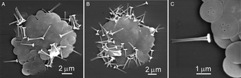
Thomseniophora spiculata pacifica ssp. nov.: (A, B) two whole cells (dried and collapsed); (C) portion of the scale covering from the specimen in (A) at higher magnification to reveal more detail of morphology of plate-scales and a single spine-scale. The proximal surface of plate-scales is unornamented (no holes); distal surface with a few small centrally-located holes.

DIAGNOSIS
Cells about 8–10 µm in diameter (as inferred from two dried and flattened specimens). Spine-scales 1.6–2.8 µm long, consist of a 4-sided shaft with thickened longitudinal ribs marking the four corners (in cross-section) ending distally in a flattened, slightly flared and bifurcate tip. Base of the spine-scale disc-like about 0.1 µm thick and 0.6–0.7 µm in diameter. Proximal 1/3 of the spine shaft ornamented with a row of perforations of the scale material between the ribs. Plate-scales nearly circular, 1.5–2.0 µm in diameter, with 4–6 small holes 0.06–0.1 µm in diameter centrally located on the distal surfaces of the scales.
ETYMOLOGY
The subspecific epithet (pacifica) refers to the Canadian Pacific Ocean habitat of this taxon.
TYPE SPECIMEN
TYPE LOCALITY
Pacific Ocean beach near Gillatt (Grassy) Island, Haida Gwaii (Queen Charlotte Islands), British Columbia, Canada (53.241°N 131.898°W); salinity = 32 ppt; sample collected 8 December 2008.
MATERIAL FROM TYPE LOCALITY
Retained by the author (aqueous dilute formalin solution) as preserved sample No. V-2111.
REMARKS
The spine-scales of Canadian Arctic specimens (Manton & Sutherland, Reference Manton and Sutherland1979) were longer than the median length of 2.2 µm in my British Columbia material. But their value (‘commonly 3 µm long’; p. 294 of Manton & Sutherland (Reference Manton and Sutherland1979)) does not agree with my calculated spine lengths of 1–2 µm for their specimens based on their electron microscopy (EM) images and the magnifications given in their figures. Thomsen (Reference Thomsen1978) illustrated a spine-scale with a length of 2.5 µm and mentioned that there was overall good agreement between the scale morphology of the only two widely separated populations known at that time (Canadian Arctic and Denmark).
Notwithstanding the small differences in spine-scale morphologies among the three known populations, it is mainly on the basis of the different plate-scale morphology that I justify separation of T. s. pacifica from the Canadian Arctic and Danish populations at the level of subspecies. Based on material from both Dybsø Fjord, Denmark (Thomsen, Reference Thomsen1978) and Resolute Bay in the Canadian Arctic (Manton & Sutherland, Reference Manton and Sutherland1979), the plate-scales of T. s. spiculata have a series of usually 5–7 centrally located large holes interspersed amongst more numerous smaller perforations. In the Pacific Ocean material reported here, the large holes are entirely absent and the plates are ornamented with just a few (3–6) small holes. The holes in T. s. spicata are about 3 times larger than the holes on the scales of T. s. pacifica.
Thomseniophora muticata sp. nov.
(Figure 9A–C)
Thomseniophora muticata sp. nov.: (A) scanning electron microscopy image of a whole cell (partly collapsed) and its investiture of plate-scales and spine-scales; (B) diagrammatic representation of the distal surface of a plate-scales and of two spine-scales; (C) detailed structure of scales.

DIAGNOSIS
Cells about 13 µm in diameter (one whole cell measured) covered in plate-scales and spine-scales. Plate-scales circular, 2.3–2.5 µm in diameter with a narrow rim and a series of 5–8 small or poorly developed holes in the distal surface. Spine-scales narrowest (about 0.3 µm) near the flared distal apex which consists of usually four, divergent, blunt-tipped projections. Spine-scale shafts widening near the base which consists of a circular pad with a concave bottom, 0.5–0.9 µm in diameter. The base of the spine shaft has a series of up to 5 small holes (about 0.7 µm in diameter) just above the circular base. Spine-scale total length is 1.4–3.0 µm.
ETYMOLOGY
The specific epithet (muticata) refers to the apex of the spine scales with their blunt, knob-like projections.
TYPE SPECIMEN
TYPE LOCALITY
Pacific Ocean beach near Gillatt (Grassy) Island, Haida Gwaii (Queen Charlotte Islands), British Columbia, Canada (53.241°N 131.898°W); salinity = 32 ppt; sample collected 8 December 2008.
MATERIAL FROM TYPE LOCALITY
Retained by the author (aqueous dilute formalin solution) as preserved sample No. V-2111.
Thomseniophora muticata acuminata ssp. nov.
(Figure 10A–C)
Thomseniophora muticata acuminata ssp. nov.: (A) scanning electron microscopy image of a whole cell with its investiture of plate-scales and spine-scales; (B) patch of scales; (C) upper-right part of (B) at higher magnification to reveal details of morphology.

DIAGNOSIS
Cells 12–16 µm in diameter. Plate-scales circular, 2.2–2.8 µm in diameter; the convex distal surface of the plate-scale with 6–10 small holes (about 0.15 µm in diameter) arranged in a more or less circular pattern about 1/4 of the diameter of the scale from the scale margin, and sometimes containing an additional 1–2 holes near the centre of the scale. Spine-scales 1.3–3.0 µm long and 0.25–0.3 µm wide at the narrowest point (near the middle of the shaft). Shaft of the spine-scale, 4-sided near its base with a flared apex having 3–4 terminal sharp-pointed teeth. Base of spine-scale consists of a circular disc about 0.7–0.9 µm in diameter and about 0.08 µm thick; it is fused near its outer rim to the flared basal end of the spine shaft. There are 1–2 holes in each of the four sides of the spine shaft just above the basal disc. Differs from the nominate species by having spine-scales with sharp-pointed teeth at the apex.
ETYMOLOGY
The sub-specific epithet (acuminata) refers to the apex of the spine scales with their sharp-pointed tooth-like projections.
TYPE SPECIMEN
TYPE LOCALITY
Pacific Ocean beach near Gillatt (Grassy) Island, Haida Gwaii (Queen Charlotte Islands), British Columbia, Canada (53.241°N 131.898°W); salinity = 32 ppt; sample collected 8 December 2008.
MATERIAL FROM TYPE LOCALITY
Retained by the author (aqueous dilute formalin solution) as preserved sample No. V-2111.
REMARKS
Scales of both T. muticata muticata and T. muticata acuminata resemble in some ways those of T. paucipora. The plate-scales of T. m. muticata have small holes forming the circular pattern typically also found in T. paucipora, but in T. paucipora scales these holes are scarcely visible or are vestigial. More importantly, the spine-scales of T. paucipora have a nail-head like basal structure reminiscent of species of Acanthocystis. In contrast, the base of the spines in both T. m. muticata and T. m. acuminata is expanded in diameter well above the base-plate and is enlarged to the nearly the full extent of the diameter of the base-plate where it merges with the base-plate near its rim. This differently structured spine-scale base, together with the prominent knob-like projections at the spine terminus, allow T. m. muticata to be classified as a species separate from T. paucipora.
Occasionally, one of the knob-like projections on the end of the spine-scales of T. m. muticata was itself bifurcate, so that the total number of these ‘knobs' was five instead of the usual four. The sharp-pointed teeth at the distal ends of T. muticata acuminata spine-scales are similar to those of T. paucipora, but its very different spine bases, as described above, distinguish it from T. paucipora.
Thomseniophora muticata acuminata might also be confused with T. tridentata. The most important differences relate to the structure of the plate-scales, which in T. tridentata, are elliptical and consistently have a series of five larger holes arranged in a circle enclosing a single smaller hole. The plate-scales of T. muticata acuminata typically have about twice as many holes and a more irregular placement on the distal surface of the scale. As well, the spine-scales of T. muticata acuminata differ in their four-sided construction of the spine shaft near its base and the flared spine terminus which has up to four ‘teeth' some of which are poorly developed. In contrast, the ends of the spines of T. tridentata consistently show three well-developed ‘teeth' (Thomsen, Reference Thomsen1978).
Had specimens of T. m. muticata and T. m. acuminata been found in different habitats, it perhaps might suggest that such small differences (sharp-pointed versus blunt ended spine terminae) might be attributable to differences in ambient growing conditions. Both taxa were, however, found together in the same collection, so such differences in morphology were more likely to be genetic, and, because they were of a relatively minor nature, they were designated here as sub-specific differences, not warranting distinction at the species level.
GENUS PINACIOPHORA
Pinaciophora rubicunda (Hertwig & Lesser, 1874)
emend. Roijackers & Siemensma, Reference Roijackers and Siemensma1988
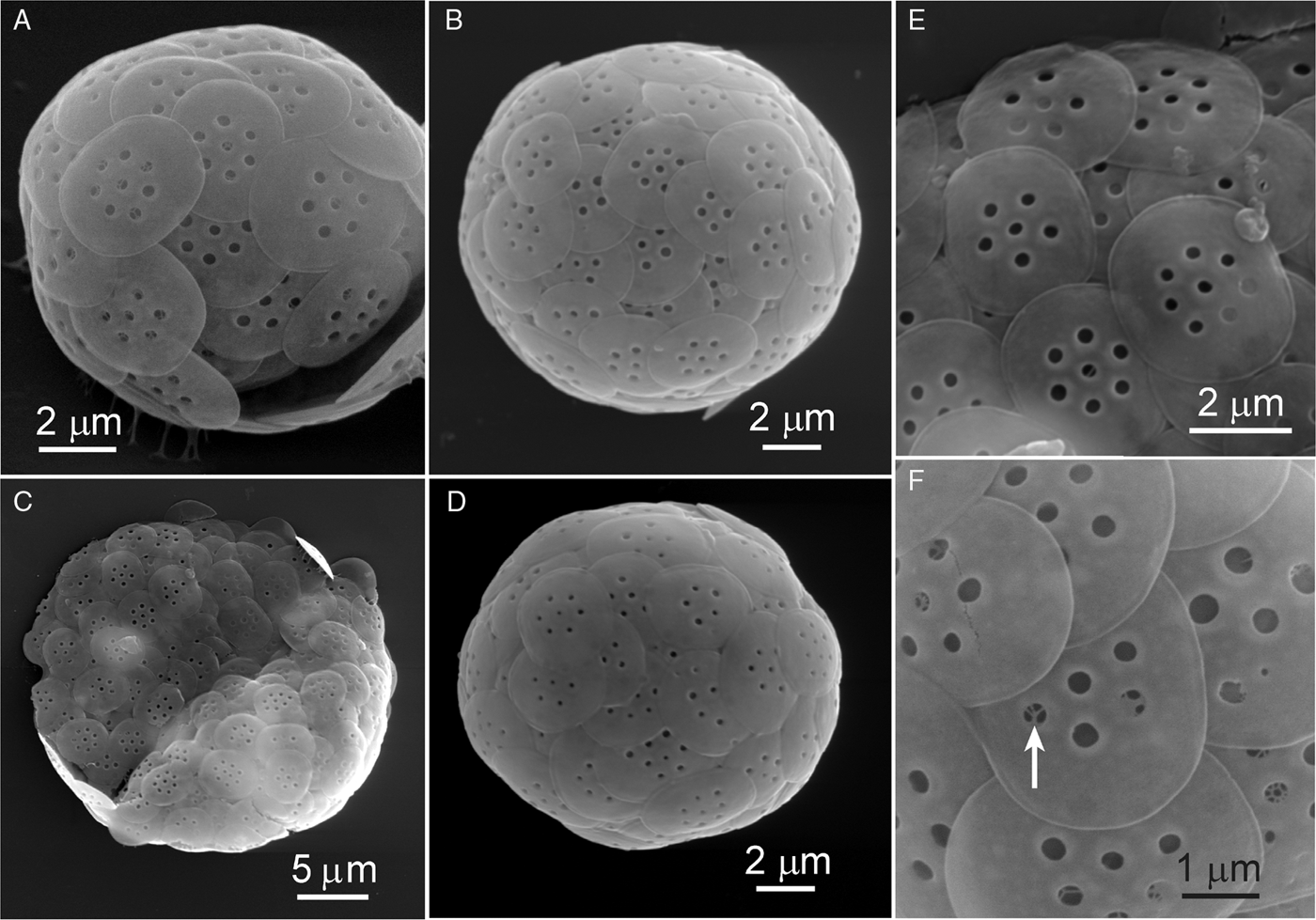
(Figure 11A–F)
The dimensions given here are representative of the morphology found in specimens best represented by the two whole cells portrayed in Figure 11A, B. Other specimens, with different cell size and scale morphology, may be representative of undescribed species, but are included here for comparisons only at this time, because only one whole cell of each of these forms has been studied (Figure 11C, D) and designation as new species is probably premature.
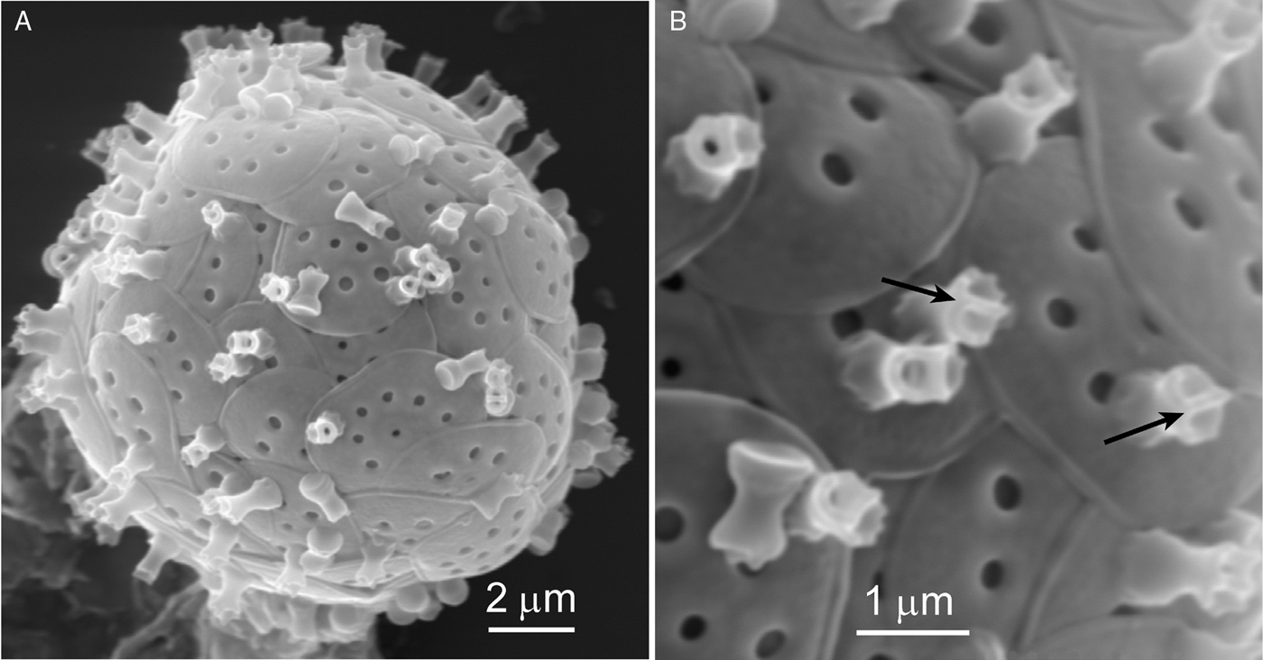
Pinaciophora rubicunda (A, B, E, F), and two unnamed ‘morphotypes' (C, D): (A, B) scanning electron microscopy images of whole cells showing their investitures of plate-scales; (E, F) details of plate-scale morphology showing the typical pattern of holes in the distal surfaces of the scales, including a ‘keyhole' view of the underlying reticulate meshwork that separated the proximal and distal plates of the scale (arrow).
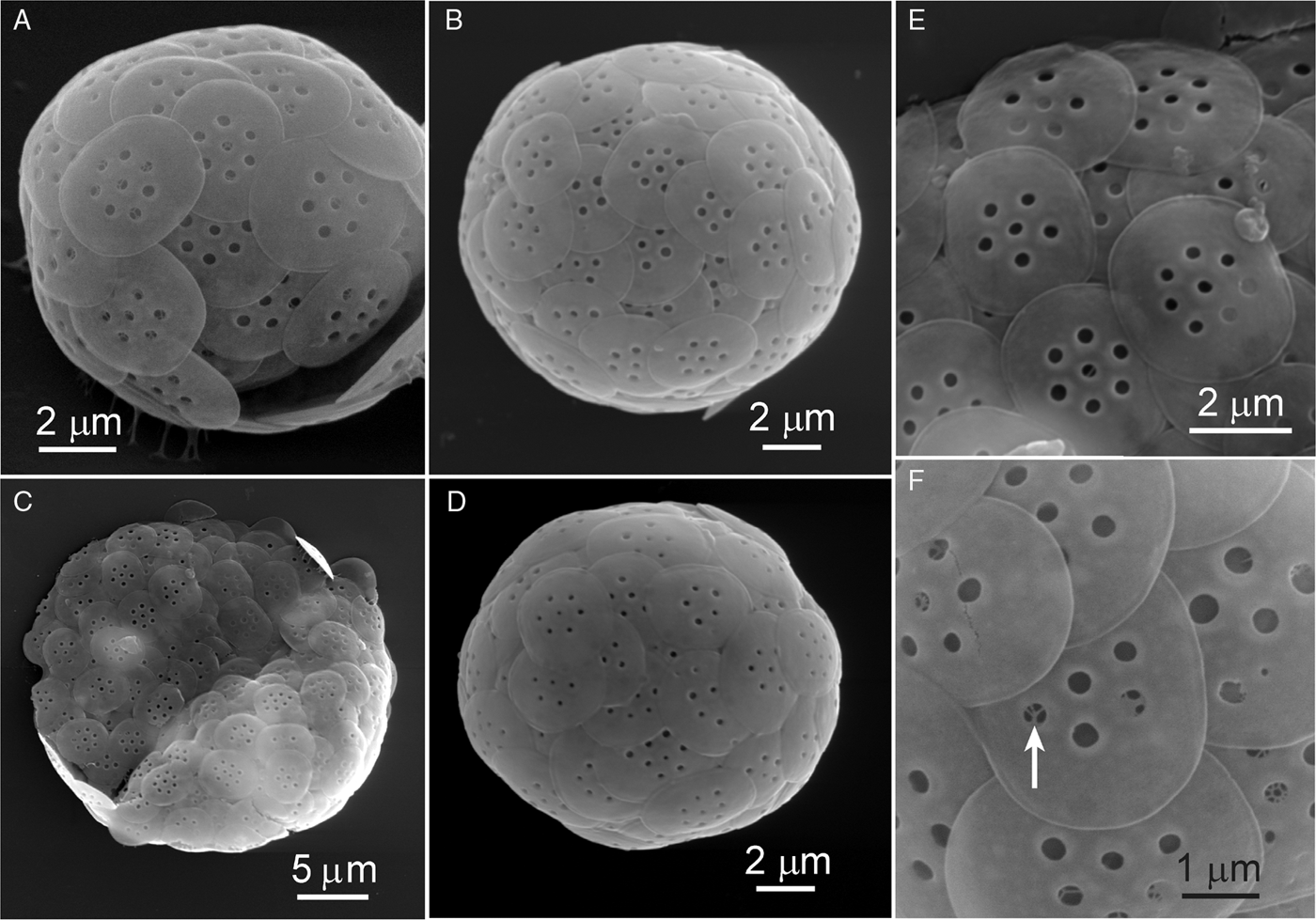
Cell size 10–15 µm in diameter. Plate-scales circular to elliptic, 3.0–3.5 µm wide × 3.5–4.3 µm long, ornamented with usually one central hole in the distal layer of the scale surrounded by 5–7 generally equally-spaced holes 0.19–0.35 µm in diameter. Short segments of the internal meshwork in the space between the outer and inner scale layers could be seen through several of the holes in the outer (distal) layer (Figure 11F). A slightly thickened rim about 0.07 µm wide was present around the scale margin, presumably where the two layers were fused.
REMARKS
The Canadian specimens agree generally with the material from the Baltic Sea (Roijackers & Siemensma, Reference Roijackers and Siemensma1988), except that cell size is smaller (mean size of the Baltic cells was 22 µm in diameter) and in scale size (up to 5.3 µm in the Baltic material)). The size of the holes on the distal surface of the plate-scales would also appear to be smaller in the Canadian specimens (judging by comparisons with Roijackers & Siemensma (Reference Roijackers and Siemensma1988; their figure 3; but measurements of hole sizes were not explicitly given for the Baltic specimens)). Notwithstanding the size differences mentioned above, I have assigned the specimens in Figure 11A, B, E and F to P. rubicunda. If, however, other specimens that agree more closely with the Baltic specimens described by Roijackers & Siemensma (Reference Roijackers and Siemensma1988) are found in the Canadian Pacific study area, then a case could be made for elevation of the specimens listed above to new species status. This is further complicated by the finding in the Canadian Pacific samples of a much larger specimen (Figure 11C) and another with much smaller holes on the plate-scales (Figure 11D). Because both of these have arrangements of holes on the scales that are like those found in P. rubicunda, they are included here for comparison purposes only, but cannot be identified as P. rubicunda or any other Pinaciophora species at this time.
I cannot agree with Mikrjukov's (1999) conclusion that P. rubicunda (sensu Roijackers & Siemensma, Reference Roijackers and Siemensma1988) is identical to Rabdiophrys thomseni Rojackers & Siemensma, 1988, which has abundant and distinctive spine-scales. Mikrjukov (Reference Mikrjukov1999) illustrated a spine-scale and a plate-scale (his figure 3) labelled as ‘P. rubicunda' that should have been labelled P. thomseni (now Thomseniophora thomseni (Roijackers & Siemensma, Reference Roijackers and Siemensma1988) comb. nov.) As Roijackers & Siemensma (Reference Roijackers and Siemensma1988) point out, there are many ‘Pinaciophora/Rabdiophrys' species with similar plate-scales. Furthermore, there is no evidence that the absence of spine-scales is a facultative phenomenon in Pinaciophora. Indeed, the absence of spine-scales in the freshwater Pinaciophora fluviatilis, arguably the best studied and most widespread of the rotosphaerids, is a well-established fact. Although the size of the plate-scales in both T. thomseni and P. rubicunda is similar, the diameter of the holes in the distal surface is about 50% smaller in P. rubicunda. Most importantly, the absence of spine-scales in the Canadian Pacific (this paper) and Swedish Baltic (Roijackers & Siemensma, Reference Roijackers and Siemensma1988) specimens, together with the discovery of a third species of Pinaciophora (P. marina; see below), further strengthens the validity of P. rubicunda and enhances the distinction between the genus Pinaciophora and the spine-scale bearing Thomseniophora.
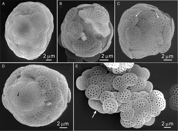
Pinaciophora marina sp. nov.
(Figure 12A–E)
Pinaciophora marina sp. nov.: (A–D) scanning electron microscopy images of whole cells with their investitures of plate-scales; (C) atypical scales (arrows) with just a single centrally-located hole in the distal surface of the scale; (D) a scale with unusually small holes (arrow); (E) a patch of loose scales including a few revealing the thin proximal layer (arrow).

DIAGNOSIS
Cells 12–22 µm in diameter covered in imbricate, generally circular or (occasionally) slightly elliptical plate-scales, 3.5–5.8 µm in diameter. Scales consist of two layers joined at the peripheral margin with a slightly thickened rim, about 0.10–0.15 µm wide. The distal layer of the scale is perforated with generally two series of holes—an outer series of 7–13 holes and an inner group of 1–7 holes. Hole size (0.22–0.42 µm in diameter) was not necessarily determined by location on the scales; the largest holes were, however, usually located in the outer series and the smallest holes were located in the inner region of the scale. Holes of the large type were never found to represent the inner series occupying the central area of the scale unless only 1–2 such holes were present in that part of the scale. Some scales included an apparently rare occurrence of 1–2 tiny holes (0.1 µm) at unpredictable positions on the scales. The underside (proximal surface) of the plate-scales appears thin and semi-transparent in SEM, often revealing the holes of the upper (distal) surface (e.g. Figure 12E).
ETYMOLOGY
The species name (marina) refers to the marine habitat in which this species has been found.
TYPE SPECIMEN
TYPE LOCALITY
Pacific Ocean beach near Gillatt (Grassy) Island, Haida Gwaii (Queen Charlotte Islands), British Columbia, Canada (53.241°N 131.898°W); salinity = 32 ppt; sample collected 8 December 2008.
MATERIAL FROM TYPE LOCALITY
Retained by the author (aqueous dilute formalin solution) as preserved sample No. V-2111.
REMARKS
Measurements were made on five intact spherical cells (four of which were illustrated—Figure 12A–D); two other partially collapsed cells were used for study of the scales and suggest that this species was relatively common in the Pacific Ocean coastal sample. It is possible that the specimen in Figure 12A represents a species distinct from the others included here in P. marina. The reasons include: (1) the cell size (22 µm) is outside the median cell size of 12.7 µm for the other four specimens; (2) minimum (4.2 µm) and maximum (5.6 µm) scale diameters of the specimen in Figure 12A were greater than the corresponding median values for the other four specimens of 3.6 µm and 4.4 µm, respectively; and (3) the largest holes in the distal surface of the scales in Figure 12A were always in the outer ring of holes and never among the group of 3–7 smaller holes occupying the central region of the scale; distribution of holes by size in the other four specimens of P. marina were more variable and included some scales with just single large hole in the centre of the scale (Figure 12C, arrows) or of a larger number of central region holes, all of about the same size as the holes in the outer ring.
Takahashi (Reference Takahashi1981) included two scales that he assigned to P. fluviatilis (we now know that cannot be the case, because they lack the large central holes combined with the peripheral ring of small holes; see below). His scales also had a few of the largest holes located in the outer ring, not unlike the distribution noted above for the specimen in Figure 12A. Takahashi's two scales were 5.8–5.9 µm × 4.8–5.2 µm (the scales in Figure 12A ranged between 4.2 and 5.6 µm). Roijackers & Siemensma (Reference Roijackers and Siemensma1988) also drew attention to the curious occurrence of the larger holes in the outer ring of holes in Takahashi's (1981) scales. The rediscovery of this consistent pattern in the scales of a whole intact cell (Figure 12A), lends considerable weight to the notion that this ‘morphotype' likely represents a distinct species. Still, because only one whole cell was studied, and because of the wide variability evident in scales of P. marina (see below), I suggest it may be premature not to include the specimen in Figure 12A among the range of forms known for P. marina.
The scale structure in P. marina was quite variable. On any individual cell, considerable differences were found among some scales in the arrangement and size of holes in the distal surfaces. Generally, when only one or two holes occupied the central region of a scale, they were larger than in those scales where the central region had three or more holes, in which case they were smaller than the holes in the outer region of the scale (see e.g. Figure 12C).
Despite this variability, transmission electron microscopy (TEM) and SEM images of scales of P. marina can be easily distinguished from those of the freshwater P. fluviatilis which has a series of smaller marginal holes surrounding the main patch of 12–18 more centrally located holes. TEM and SEM images of scales of P. fluviatilis were illustrated by Belcher & Swale (Reference Belcher and Swale1978; their figure 1), and by Roijackers & Siemensma (Reference Roijackers and Siemensma1988; their figures 1 and 2). Incidentally, because of the low magnification, Roijackers & Siemensma's (1988) figure 1 does not reveal the outer ring of small holes on the scales, but figure 64c in Page & Siemensma (Reference Page and Siemensma1991) is clearly taken from the upper right side of figure 1 in the 1988 paper and the higher magnification of their figure 64c clearly shows the outer ring of small holes in the scales. Other EM images of P. fluviatilis scales from freshwater locations include figure 5 in Kalb (1979; Borgwallsees, near Stralsund, Germany) and figure 2c in Esteban et al. (Reference Esteban, Gooday and Clarke2007; Priest Pot, UK).
Pinaciophora marina would appear to have a wide geographical distribution. In addition to the Pacific Ocean (Canada) location reported here, EM images of scales that might be assigned to this species include: figure 14 in Thomsen (Reference Thomsen1978; Isefjorden, Denmark), figures 11a, b in Gaarder et al. (Reference Gaarder, Fryxell and Hasle1976; Gulf of Mexico), and three reports from the Antarctic Ocean: figure 7 in Croome (Reference Croome1987a); figure 2a in Esteban et al. (Reference Esteban, Gooday and Clarke2007); and figures 27–28 in Takahashi (Reference Takahashi1981). All of these earlier reports were based on findings of single scales and in each case these specimens were erroneously assigned to P. fluviatilis. Several reports of ‘P. fluviatilis' from marine habitats have included images of scales that are similar to those of P. marina, but have also included images of other scales assigned to P. fluviatilis that are now known to be the scales of other rotosphaerids. For example, all of the scales illustrated by Gaarder et al. (Reference Gaarder, Fryxell and Hasle1976), with the likely exception of their figure 9, are now known to originate in taxa other than P. fluviatilis, including Rabdiaster reticulata (Thomsen, Reference Thomsen1979) Mikrjukov Reference Mikrjukov1999, Thomseniophora denticulata, T. spiculata and T. tridentata (and P. marina per their figure 11, mentioned above). Similarly, of the four scales attributed to P. fluviatilis by Kalbe (Reference Kalbe1979), only one can correctly be assigned to this taxon; all others were collected from marine habitats and their structures do not correspond to the typical freshwater P. fluviatilis (per Belcher & Swale, Reference Belcher and Swale1978; Page & Siemensma, Reference Page and Siemensma1991). Nicholls (Reference Nicholls1983) included a TEM image of a single scale from Lake Erie that he identified as P. fluviatilis. In retrospect, this scale now likely should be assigned to P. marina. Its occurrence in Lake Erie might be explained by the fact that its origin could well have been ship ballast water discharge, an activity responsible for the introduction of large volumes of seawater with their associated marine protists into the Laurentian Great Lakes. Only one scale, somewhat degenerate in appearance (Nicholls, Reference Nicholls1983; his figure 8) was found, and would suggest that it was not of recent biological origin, likely explaining its occurrence in fresh water.
It should be mentioned that although Takahashi's (1981) two scales (mentioned above) could be included in P. marina, there are some differences with the type material from Canada: these include a larger number of small holes in the central region of the scales of the Canadian material (up to 7) and the presence of a few ‘extra' small holes at apparently random locations on the scales of Takahashi's specimens. Should whole cells with plate-scales in better agreement with the two loose scales illustrated by Takahashi (Reference Takahashi1981) ever be found, then a case might be made for splitting this P. marina into two separate species.
Roijackers & Siemensma (Reference Roijackers and Siemensma1988) have pointed out the risks in attempting to assign single scales to any particular taxon, owing to the highly similar morphology of the plate-scales of many species. Some loose plate-scales of P. marina could resemble certain loose plate-scales of the spine-scale bearing T. denticulata, for example. The difficulty in identifying single isolated plate-scales is further emphasized here with the illustration of scale variability within a single cell of P. marina. The SEM investigation of several whole cells of P. marina, none of which were associated with spine-scales reinforces the dissimilarity between the genera Pinaciophora and Thomseniophora, especially since whole cells of P. rubicunda (sensu Roijackers & Siemensma, Reference Roijackers and Siemensma1988) were also present in these Canadian Pacific collections and revealed no morphological integration among any of the three known Pinaciophora species (including the freshwater P. fluviatilis).
GENUS RABDIASTER MIKRJUKOV, 1999
Rabdiaster reticulata (Thomsen, Reference Thomsen1979) Mikrjukov, Reference Mikrjukov1999. emend.
(Figures 13A–H; 14A, B)
[=Pinaciophora reticulata Thomsen, 1979]
[=Rabdiophrys reticulata (Thomsen, Reference Thomsen1979) Roijackers & Siemensma, 1988]
Rabdiaster reticulata: (A, C, D) whole cells with investitures of plate-scales and spine-scales; (B, G, H) higher magnification transmission electron microscopy images of scale structure; (E, F) scanning electron microscopy images of a single plate-scale with the contrast optimized in (F) to reveal the meshwork structure of scale material located between the unornamented distal and proximal surfaces of the scale. Bar in F (also applies to E) = 1μm.

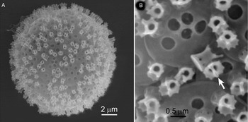
Rabdiaster reticulata: (A) scanning electron microscopy (SEM) image of a collapsed whole cell with its investiture of plate-scales and spine-scales; (B) higher magnification SEM image showing distal surfaces of plate-scales revealing (as small bumps and cavities on the external surfaces (arrows)) the underlying reticular material between the two scale layers.

AMENDED DIAGNOSIS
Cells 15–25 µm in diameter, with tubular spine-scales 2–6 µm long with a solid disc-like base 0.5–0.7 µm in diameter and about 0.1 µm thick. The middle region of the spine shaft with parallel walls in optical cross-section, about 0.35 µm in diameter; some spines with internal short, curved rib-like thickenings at irregular intervals throughout the length of the spine shaft. Distal end of the spine-scales flared to about 0.35–0.65 µm in diameter with a scalloped rim ornamented with 3–5 tooth-like projections. Plate-scales, 2.5–3.2 µm in diameter, comprised a double layer of siliceous sheeting, unornamented or with a few small depressions in the distal sheet (visible in TEM images of obliquely oriented scales). The space between the double layer is filled with a maze-like pattern of ribs and/or hexagonal or polygonal meshes revealed in TEM images as electron-dense dark lines and forming an elaborate ribbed network with the largest and more completely structured meshes located near the centre of the scale. Aberrant plate-scales with non-circular outlines (smaller with rounded polygonal outlines) occasionally present.
REMARKS
Rabdiaster reticulata was found in the 8 December 2008, Pacific Ocean beach sample referenced above. This species is close to Rabdiaster pertzovi (Mikrjukov, Reference Mikrjukov1994) Mikrjukov, Reference Mikrjukov1999 known only from Kandalaksha Bay of the White Sea, Russia. The differences include the presence of a cluster of small tubercules on the surface of the swollen basal portion of the spine shaft in R. pertzovi which was not found in the R. reticulata. Mikrjukov (Reference Mikrjukov1994) reported separate short and long spine-scale lengths in R. pertzovi; the total range spanning both length categories corresponded to the range in spine-scale lengths reported here for R. reticulata, but lengths were continuous, and did not fall into two distinct size-categories.
Until the discovery of whole cells of this taxon reported here (Figures 13A, C, D & 14A), only occasional loose scales had been reported (Vørs, Reference Vørs1993; Esteban et al., 2007)—a statement that also applies to the type material (Thomsen, Reference Thomsen1979). None of those loose scales had been investigated by SEM, which, in this study has revealed the presence of double walled plate-scales, and is in contrast to the earlier presumption based on TEM that plate-scales in this species consisted of a single layer. TEM is, however, necessary to visualize the inter-layer structure of polygonal meshes and discontinuous concentric patterns (Figure 13B, G, H). SEM has some utility in revealing the inter-layer complexity, however, if the images are optimized by digitally adjusting brightness and contrast (Figure 13E, F). This inter-layer infrastructure was also revealed in SEM images as a ‘lumpy' surface, especially in the central regions of the distal surface of the scales (Figure 14B).
Rabdiaster reticulata should remain in Rabdiaster, at least for the present, owing to the lack of any holes in the distal surface of their plate-scales; further molecular–genetic assessments may show this to be an artificial criterion for separation from Thomseniophora.
I do not believe that R. apora and R. reticulata are synonymous, as claimed by Roijackers & Siemensma (Reference Roijackers and Siemensma1988). Rabdiaster reticulata differs from R. apora (Croome, Reference Croome1987a) by: (i) the markedly smaller ‘scallops' and the more numerous and smaller marginal ‘teeth' on the distal margin of the spine-scales of R. apora; (ii) shorter spine-scales with a larger basal diameter (the ratio of spine length to diameter of the base is about 4.3 in R. apora, but 7.9 in R. reticulata); (iii) the fine perforations (micro-pores) on the plate-scale surfaces of R. apora are of larger diameter and more closely arranged than those in R. reticulata (in R. reticulata, the micro-pores are separated by distances of about 4–5 micro-pore diameters, while in R. apora, inter-pore distances are only about one micro-pore diameter); and (iv) plate-scales in R. reticulata are about 25% larger than those in R. apora. There is some risk in making detailed comparisons between these two species since it is possible that Croome (Reference Croome1987a) based his R. apora measurements on a single spine-scale and a single plate-scale (his figure 12) since he points out that whole intact cells were not found. Significantly, R. apora was found in a dilute, soft water habitat (specific conductance = 30 µmhos cm−1), while the habitat for R. reticulata reported here was seawater with salinity of 32 ppt. Incidentally, another point of confusion among taxa of this group includes the fact that Mikrjukov's (1994) drawing of the plate-scale of Rabdiaster pertzovi was used by him (Mikrjukov, Reference Mikrjukov1999) to illustrate both R. apora and R. pertzovi; the image has been rotated about 170° and differently scaled, but is otherwise identical. Clearly, more specimens of R. apora are needed to assess its relationship to both R. reticulata and R. pertzovi Mikrjukov, Reference Mikrjukov1994.
Rabdiaster multicosta (Thomsen, Reference Thomsen1978) comb. nov.
(Figure 15A, B)
[=Pinaciophora multicosta Thomsen, 1978]
[=Rabdiophrys multicosta (Thomsen, Reference Thomsen1978) Roijackers & Siemensma, 1988]
This species was previously known only from a patch of loose scales from Dybsø Fjord, Denmark (Thomsen, Reference Thomsen1978). A single whole cell of this taxon was found in the Haida Gwai collection of 8 December 2008. Two features of its morphology allowed the specimen to be readily identified: the nail-head like structure of the spine-scale bases, 1.4–1.6 µm in diameter, and the distinctively flattened and flared spine apices. The spine apex consists of two splayed edges of the spine with a series of stacked curved ribs that bridge the two edges. This is best observed in TEM (see Thomsen's (1978) figure 35). In SEM it is revealed by a rounded dome in the middle of the spine-scale apex (arrows in Figure 15A). The spine-scale lengths of the Canadian specimen were 1.8–4.5 µm long including the disc-like base. The spine shafts at about the mid-length point were about 0.45 µm in diameter. The plate-scales were circular, about 3–3.5 µm in diameter and were devoid of holes in the distal surface (Figure 15B), thus providing justification for transferring this taxon from Rabdiophrys to Rabdiaster, consistent with Mikrjukov's (1999) treatment of this group.
Rabdiaster multicosta: (A, B) two scanning electron microscopy images of the same whole cell with its investiture of plate-scales and spine-scales; (A) contrast adjusted to observe the minute ‘bud' produced in the middle of the flared apex of the spine-scale attributed to the stacked costae spanning the two margins of the scale apex; (B) contrast adjusted to observe the overlapping circular plate-scales.

DISCUSSION
Mikrjukov's (1999) reasons for justifying the reunification of pinaciophorids possessing only plate-scales with those species possessing both spine-scales and plate-scales were: firstly, he believed that Rabdiophrys thomseni Roijackers & Siemensma, Reference Roijackers and Siemensma1988 (now Thomseniophora thomseni) was a junior synonym of Pinaciophora rubicunda Hertwig & Lesser, 1874 emend. Roijackers & Siemensma, Reference Roijackers and Siemensma1988, because their plate scales were ‘identical' and because the lack of spine-scales in P. rubicunda was a facultative character; and secondly, as a result of this supposed synonymy, only one species lacking spine-scales was left (P. fluviatilis), about which Mikrjukov's argument about facultative loss of spine-scales was applied, thus rendering the spinelessness of P. fluviatilis an artificial generic character.
There are several problems with these arguments: Mikrjukov (Reference Mikrjukov1999) confusingly included P. rubicunda twice in his key to Pinaciophora—the first time among those species with plate-scales only, and then among a second group of species possessing both plate-scales and spine-scales. There is no question that Roijackers & Siemensma (Reference Roijackers and Siemensma1988) recognized the high degree of similarity between the plate-scales of several Pinaciophora (sensu latto) species (e.g. Rojackers & Siemensma (1988) state (p. 244) that P. rubicunda plate-scales ‘are identical' to those of Pinaciophora denticulata Thomsen, Reference Thomsen1978, and ‘almost identical' (p. 239), or ‘are identical' (pp. 241 & 244), or ‘are nearly identical' (p. 245) to those of their Rabdiophrys thomseni). It is, therefore, not clear why Mikurukov (1999) identified synonymy of P. rubicunda with one but not the other of these two species (R. thomseni and P. denticulata) which have plate-scales similar (or ‘identical') to those of P. rubicunda. As was reinforced by the rediscovery of P. rubicunda reported here from Canada's Pacific coast, its plate-scales are not ‘identical' to those of Thomseniophora thomseni—the main difference being the size of holes on the distal surface of the plate-scales of P. rubicunda being up to 50% smaller than those in T. thomseni (the Swedish specimens illustrated in Roijackers & Siemensma (Reference Roijackers and Siemensma1988) and from the south-west coast of Finland). I agree with Roijackers & Siemensma's (1988) statement that Thomsen's (1979) figure 53 of ‘an aberrant P. denticulara scale' from the south-west coast of Finland was likely from R. thomseni (now Thomseniophora thomseni).
Mikrjukov (Reference Mikrjukov1999) said ‘Thomsen (Reference Thomsen1978) points out some species now considered as Rabdiophrys might have 2–3 spicules only, and hence the presence of spicules could be a facultative feature. Thus there is no clear border between Pinaciophora and Rabdiophrys'. In fact, Thomsen (Reference Thomsen1978) did not say that the absence of spine-scales was a facultative phenomenon; neither was there any mention of Rabdiophrys, and Rainer (Reference Rainer and Dahl1968) was not cited in Thomsen (Reference Thomsen1978, Reference Thomsen1979). Had Thomsen been aware of Rainer's genus Rabdiophrys, he might have considered it as the logical destination of the several spined Pinaciophora he described in his 1978 paper (as Roijackers & Siemensma (Reference Roijackers and Siemensma1988) did 10 years later). Thomsen (Reference Thomsen1978) did point out that ‘… P. fluviatilis appears to be covered with only one type of scale; so far this is a unique feature within the genus'. The facts that both species (T. thomseni and T. denticulata) have spine-scales and the P. rubicunda described by Roijackers & Siemensma (Reference Roijackers and Siemensma1988) does not (their figure 4, and confirmed by the Canadian material presented in this paper), is grounds for reinstating R. thomseni (but as Thomseniophora thomseni).
Roijackers & Seimensma (1988) considered Pinaciophora apora Croome, 1987 to be identical to P. reticulata Thomsen, Reference Thomsen1978 (see their p. 240). Mikrjukov (Reference Mikrjukov1999) kept them separate, but under the new genus Rabdiaster, which he defined as rotosphaerids with both spine-scales and plate-scales, the latter lacking the large holes that characterize the distal surface of the plate-scales of Pinaciophora. Although R. apora has been reported only once (Croome Reference Croome1987a), scales of Rabdiaster reticulata, first described by Thomsen (Reference Thomsen1979) were also reported by Vørs (1992; her figure 19), Manton & Sutherland (Reference Manton and Sutherland1979; their figure 3, identified as an aberrant scale of P. fluviatilis), and by Esteban et al. (Reference Esteban, Gooday and Clarke2007). Until now, the lack of a secondary scale layer of the type found in Pinaciophora was considered to be an important distinguishing feature of R. reticulata (Thomsen, Reference Thomsen1979). Rojackers & Siemensma (1988) considered this an artefact of either incomplete scale formation or of scale deterioration in R. apora, but they may have intended to say this about R. reticulata rather than R. apora, because R. apora has been reported only once (Croome, Reference Croome1987a) where it was clearly stated that the plate-scales of this species comprised both proximal and distal plates. There is no strong evidence for loss of a scale layer by deterioration or incomplete formation.
The Canadian material reported here for R. reticulata includes the most complete examples of scales for this species (whole intact cells were found with complete complements of spine- and plate-scales examined with both TEM and SEM). These images confirm the double-layered construction of the plate-scales. Evidently, the earlier conclusion about existence of a single scale layer only in R. reticulata plate-scales arose from the availability of TEM images of scales only; SEM clearly shows the double scale layer (but is inadequate for resolution of the reticular meshed pattern of inter-layer scale material). On this basis, the definition of the genus Rabdiaster is simplified to the single difference being that the three known members (R. reticulata, R. apora and R. pertzovi) differ from Thomseniophora and Rabdiophrys in their absence of holes on the distal surfaces of their plate-scales.
Based on the single 8 December 2008 collection, it would appear that the Pacific Ocean Haida Gwai area harboured a diverse assemblage of rotosphaerids. Additionally, there was consistency in scale morphology within several species (many whole cells with intact scaly periplasts were investigated). Some of the morphological differences were small among small groups of taxa with similarities in scale structure (e.g. the T. emarginata–T. brevispina–T. minima group, and the T. m. muticata–T. m. acuminata group) and under different circumstances (different samples in space and time) such differences might have been considered taxonomically trivial because of possible environment influences on scale morphology. In this case, however, these taxa all existed under the same environmental conditions of temperature, salinity, etc. I conclude therefore, that genetic differences were more likely the cause of the diversity in silica-scale morphology and provided the justification for description of several new taxa. Recent molecular biological investigations have generally confirmed that far more taxonomic diversity likely exists among some protists than previously believed on the basis of classical morphological descriptors alone (Howe et al., Reference Howe, Bass, Scoble, Lewis, Vickerman, Arndt and Cavalier-Smith2011). Although that approach was well outside the scope of the present study, it would be instructive to include the taxa identified here in such studies in the future.
ACKNOWLEDGEMENTS
Doug Burles (Gwaii Haanas National Park Reserve and Haida Heritage Site) and Zoe Lucas collected samples for me from the Canadian Pacific and Atlantic coasts, respectively. Karen Rethoret, Biology Department, York University, assisted with the scanning electron microscopy and transmission electron microscopy operations.