Introduction
Biomineralization in bacterial systems
Bacteria are tiny organisms; however, their role in shaping our environment has been anything but small. Microorganisms are the oldest form of life on Earth and therefore have had a long time to interact with their surroundings, where they have left their mark. Their imprint can be found in many places, for example, in banded iron formations, which still serve as a source of iron ore today.Reference Widdel, Schnell, Heising, Ehrenreich, Assmus and Schink1 In addition, a microorganism rarely exists alone; they are found in colonies composed of billions of cells in close proximity.
As such, it is not surprising that due to their high capacity of adaptation, microorganisms have been able to evolve different strategies to cope with their environment. This encompasses the possibility to use a large variety of chemical elements for physiological support and as sources of energy and nutrition for growth. In some instances, this process can be accompanied by the mineralization of the element as a byproduct of respiration.Reference Croal, Gralnick, Malasarn and Newman2–Reference Frankel and Bazylinski4 As such, there is little control over the materials formed by such reactions; therefore, the interest in using them as functional materials for specific applications is dampened.
Alternatively, some microorganisms are able to intracellularly form minerals.Reference Lin, Benzerara, Faivre and Pan5,Reference Bazylinski and Frankel6 In such cases, the material is typically formed as the result of genetic information, and the subsequent process enables the organisms to control the properties of the outcomes. The materials formed offer several properties, which are not necessarily matched by human-made materials synthesized with the same element. Several elements are the subject of such transformations. One microorganism that has evolved as a model organism in this regard is magnetotactic bacteria.
This article focuses on this type of prokaryote (a single-celled organism that lacks membrane-bound organelles or nucleus as opposed to eukaryotes) since their biomineralizing capabilities have long been recognizedReference Blakemore7 and have spawned a multidisciplinary research effort in molecular biology, genetics,Reference Schüler8,Reference Cornejo, Abreu and Komeili9 geobiology, bio- and nanotechnologies,Reference Lang, Schüler and Faivre10,Reference Matsunaga, Suzuki, Tanaka and Arakaki11 as well as materials science.Reference Carillo, Vach, Faivre, Fratzl, Dunlop and Weinkamer12 This holds potential implications for understanding the origin of life on Earth and our search for life on Mars.Reference McKay, Gibson, Thomas-Keprta, Vali, Romanek, Clemett, Chilier and Maechling13
Magnetotactic bacteria
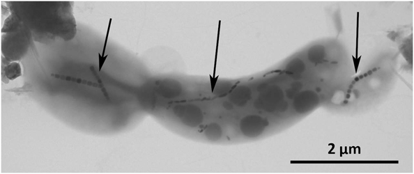
Magnetotactic bacteria, shown in Figure 1, are ubiquitous microorganisms found in any aqueous environments. Magnetotactic bacteria share a common trait in that they are capable of forming intracellular magnetic nanoparticles in vesicles called magnetosomes.Reference Balkwill, Maratea and Blakemore14 In addition to existing in a wide variety of environments, magnetotactic bacteria also exist with varying morphologies, exhibit varying flagellar apparatusReference Lefèvre and Bazylinski15 and can synthesize different mineral phases. The magnetosome crystals can be made of iron oxide magnetite (Fe3O4)Reference Mann, Frankel and Blakemore16–Reference Baumgartner, Morin, Menguy, Perez Gonzalez, Widdrat, Cosmidis and Faivre20 or iron sulfide greigite (Fe3S4),Reference Mann, Sparks, Frankel, Bazylinski and Jannasch21–Reference Pósfai, Buseck, Bazylinski and Frankel23 with some cells capable of forming both types of mineral.Reference Bazylinski, Heywood, Mann and Frankel24,Reference Lefèvre, Menguy, Abreu, Lins, Pósfai, Prozorov, Pignol, Frankel and Bazylinski25 This variety also extends to particle morphology, for example, while a given strain always forms particles with the same morphology, the morphology can differ from strain to strain.Reference Devouard, Posfai, Hua, Bazylinski, Frankel and Buseck26 Magnetosomes are typically arranged in a linear fashion along the long axis of the organism to form a magnetosome chain, which is a mechanically stable structure,Reference Körnig, Dong, Bennet, Widdrat, Andert, Müller, Schüler, Klumpp and Faivre27 primarily due to the role of two particular magnetosome membrane (MAM) genes mamJ and mamK.Reference Komeili, Li, Newman and Jensen28,Reference Scheffel, Gruska, Faivre, Linaroudis, Plitzko and Schüler29 The magnetosome organization can vary from a single chain to several parallel chains of inorganic nanoparticles and to clusters.Reference Faivre and Schüler30
Transmission electron microscopy (TEM) image of a sample of magnetotactic bacteria (obtained from Camargue, France). The image shows the coexistence of three cells with different sizes and morphologies. In addition, the magnetosomes also exhibit different morphological features and organization. Black arrows point toward the magnetosome chains. TEM image courtesy of Christopher Lefèvre.

Genetic background
The magnetosome membrane separates the magnetosome organelle from the cytoplasm (the inner part of the bacteria) and shares some common properties with the inner membrane of the bacterial cell wall. Transiently, the membrane is an invagination of the inner membrane, which means that the magnetosome vesicles originates from and is bound to the bacterial inner membrane.Reference Komeili, Li, Newman and Jensen28,Reference Gorby, Beveridge and Blakemore31 Primary biochemical and proteomic analyses have revealed the primordial role of the magnetosome membrane in the biomineralization process.Reference Grünberg, Müller, Otto, Reszka, Linder, Kube, Reinhardt and Schüler32,Reference Matsunaga, Nemoto, Arakaki and Tanaka33
The development of sequencing techniques and molecular tools for the genetic manipulation of a restricted amount of strains has started to highlight the role of dedicated genes and proteins in the biomineralization process. In particular, it has been shown that the genes responsible for the formation of the magnetic materials and their organization are clustered in the so-called “magnetosome gene island.”Reference Grünberg, Wawer, Tebo and Schüler34–Reference Jogler and Schüler38 Separate sets of genes have been discovered for magnetite-building cells and greigite-building cells.Reference Lefèvre, Menguy, Abreu, Lins, Pósfai, Prozorov, Pignol, Frankel and Bazylinski25,Reference Abreu, Cantao, Nicolas, Barcellos, Morillo, Almeida, do Nascimento, Lefevre, Bazylinski, de Vasconcelos and Lins39
The magnetosome membrane (mam) and magnetosome membrane specific (mms) genes have been shown to be particularly important, as varying phenotypes have been described as a result of their deletion. Consequently, their role is becoming clearer in magnetosome formation and organization and associated mechanisms. Using gene mutation, an encompassing role for mam and mms genes in control of magnetosome size and morphology, iron uptake, and magnetosome filament formation has been unraveled.Reference Murat, Falahati, Bertinetti, Csencsits, Körnig, Downing, Faivre and Komeili40–Reference Komeili, Vali, Beveridge and Newman46
Recently, through deletion of a larger genomic region, it has been shown that about 20 genes were sufficient to form magnetic particles when the rest of the magnetosome genomic island was absent.Reference Murat, Falahati, Bertinetti, Csencsits, Körnig, Downing, Faivre and Komeili40,Reference Lohße, Ullrich, Katzmann, Borg, Wanner, Richter, Voigt, Schweder and Schüler43 Furthermore, the expression of part of this genomic island in a host organism has revealed not only the essential role played by the genes of the mamAB cluster, but it has also opened the door to synthetic biology approaches for genetically equipping unicellular and then multicellular organisms with magnetization properties.Reference Kolinko, Lohsze, Borg, Raschdorf, Jogler, Tu, Posfai, Tompa, Plitzko, Brachmann, Wanner, Muller, Zhang and Schuler47
The magnetotactic function
The current theory is that magnetotactic bacteria use the magnetosome chain as a compass needle. The cells passively align with the external Earth’s magnetic field lines and actively swim by the means of their flagellar rotation toward their preferred habitat in a process controlled by aerotaxis, which is the search for a given oxygen concentration.Reference Frankel, Bazylinski, Johnson and Taylor48–Reference Bazylinski and Frankel50 However, in a study published in 2014, magnetotactic bacteria in sediments have a behavior more complex than initially thought, because they cannot simply swim along field lines in a granular environment.Reference Mao, Egli, Petersen, Hanzlik and Liu51 In addition, bacteria were initially supposed to respond either to the magnetic field or the oxygen gradient. When confronted with a contradicting stimulus (change in the direction of the field),Reference Frankel, Bazylinski, Johnson and Taylor48 a third mechanism of magneto-aerotaxis was discovered, where bacteria respond to an inversion of the magnetic field direction differently at high and low oxygen concentrations.Reference Lefèvre, Bennet, Landau, Vach, Pignol, Bazylinski, Frankel, Klumpp and Faivre52
Using a multi-modal microscopy platform that employs high-speed fluorescence imaging in conjunction with diffusion and aerotactic models, an improved understanding of how oxygen consumption is linked to the field orientation has been developed.Reference Bennet, McCarthy, Fix, Edwards, Repp, Vach, Dunlop, Sitti, Buller, Klumpp and Faivre53 This method permits simultaneous mapping of the chemical environment and response of the organisms under a controlled and physiological magnetic field. The influence of the magnetic field on the formation and dynamics of a micro-aerotactic band, which is the region where cells preferentially live, showed that neither a tenfold increase of the field strength nor a tilt of 45° resulted in a significant change of aerotactic efficiency. In contrast, when the field strength is zeroed or when the field angle is tilted to 90°, the magneto-aerotaxis’ efficiency is drastically reduced. Since the classical model of magneto-aerotaxis assumes a response proportional to the cosine of the angle difference between the directions of the oxygen gradient and that of the magnetic field, such results point toward alternative mechanisms currently being tested, such as active field sensing.
In addition, our understanding of the molecular mechanism of magneto-aerotaxis has also been advanced. Cells of Magnetospirillum gryphiswaldense, for example, display swimming properties dependent on aerotactic signal transduction through one of its four chemotaxis operons (cheOp1). Additionally, the growth of cells in magnetic fields superimposed on oxygen gradients results in a gradual inherited bias of swimming runs with one of the cell poles leading, such that the resulting overall swimming direction of entire populations can be reversed by changes in oxygen concentration. These findings led to the conclusion that there is a direct molecular link between aerotactic sensing and the determination of magnetotactic polarity through the sensory pathway cheOp1.Reference Popp, Armitage and Schüler54
One-dimensional magnetic nanostructures
Since it is not obvious why the materials community should be interested in biomineralization by magnetotactic bacteria, a few points highlighting the relevance of biological magnetic materials are presented here. For instance, magnetosomes have been shown to have potential applications in hyperthermiaReference Alphandéry, Faure, Raison, Duguet, Howse and Bazylinski55,Reference Hergt, Hiergeist, Zeisberger, Schüler, Heyen, Hilger and Kaiser56 and as contrast agents for magnetic resonance imaging (MRI).Reference Hartung, Lisy, Herrmann, Hilger, Schüler, Lang, Bellmann, Kaiser and Reichenbach57,Reference Lisy, Hartung, Lang, Schüler, Richter, Reichenbach, Kaiser and Hilger58 Biomineralization of magnetosomes can serve as an inspiration for the development of novel magnetic nanomaterials for the aforementioned applications, which are more biocompatible.Reference Prozorov, Bazylinski, Mallapragada and Prozorov59 Magnetotactic bacteria are not only able to control magnetosome dimensions, they can also control the morphology of the particles they synthesize and arrange them in a chain. The materials and physics communities are interested in such 1D magnetic structures due to their high anisotropic properties.Reference Yuan, Xu and Müller60,Reference Wang, Yu, Sun and Chen61 For example, it is known that a change in morphology can dramatically impact the self-assembly of magnetic particles.Reference Ahniyaz, Sakamoto and Bergström62,Reference Singh, Chan, Baskin, Gelman, Repnin, Krat and Klajn63 Synthetic, green chemistry techniques have been developed that enable the formation of synthetic magnetic nanoparticles of similar size to magnetosomesReference Baumgartner, Bertinetti, Widdrat, Hirt and Faivre64 and that show a retention of magnetic properties even upon aging.Reference Widdrat, Kumari, Tomps, Posfai, Hirt and Faivre65
Motivated by these recent trends, this article presents the properties that make the magnetosome chain so specific and explains how these properties arise. This, in turn, may spark significant interest in the materials research community to explore and develop new strategies for obtaining 1D magnetic nanostructures so that their potential can be realized.
All for magnetism: The hierarchically structured magnetosome chain
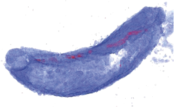
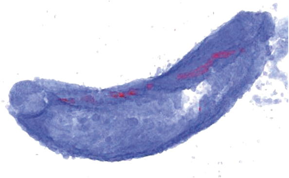
The magnetosome chain is a paradigm of a biological 1D nanostructure (Figure 2) and an assembly over several hierarchical levels. It starts with the formation of pure magnetite and ends with the positioning of several magnetosomes in a row to form a single magnetic dipole. Between these steps, not only are the dimensions (and morphology) of the particles controlled, so is their crystallographic orientation. These different stages leading to the magnetosome chains are detailed in the following sections.
Three-dimensional (3D) rendering of cryo-focused ion-beam sectioning and scanning electron microscopy imaging of Magnetospirillum gryphiswaldense. The magnetosomes are plotted in red while the magnetosome membrane is presented in blue. The magnetosomes are clearly aligned along the long axis of the cell. A scale bar is not shown since the image is a snapshot of a 3D rendering with perspective (the pixel size can change as a function of the position on the image), but the diameter of the cell is ca. 500 nm. Image courtesy of L. Bertinetti and A. Schertel.
The magnetosome ultrastructure: Pure magnetite
It was recognized early on that magnetotactic bacteria form magnetite as a mineral inclusion in their magnetosome organelles.Reference Mann, Frankel and Blakemore16,Reference Frankel, Papaefthymiou, Blakemore and O’Brien17,Reference Matsuda, Endo, Osakabe, Tonomura and Arii66 However, the presence of maghemite (α−Fe2O3), an oxidized spinel phase analog to magnetite where vacancies replace Fe(II), has been the subject of debate.Reference Frankel, Papaefthymiou, Blakemore and O’Brien17,Reference Faivre, Böttger, Matzanke and Schüler18,Reference Kopp and Kirschvink67 The magnetic properties of iron oxide nanoparticles typically depend on the mineral phase, in addition to numerous other factors. In particular, maghemite has different magnetic properties when compared to magnetite.Reference Dunlop and Özdemir68
The reduced precision of electron diffraction for lattice parameter determination has long prevented quantitative characterization of the magnetosomes mineral phase and its differences with respect to synthetic particles of similar sizes. However, with the development of synchrotron sources with high brilliance, high-resolution x-ray diffraction (XRD) has developed into a powerful technique for such experiments.
Using high-resolution XRD, significant differences in the lattice parameter were found between intracellular magnetosomes from cultured magnetotactic bacteria when measured in whole cells, which exhibited the perfect expected value of magnetite (8.3969 Å),Reference Fleet69,Reference Fleet70 and those measured after isolation from the bacteria, which exhibited slightly lower values.Reference Fischer, Schmitz, Aichmayer, Fratzl and Faivre71 Synthetic materials of similar size used as control and for comparison showed that this difference is related to different oxidation states, and the biogenic nanomagnetite found in the whole cells is stoichiometric, or structurally pure. Isolated magnetosomes and synthetic particles are slightly oxidized (an oxidation parameter of 21% was calculated when using the formalism of Dunlop and Özdemir,Reference Dunlop and Özdemir68 meaning that such particles are a mixture of about .79/.21 magnetite/maghemite). This stoichiometric magnetite is ferrimagnetic with a magnetic moment (4 µB) larger than that for maghemite (3 µB).Reference Dunlop and Özdemir68 By fitting the relationship between the magnetic moment and the oxidation parameter of Dunlop and Özdemir (68), Fischer et al.Reference Fischer, Schmitz, Aichmayer, Fratzl and Faivre71 showed that a loss of about 4% of the saturation moment was expected for the oxidized synthetic particles as compared to the genuine magnetosomes; therefore, a reduced efficacy can be expected when they are used for actuation. Since iron is very insoluble in a natural aqueous environment, it might be important for the bacteria to carefully control the fate of the iron in the biomineralization process. Besides the role of the bacterial membrane, it is currently not clear how such control is achieved at the molecular level.
The magnetosome dimension: Stable single domain
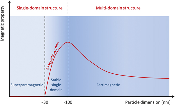
Magnetotactic bacteria of a given strain always form magnetosomes of the same dimensions of the mature crystals of a given strain, but a variety particle sizes are observed for different strains.Reference Devouard, Posfai, Hua, Bazylinski, Frankel and Buseck26,Reference Thomas-Keprta, Bazylinski, Kirschvink, Clemett, McKay, Wentworth, Vali, Gibson and Romanek72,Reference Faivre and Ukmar Godec73 The magnetic properties of magnetite nanoparticles typically depend, besides numerous other factors, on their size. Particles smaller than ∼25–30 nm are superparamagnetic, that is, they only have a measurable magnetic signal if placed in an external magnetic field (Figure 3). Larger nanoparticles have intrinsic and permanent magnetic moments. Particles with isometric shapes from 25–30 nm to ∼80–100 nm fall within the stable single-domain category. Finally, particles larger than this threshold of 80–100 nm are called multi-domain, in which several magnetic domains coexist, reducing the magnetostatic energy.Reference Dunlop and Özdemir68 The magnetosome size usually falls within the stable single-domain region, where the volume magnetization is the highest.Reference Muxworthy and Williams74,Reference Muxworthy and Williams75
Magnetic property (e.g., coercivity) as a function of the particle dimension (here, diameter). Magnetosomes are found in the stable single-domain region.

Although the chemical mechanism leading to magnetite has now been unraveled with some detail for the magnetospirilla and includes an intermediate ferrihydrite phase that is poorly crystalline,Reference Fdez-Gubieda, Muela, Alonso, García-Prieto, Olivi, Fernández-Pacheco and Barandiarán19,Reference Baumgartner, Morin, Menguy, Perez Gonzalez, Widdrat, Cosmidis and Faivre20 it is still not fully clear what factor controls the particle size. By deleting specific genes and then analyzing the bacterial phenotype, the role of several proteins in the formation of magnetosomes has been exposed. This is a somewhat indirect methodology, since other genes present in the genome of the bacteria might compensate for the loss of function upon deletion of a particular gene, and the level of expression of other related genes and proteins might also be altered. Such genetic compensation has already been observed in the case of the magnetotactic bacteria.Reference Rioux, Philippe, Pereira, Pignol, Wu and Ginet76,Reference Draper, Byrne, Li, Keyhani, Barrozo, Jensen and Komeili77 Still, this is currently the best available technique and has permitted the discovery of several genes and proteins of interest, even if the mechanism by which those biomolecules act remains unclear.
For example, the mamGFDC (the genes were called from the letter of the alphabet) cluster that encodes for membrane proteins was one of the first to be discovered.Reference Scheffel, Gärdes, Grünberg, Wanner and Schüler45 When mamGFDC are absent, the magnetosomes formed by the bacteria are smaller, in turn, when the proteins are overexpressed in the cells, the magnetosomes are larger than typical wild-type magnetosomes.Reference Scheffel, Gärdes, Grünberg, Wanner and Schüler45 The size of the empty magnetosome vesicle is also changed accordingly. But as the study points out, even for mature magnetosomes, apparently the magnetosome organelle is never fully filled by the magnetosome crystals, ruling out the possibility of simple size control by the organelle size.Reference Scheffel, Gärdes, Grünberg, Wanner and Schüler45
Another extensively studied protein is mms6,Reference Arakaki, Webbs and Matsunaga78 an acidic protein that is tightly attached to the magnetosome particles and is thought to help in the control of the magnetosome size elucidated in vitro and in vivo.Reference Tanaka, Mazuyama, Arakaki and Matsunaga44 However, in the in vivo studies, since the deletion was done in a way in which genes in proximity to mms6 were also altered, it is not fully clear if the change in magnetosome size is due to the mms6 alone. A more recent study without this limitation points toward the role of an alternative gene in size control, mmsF, which is present and expressed in the neighboring region of mms6.Reference Murat, Falahati, Bertinetti, Csencsits, Körnig, Downing, Faivre and Komeili40 Advancements in structural biology may help unravel this mechanism parallel to that of magnetosome formation more generally.Reference Nudelman and Zarivach79–Reference Siponen, Legrand, Widdrat, Jones, Zhang, Chang, Faivre, Arnoux and Pignol81 While the main molecular players are summarized here, a more detailed analysis is available of the molecular players involved in the biomineralization of magnetite in Magnetospirillum gryphiswaldense Reference Lohße, Borg, Raschdorf, Kolinko, Tompa, Posfai, Faivre, Baumgartner and Schüler42,Reference Lohße, Ullrich, Katzmann, Borg, Wanner, Richter, Voigt, Schweder and Schüler43 and Magnetospirillum magneticum. Reference Murat, Falahati, Bertinetti, Csencsits, Körnig, Downing, Faivre and Komeili40,Reference Murat, Quinlan, Vali and Komeili41
The crystal orientation: Favor the easy axis
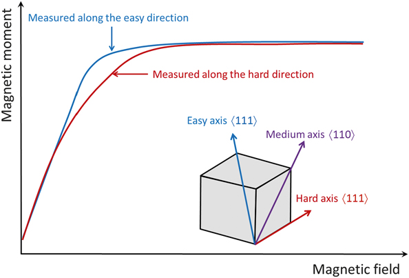
Magnetic anisotropy in magnetite nanoparticles is expressed by a squarer hysteresis loop for a magnetic field applied parallel to the direction of the alignment than for a magnetic field applied perpendicular to that direction. In the case of the magnetosome chain, this magnetic anisotropy is the combination of two parameters: the preferred orientation of the magnetosomes along their easy axis of magnetization (〈111〉) with respect to that of the hard axis (〈100〉) (Figure 4), and from dipolar interactions due to the assembly in the chain.Reference Alphandéry, Ding, Ngo, Wang, Wu and Pileni82
Schematic of magnetic moment as a function of the applied field. In a small region of the field, the magnetic moment is larger when measured along the easy axis than when measured along the hard axis of magnetite.

Early transmission electron microscopy (TEM) studies have shown that the cubooctahedral crystals of Magnetospirillum magnetotacticum are preferentially aligned with the [111] direction parallel to the chain axis of the cell, which is one of the 〈111〉 directions corresponding to the easy axis of magnetization. This result was confirmed later by electron holographyReference Dunin-Borkowski, McCartney, Frankel, Bazylinski, Pósfai and Buseck83 and the palette of bacterial strains has expanded with further high-resolution TEM studies.Reference Alphandéry, Ding, Ngo, Wang, Wu and Pileni82 In turn, particles from strains producing magnetosomes elongated along the [100] direction such as Desulfovibrio magneticus or strains MYR-1, LO-1, and AV-1 have been suggested to have their easy axis switched to that direction,Reference Pósfai, Moskowitz, Arato, Schüler, Flies, Bazylinski and Frankel84–Reference Lefèvre, Pósfai, Abreu, Lins, Frankel and Bazylinski86 possibly as a result of the shape anisotropy and magnetostatic interactions. Even particle elongation along [110] for strain HSMV-1 has been reported,Reference Lefèvre, Pósfai, Abreu, Lins, Frankel and Bazylinski86 which represents the intermediate direction of magnetization.Reference Dunlop and Özdemir68 All of these results, however, are based on a limited number of samples used in microscopy studies.
To gain further insight into the organization of magnetosome nanomaterials, two-dimensional synchrotron XRD was recently applied to cells of magnetotactic bacteria that were pre-aligned with an externally applied magnetic field to determine the crystallographic orientation of bulk magnetosomes relative to the chain axis.Reference Körnig, Winklhofer, Baumgartner, Gonzalez, Fratzl and Faivre87 This analysis revealed a [111] fiber texture along the chain direction for Magnetospirillum gryphiswaldense and Magnetospirillum magneticum, and a [100] fiber texture for Desulfovibrio magneticus. These bulk results confirm those obtained earlier for individual particles. The [100] axis is primarily energetically unfavorable because it represents a magnetic hard axis in magnetite, but can be turned into an effective easy axis by particle elongation along [100] for aspect ratios higher than 1.25, consistent with aspect ratios in RS-1 magnetosomes.Reference Pósfai, Moskowitz, Arato, Schüler, Flies, Bazylinski and Frankel84 The pronounced fiber textures can be explained possibly by strain-specific biological control of crystal orientation at the chain level, as already observed for the strain-specific size and organization of the magnetosome and of the magnetosome chain respectively. Even if this is the case, it remains questionable how such an accomplishment can be achieved biologically. Another possibility is that the physical alignment results from intra-chain magnetic interactions. In this case, biological control of the axis of elongation would be sufficient to influence the crystallographic texture of the magnetosome chain.
The magnetosome alignment: Dipoles sum up
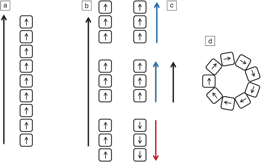
The final level of hierarchy observed in the magnetosome chain is the formation of the chain itself. The magnetic anisotropy was shown to arise mainly from the dipolar interactions between the particles. However, free magnetic particles would have the tendency to collapse and form a ringReference Philipse and Maas88,Reference Kiani, Faivre and Klumpp89 (Figure 5). Therefore, biological determinants probably play a critical role in the formation and stabilization of magnetosome chains.Reference Körnig, Dong, Bennet, Widdrat, Andert, Müller, Schüler, Klumpp and Faivre27 The molecular players involved in the process have been extensively studied and mainly involve the proteins MamJ and MamK.Reference Komeili, Li, Newman and Jensen28,Reference Scheffel, Gruska, Faivre, Linaroudis, Plitzko and Schüler29,Reference Rioux, Philippe, Pereira, Pignol, Wu and Ginet76,Reference Draper, Byrne, Li, Keyhani, Barrozo, Jensen and Komeili77,Reference Abreu, Mannoubi, Ozyamak, Pignol, Ginet and Kimeili90–Reference Katzmann, Scheffel, Gruska, Plitzko and Schüler98 Briefly, MamK is involved in the formation of the magnetosome filamentReference Komeili, Li, Newman and Jensen28,Reference Katzmann, Scheffel, Gruska, Plitzko and Schüler98 to which magnetosomes are attached via MamJ.Reference Scheffel, Gruska, Faivre, Linaroudis, Plitzko and Schüler29 However, MamJ is only present in magnetospirilla, so that the formation of the magnetosome chain in other strains is still unclear.
Sketch of the magnetic property of an assembly of individual dipoles. (a) Magnetosomes arranged in chains as in wild-type cells. The cellular dipole is the sum of the individual dipoles of each magnetosome. (b) Shorter chains can be formed as for example, in the mamK deletion mutant. The magnetic property is unchanged when compared to (a). However, the dipole of one of the shorter chains can be switched to the opposite direction (as in (c), red arrow versus blue arrows) and reduces the resulting cellular dipole (black arrow). This was also observed in Magnetovibrio blakemorei.Reference Kalirai, Bazylinski and Hitchcock99 (d) Finally, in the absence of a stabilizing biological template, isolated magnetosomes form a closed ring structure.

The formation of a magnetic dipole large enough to orient the cell in the Earth’s magnetic field is a combination of biological and magnetic forces.Reference Klumpp and Faivre100,Reference Bennet, Bertinetti, Neely, Schertel, Kornig, Flors, Muller, Schuler, Klumpp and Faivre101 First-order-reversal-curve diagrams showed that nucleation and growth of magnetosome crystals were a continuous process in Magnetospirillum gryphiswaldense,Reference Carvallo, Hickey, Faivre and Menguy102 meaning that crystals in the superparamagnetic size range were cohabiting with particles in the stable single-domain size range along the time of the chain formation. The role of the first nucleating magnetite crystal has been proposed to be critical, as it is thought to be employed as the docking station for the further growth of neighboring particles.Reference Faivre, Fischer, Garcia-Rubio, Mastrogiacomo and Gehring103 Therefore, if biological forces play a major role in the formation of a stable chain from non-magnetic bacteria,Reference Klumpp and Faivre100 when a magnetic bacterium divides, its chain is split into two identical parts, which are inherited by the daughter cells.Reference Katzmann, Müller, Lang, Messerer, Winklhofer, Plitzko and Schüler104,Reference Lefèvre, Bennet, Klumpp and Faivre105 In this case, the biological apparatus is already present, and the newly formed magnetosome particles are strongly influenced by their neighbors,Reference Körnig, Dong, Bennet, Widdrat, Andert, Müller, Schüler, Klumpp and Faivre27,Reference Körnig, Hartmann, Teichert, Fratzl and Faivre106,Reference Kirschvink107 having no other choice than to align with the right orientation as the rest of the particles forming the magnetic dipole. This will occur as soon as they are close enough.
Summary
The formation of a magnetosome chain to form a functional one-dimensional magnetic structure in microorganisms has been discussed. In the first step, the preferential formation of magnetite over the more oxidized but less magnetic maghemite occurs. In the second step, specific proteins play an important role in controlling the size of the magnetosome so as to obtain particles within the stable single-magnetic domain. The particles exhibit preferential alignment along their easy axis of magnetization. Finally, the assembly of the stable magnetosome chain occurs under the influence of both biological and magnetic components.
As a lesson from nature, it is clear that the formation of such anisotropic magnetic nanostructures in particular requires the use of stable single-domain particles and the addition of a template to stabilize the dipole and prevent collapse in close-ring structures. Under such conditions, magnetic interactions most probably promote particle orientation in a concerted fashion. However, the formation of synthetic, highly monodispersed magnetite nanoparticle within the stable single domain and the template that is necessary for the stabilization of the particles are unavailable and are therefore important research areas for future work.
Understanding magnetosomes and magnetosome chain formation is of particular relevance for the materials research community as these materials offer new insights into the assembly processes of inorganic materials in living organisms but they also offer advantages over conventional materials in applications such as MRI contrast agents.Reference Alphandéry, Faure, Seksek, Guyot and Chebbi108 More recently, by combining nanoliposomes with magnetotactic bacteria, therapeutics systems with self-propulsion capacity have been realized.Reference Taherkhani, Mohammadi, Daoud, Martel and Tabrizian109 More such advances can be expected in the near future.
Acknowledgments
D.F. thanks the past and present members of his group as well as all collaborators for their help with the research on magnetotactic bacteria. Arash Komeili and Dirk Schüler are acknowledged for their introduction to the world of magnetotactic bacteria. Thanks to Peter Fratzl for his help in the establishment of the author’s research group at the MPI of Colloids and Interfaces. Financial support from the Max Planck Society, the Deutsche Forschungsgemeinschaft (SPP 1420 (FA 835/2), SPP 1569 (FA 835/5), SPP 1726 (FA 835/7), and individual projects (BO 1762/5, FA 835/9, FA 835/10)), from the European Union (Project Bi02MaN4MRI n°245542 and Nanoathero n°305312), from COST (n°0902), and the European Research Council (Starting Grant MB2 n°256915) is acknowledged.