Introduction
The genus Sticta is defined by a unique trait: the cyphellae, well-differentiated ventral pores that facilitate gas exchange (Green et al. Reference Green, Snelgar and Brown1981; Moncada et al. Reference Moncada, Lücking and Betancourt2013a ). Its species are characteristic of humid, cool-temperate environments; in tropical latitudes of South America, where they grow luxuriously in easily accessible habitats such as andine páramos (Sipman Reference Sipman and Luteyn1999). Most species are associated with nitrogen-fixing cyanobacteria as primary photobiont and contribute substantially to local nitrogen input (Forman Reference Forman1975; Green et al. Reference Green, Horstmann, Bonnett, Wilkins and Silvester1980, Reference Green, Büdel, Heber, Meyer, Zellner and Lange1993; Green & Lange Reference Green and Lange1991). Also important is their role in water cycles, since these lichens are capable of storing up to over 500% of their dry weight as water and can release it over several hours into the neighbouring atmosphere (Green et al. Reference Green, Snelgar, Wilkins and Brown1985; Guzmán et al. Reference Guzmán, Quilhot and Galloway1990; Green & Lange Reference Green and Lange1991; Beckett Reference Beckett1995; Zotz et al. Reference Zotz, Büdel, Meyer, Zellner and Lange1998; Lange et al. Reference Lange, Büdel, Meyer, Zellner and Zotz2004). Certain species of Sticta, in particular green algal species characteristic of tropical montane rainforests, might be useful as indicators of ecological continuity and environmental health, similar to the Lobariaceae, in particular Lobaria pulmonaria, in Europe and North America (Scheidegger et al. Reference Scheidegger, Frey and Zoller1995; Zoller et al. Reference Zoller, Frey and Scheidegger2000; Radies et al. Reference Radies, Coxson, Johnson and Konwicki2009). The species-rich communities at high altitudes that are mostly associated with cyanobacterial photobionts also appear to be affected by the ongoing destruction of páramo habitats (Luteyn Reference Luteyn1999; Crespo et al. Reference Crespo, Célleri, Buytaert, Feyen, Iñiguez, Borja, De Bièvre, Hermann and Schumann2010; De Brievre & Calle Reference De Brievre and Calle2011).
In his treatment of Sticta weigelii s. lat. in eastern North America, Harris (Reference Harris1984) anticipated that the systematics of this supposedly ‘easy’ genus would become more difficult. While Harris (Reference Harris1984) chiefly referred to extratropical taxa, his assessment also applies to tropical material, since names such as S. fuliginosa and S. weigelii are commonly applied to tropical montane species. Other groups of species are either restricted to, or are most diverse in, the tropics, such as green-algal species with scrobiculate-faveolate thalli, cyanobacterial forms with apothecia, in particular those with tomentum on the upper surface, and cyanobacterial taxa with isidia and a pale underside (Delise Reference Delise1825; Nylander Reference Nylander1859, Reference Nylander1865; Joshi & Awasthi Reference Joshi and Awasthi1982; Swinscow & Krog Reference Swinscow and Krog1988; Chen et al. Reference Chen, Wang and Wang1994; Galloway Reference Galloway, Marcelli and Ahti1998a ). Yet recent discoveries of new species are rare (e.g., Aptroot Reference Aptroot2008; Lumbsch et al. Reference Lumbsch, Ahti, Altermann, Amo De Paz, Aptroot, Arup, Bárcenas Peña, Bawingan, Benatti and Betancourt2011). Currently, c. 120 species are accepted (Kirk et al. Reference Kirk, Cannon, Minter and Stalpers2008). This is due to the fact that revisionary treatments have focused on extratropical regions (Galloway Reference Galloway1994, Reference Galloway1997, Reference Galloway1998b , Reference Galloway and McCarthy2001; Galloway et al. Reference Galloway, Stenroos and Ferraro1995; Galloway & Thomas Reference Galloway, Thomas, Nash, Ryan, Diederich, Gries and Bungartz2004; Jørgensen & Tønsberg Reference Jørgensen, Tønsberg, Ahti, Jørgensen, Kristinsson, Moberg, Søchting and Thor2007; Makryi Reference Makryi2008), whereas only a few studies exist for tropical taxa (Joshi & Awasthi Reference Joshi and Awasthi1982; Swinscow & Krog Reference Swinscow and Krog1988; Galloway Reference Galloway, Marcelli and Ahti1998a ; Büdel et al. Reference Büdel, Meyer, Salazar, Zellner, Zotz and Lange2000; Aptroot et al. Reference Aptroot, Sparrius and Lai2002; Farkas Reference Farkas2003).
While several recent papers address the phylogeny and genus concepts in Lobariaceae (Miądlikowska et al. Reference Miądlikowska, McCune and Lutzoni2002; Thomas et al. Reference Thomas, Ryan, Farnden and Galloway2002; Wiklund & Wedin Reference Wiklund and Wedin2003; Stenroos et al. Reference Stenroos, Stocker-Wörgötter, Yoshimura, Myllys, Thell and Hyvönen2003; Miądlikowska & Lutzoni Reference Miądlikowska and Lutzoni2004; Takahashi et al. Reference Takahashi, Wang, Tsubota and Deguchi2006; Högnabba et al. Reference Högnabba, Stenroos and Thell2009; Magain et al. Reference Magain, Goffinet and Sérusiaux2012; Moncada et al. Reference Moncada, Lücking and Betancourt2013a ), only one study tested morphological species concepts in Sticta, focusing on the S. weigelii complex (McDonald et al. Reference McDonald, Miądlikowska and Lutzoni2003). Reconstruction of phylogenetic affinities focusing on Colombian species, but within a global framework, suggested that phenotypes traditionally treated as within-species variants (Galloway Reference Galloway1994, Reference Galloway1997, Reference Galloway1998b ) represent many distinct species (Moncada et al. Reference Moncada, Lücking and Suárez2013b ). As a consequence, the actual number of species to be recognized in Sticta at a worldwide level should be revised from 120 up to possibly 500. Many of these are species new to science (Moncada & Lücking Reference Moncada and Lücking2012; Suárez & Lücking Reference Suárez and Lücking2013) and in the present paper six of these are described, comprising taxa forming apothecia, mainly in the S. dilatata and S. humboldtii groups.
Material and Methods
Fresh material for this study was collected during fieldwork in Colombia, Costa Rica and Ecuador, mostly within the framework of a systematic revision of the lichen genus Sticta in Colombia (Moncada Reference Moncada2012) and a neotropical workshop project (International Innovation 2012). In addition, we revised a large number of herbarium specimens housed at the Universidad Distrital Francisco José Caldas (UDBC), the National Herbarium of Colombia at the Universidad Nacional (COL), as well as other Colombian herbaria (ANDES, CUBC, FAUC, HUA, SINCHI), the collection at the Instituto Nacional de Biodiversidad in Costa Rica (INB), and collections from several parts of the Neotropics at the Field Museum of Natural History (F), the Botanical Garden & Botanical Museum Berlin-Dahlem (B), and the US National Herbarium at the Smithsonian Museum of Natural History (US). We also examined historical type material at or from BM, F, G, H, and PC. Most specimens were studied at the Universidad Distrital Francisco José Caldas and The Field Museum, using standard techniques of light microscopy and thin-layer chromatography (Orange et al. Reference Orange, James and White2001; Lumbsch Reference Lumbsch, Kranner, Beckett and Varma2002). We used a standardized protocol of 163 morphological, anatomical, and chemical characters to describe each specimen (Moncada Reference Moncada2012; Moncada et al. Reference Moncada, Lücking and Suárez2013b ). For definition of certain characters used in species delimitation, such as branching pattern, kind of vegetative propagules, tomentum, cyphellae morphology and anatomy, and upper cortex, see Moncada & Lücking (Reference Moncada and Lücking2012).
Results
Sticta atroandensis Moncada & Lücking sp. nov.
MycoBank No.: MB804394
GenBank ITS barcoding sequence: KC732533
Differing from Sticta andensis in the dark, brown-black lower tomentum and the glabrous apothecial margin.
Type: Colombia, Boyacá, Mun. El Cocuy, Parque Nacional Natural El Cocuy, Valle de las Lagunillas, Cabaña Sisuma, 6°26′N, 72°22′O, 3800 m, 19 April 2011, D. Fonseca & F. Martínez 23 (UDBC—holotype; COL—isotype).
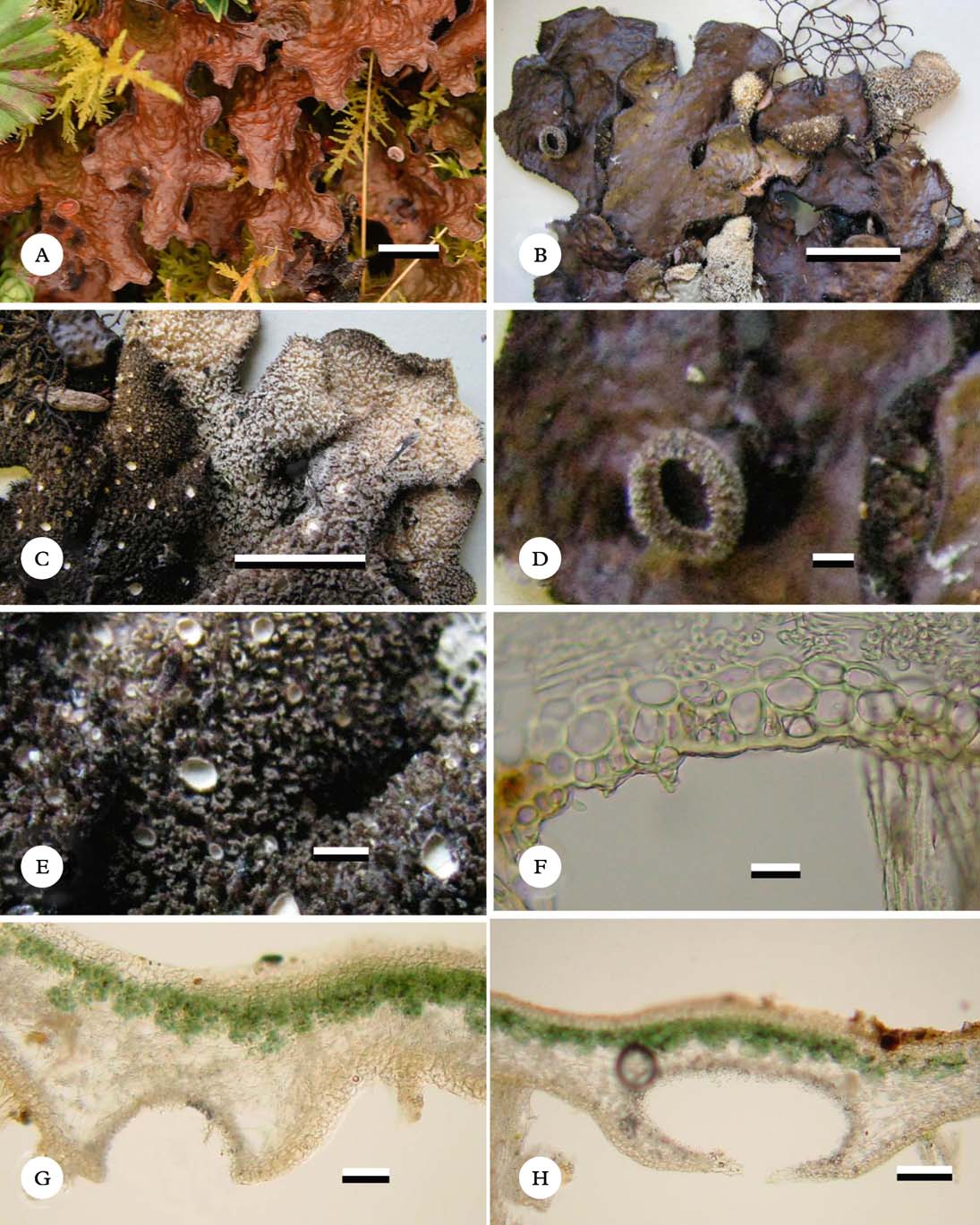
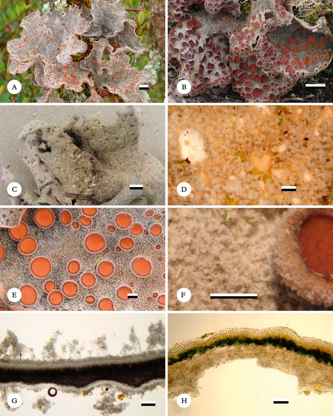
(Fig. 1)
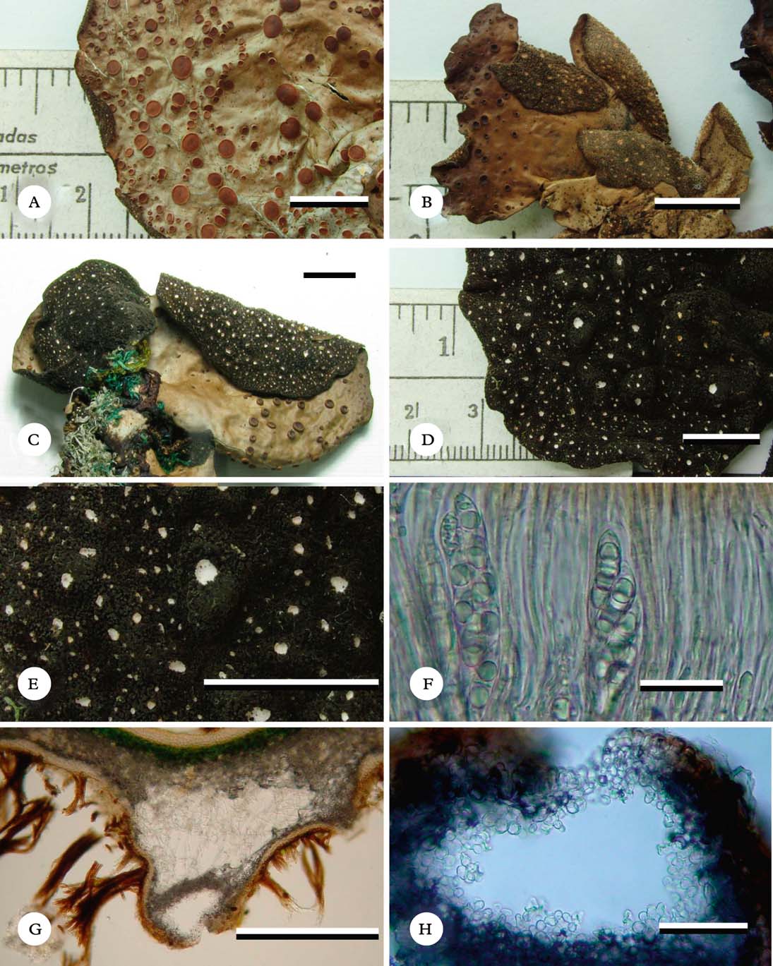
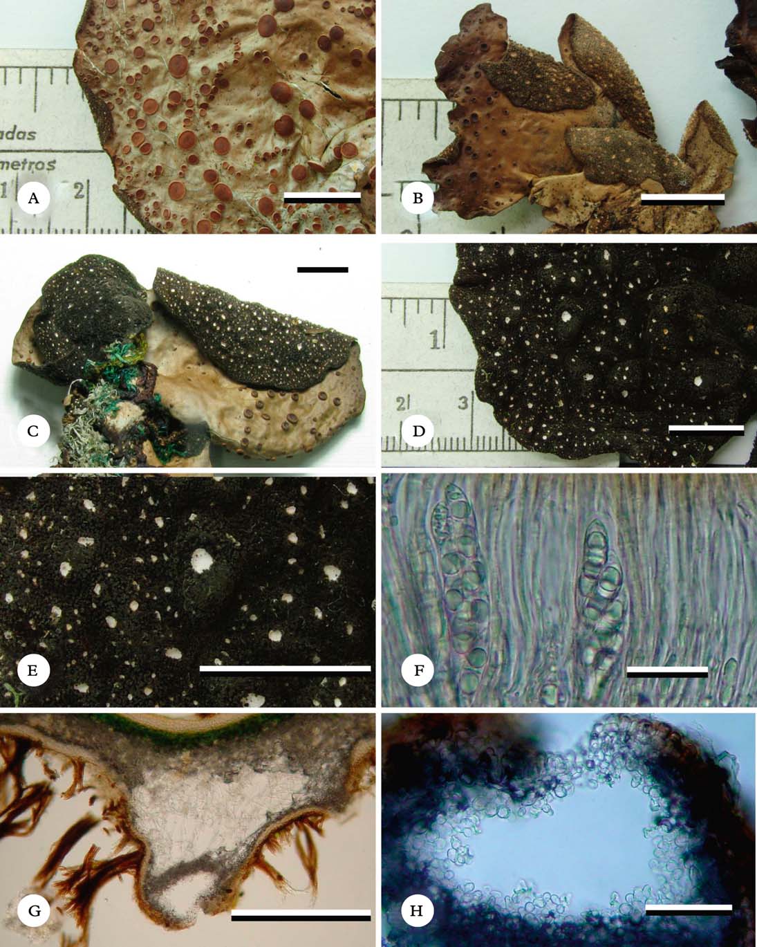
Sticta atroandensis (holotype). A, dorsal view of lobe with numerous apothecia; B & C, thallus lobes with ventral and dorsal view; D & E, ventral view of undulate surface and dark tomentum with suprasessile cyphellae; F, hymenium and asci with ascospores; G, section through suprasessile cyphella; H, section through cyphella cavity showing details of cells of basal membrane. Scales: A–E=10 mm; F=20 µm; G=500 µm; H=100 µm. In colour online.
Primary photobiont cyanobacterial (Nostoc). Basal stipe absent. Thallus orbicular, up to 10 cm diam., sparsely branched, with 0–2 branches for every 5 cm of radius, branching pattern polytomous. Lobes suborbicular, horizontal, imbricate, plane to slightly involute, with their tips rounded and plane to slightly involute and their margins entire, not thickened; lobe internodes (10–)20–30(–40) mm long, (20–)25–70(–100) mm wide, coriaceous. Upper surface scrobiculate to strongly scrobiculate, olive-green when fresh, brown-grey to dark brown in the herbarium, glossy, the marginal line concolourous to slightly paler; surface glabrous, without papillae, without pruina, with irregular, sparse, cream-coloured maculae. True marginal cilia absent but projections of lower tomentum resembling cilia usually present. Vegetative propagules absent. Medulla compact, white, K−, C−, KC−, P−. Lower surface undulate, beige to dark brown to almost black towards the centre. Primary lower tomentum dense and thick, sparse and thinner towards the margin, spongy, soft, brown-black. Secondary lower tomentum pubescent to arachnoid, pale. Rhizines absent. Cyphellae abundant, 1–20 per cm2 towards the thallus centre and 41–60 per cm2 towards the margin, dispersed, rounded, urceolate, sessile to suprasessile, below the level of the primary tomentum, with the margin raised and involute to revolute, brown to brown-black, without tomentum; pore (0·2–)0·7–1·0(–1·5) mm diam. towards the thallus centre; basal membrane pubescent, white to cream-coloured or yellowish, K−, C−, KC−, P−.
Upper cortex paraplectenchymatous, 30–40 µm thick, uniform, formed of 4–5 cell layers, 5–10 µm diam., cell walls 1·25–2·50 µm thick and cell lumina rounded to isodiametric, 4·0–7·5 µm diam. Photobiont layer 45–75 µm thick, cells 10–15 µm diam. Medulla 40–150 µm thick, medullary hyphae 2·5 µm wide, without crystals. Lower cortex paraplectenchymatous, 20–35 µm thick, formed of 3–4 cell layers; cells 4–12 µm diam., cell walls 1·25–2·50 µm thick. Lower primary tomentum 240–550 µm long, composed of fascicles formed of 12–20 hyphae, hyphae branched, septate with their tips interwoven. Lower secondary tomentum 17–25 µm long, composed of simple, septate hyphae with their tips free. Cavity of the cyphellae 40–220 µm high; cells of basal membrane without papillae.
Apothecia abundant, laminal, dispersed, subpedicellate, basal invagination pronounced, up to 3 mm diam., biatorine, up to 370 µm high, with indistinct stipe; excipulum up to 115 µm thick; disc orange-red to red-brown, glossy, K−; margin entire to slightly verrucose, cream-coloured to beige. Hymenium up to 105 µm high; epihymenium up to 2·5 µm high, orange, K−. Ascospores hyaline, 30–35×7–8 µm, fusiform, with 1–3 septa.
Pycnidia immersed.
Chemistry
No substances detected by TLC.
Etymology
The epithet of this new species refers to the similarity with Sticta andensis, from which it differs chiefly in the dark lower tomentum, as well as the glabrous apothecial margins.
Ecology and distribution
Colombian Andes. Sticta atroandensis appears to be restricted to the páramo and superpáramo life zones and was found on the western slopes of the Cordillera Central and in the Cordillera Oriental, between 3600 and 4800 m (Rangel-Ch. Reference Rangel-Ch.1995, Reference Rangel-Ch. and Rangel-Ch.2000; Rodríguez et al. Reference Rodríguez, Armenteras, Morales and Romero2006; Moncada et al. Reference Moncada, Aguirre-C. and Lücking2013c ). It is epiphytic in semi-exposed to exposed microsites and is generally associated with liverworts of the genus Metzgeria and the family Lejeuneaceae.
Notes
Sticta atroandensis is a broad-lobed, cyanobacterial species with scrobiculate, otherwise glabrous, upper surface lacking true marginal cilia, conspicuous cyphellae, abundant apothecia, lacking vegetative propagules, and is best characterized by its dark underside. No other species in Colombia has this combination of characters. Most similar are S. andensis Nyl. and S. ambavillaria (Bory) Ach. (Acharius Reference Acharius1810; Galloway Reference Galloway1995). Sticta andensis, also described from Colombia (Nylander in Stizenberger Reference Stizenberger1895), is mainly distinguished by the pale lower tomentum and the pubescent apothecial margins, whereas the African S. ambavillaria (Swinscow & Krog Reference Swinscow and Krog1988; Galloway Reference Galloway1995) has a generally thinner, fragile thallus (when dry) with pale lower tomentum and marginally ciliate apothecia (at least when young). Other cyanobacterial species with apothecia and a dark brown lower side, such as S. paramuna Moncada & Lücking and relatives, can be distinguished by their narrower, irregular lobes with mostly smooth surfaces, corresponding to apotheciate forms of the S. weigelii morphodeme (B. Moncada, A. Suárez, A. & R. Lücking, unpublished data). The difference in the colour of the lower tomentum between S. andensis and S. atroandensis is consistent in the material studied and is supported as a diagnostic character by the phylogenetic placement of both species, as well as by the fact that larger clades within Sticta are often uniformly characterized by an either pale or dark lower tomentum (Moncada et al. Reference Moncada, Lücking and Suárez2013b ).
Additional specimens examined. Colombia: Boyacá: Mun. El Cocuy, Parque Nacional Natural El Cocuy, Valle de las Lagunillas, Cabaña Sisuma, 06°26′24″N, 72°22′18·16″W, 3800 m, 2 vii 2010, F. Martínez s. n. (UDBC); ibid., 3800 m, 2011, D. Fonseca & F. Martínez 21, 23, 29a, 29b, 30, 32, 35, 40, 42, 45, 59, 61, 79, 85 (UDBC); sendero Cabaña Sisuma to Pico Pan de Azúcar, 3800–4800 m, 2011, D. Fonseca & F. Martínez 97, 99, 112 (UDBC); Alto de la Cueva, 3800 m, 2011, D. Fonseca & F. Martínez 135, 179, 183, 188b, 238 (UDBC). Cundinamarca: Mun. Bogotá D.C., Parque Nacional Natural Sumapaz, Corregimiento Nazareth, sector Andabobos, 3610 m, 2004, B. Moncada & R. Dávila 2266 (UDBC); ibid., 3610 m, 2005, B. Moncada & R. Dávila 2353 (UDBC). Risaralda: Mun. Pereira, Parque Nacional Natural Los Nevados, Laguna de Otún, 3975 m, 1980, T. Boekhout 10 (B, COL); ibid., 3930 m, 1980, T. Boekhout 175 (COL); Mun. Santa Rosa de Cabal, Volcán de Santa Rosa, 3975 m, 1980, T. Boekhout 26 (COL).
Sticta brevior Moncada & Lücking sp. nov.
MycoBank No.: MB804395
Differing from Sticta impressula in the tomentose-pubescent rather than hirsute apothecial margins with a beige to pale brown colour.
Type: Colombia, Huila, Mun. La Plata, Vereda La Candelaria, cascada del Río La Candelaria, 2300 m, 6 October 1984, J. Aguirre & H. Sipman 6134 (B—holotype; COL—isotype).
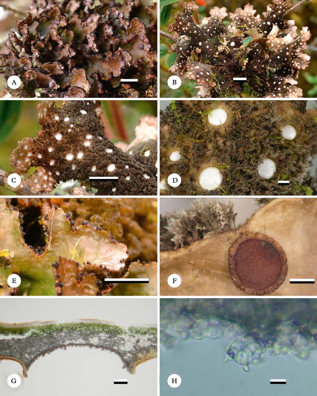
(Fig. 2)
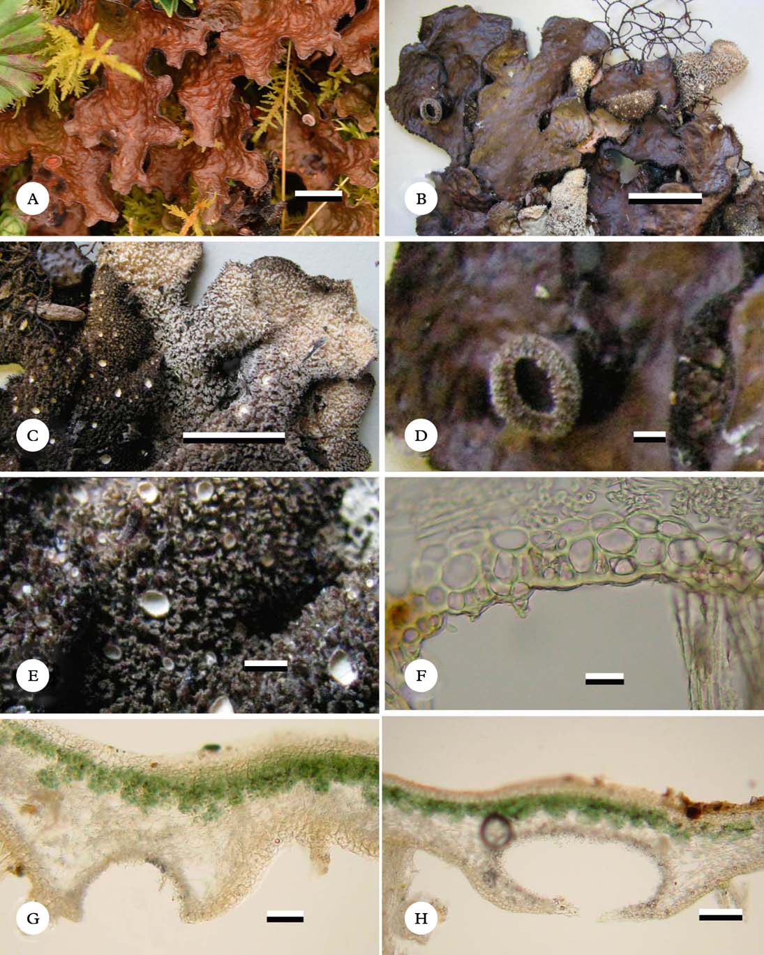
Sticta brevior (holotype except A). A, thallus in situ (Lücking 34041); B, dorsal view of thallus in the herbarium; C, ventral view of thallus with tomentum; D, detail of apothecium with tomentose margin; E, detail of ventral tomentum and cyphellae; F, section through lower cortex and tomentum; G & H, sections through cyphellae. Scales: A–C=10 mm; D & E=1 mm; F=10 µm; G=50 µm; H=100 µm. In colour online.
Primary photobiont cyanobacterial (Nostoc). Basal stipe absent. Thallus irregular to palmate, up to 5 cm diam., moderately branched, with 3–5 branches per 5 cm of radius, branching pattern polytomous to pleurotomous. Lobes flabellate, horizontal to slightly ascendent, imbricate, undulate to involute, with their tips rounded and plane to undulate and their margins entire to sinuous and slightly thickened; lobe internodes (6–)10–13(–15) mm long, (2–)10–25(–30) mm wide, coriaceous. Upper surface pitted (foveolate) to centrally rugose, dark olive when fresh, pale brown to reddish brown and darker towards the tips in the herbarium, glossy, the marginal line darker; surface glabrous, without or with sparse papillae and sometimes with papillotrichomes towards the tips, without pruina, with irregular, abundant, cream-coloured maculae. True marginal cilia abundant, sometimes resembling projections of the lower tomentum, fasciculate, golden beige-brown, up to 0·5 mm long. Vegetative propagules absent. Medulla slightly compacted, white to cream-coloured, K+ pale yellow, C−, KC−, P−. Lower surface undulate, white to cream-coloured to pale brown. Primary lower tomentum dense up to the margin, thick but thinner towards the margin, spongy, soft, greyish beige to chocolate brown towards the centre. Secondary lower tomentum absent. Rhizines absent. Cyphellae abundant, 21–40 per cm2 towards the thallus centre and 61–100 per cm2 towards the margin, dispersed, rounded to angular, urceolate with wide pore, erumpent to suprasessile, below the level of the primary tomentum, with the margin raised and involute to erect, white to cream-coloured to dark brown, without tomentum; pore (0–3–)0·5–0·8(–1·25) mm diam. towards the thallus centre; basal membrane pubescent, white, K+ pale yellow, C−, KC−, P−.
Upper cortex paraplectenchymatous, 20–28 µm thick, uniform, formed of 2–3 cell layers, cells 4–14 µm diam., cell walls 1·25 µm thick and cell lumina rounded to isodiametric, 2·5–12·0 µm diam. Photobiont layer 45–65 µm thick, cells 10–18 µm diam. Medulla 35–125 µm thick, medullary hyphae 2·5 µm wide, without crystals. Lower cortex paraplectenchymatous, 17–30 µm thick, with 2–3 cell layers; cells 5–14 µm diam., cell walls 0·6–2·5 µm thick. Lower primary tomentum 80–600 µm long, formed of fascicles of more than 20 hyphae, hyphae branched, septate with their tips interwoven. Cavity of the cyphellae 100–180 µm high; cells of basal membrane with one papilla each.
Apothecia abundant, principally laminal, dispersed, pedicellate, basal invagination pronounced, up to 3 mm diam.; biatorine, up to 600 µm high, with distinct stipe; disc reddish orange to reddish brown, glossy, K−; margin tomentose, pubescent to shortly hirsute, with abundant, cream-coloured trichomes that remain in mature apothecia except for the upper margin bordering the disc which appears crenulate and glabrous. E xcipulum up to 125 µm wide, with tomentum. Hymenium up to 125 µm high; epihymenium up to 2·5 µm high, orange-brown, K−. Ascospores hyaline, 30–40×7–8 µm, fusiform, with 1–3 septa.
Pycnidia immersed.
Etymology
This new species includes a specimen (Lindig 2548) previously studied by Nylander (Reference Nylander1863) and compared to S. lenormandii as follows: “Forma brevior, thallo breviore, datur and Choachí, altit. 2600 metr., in coll. Lindig. 2548”. We took the adjective ‘brevior’ up as epithet. One could possibly interpret Nylander's (Reference Nylander1863: 18) text as a formal description of a new taxon, in which case the epithet ‘brevior’ could be elevated to species level, with Nylander as basionym author. However, on the same page, Nylander (Reference Nylander1863: 18) establishes f. laevis of S. lenormandii, with a very different formatting style (including setting the epithet laevis in italics), suggesting that “Forma brevior…” denotes an informal description of a deviating specimen and not a formal taxon. A different interpretation of Nylander's statement does not affect the validity of the new species name introduced here.
Ecology and distribution
Colombian Andes. The species was found between 2300 and 3600 m in (sub-)andine forests bordering páramo vegetation in usually semi-exposed microsites. It is known from the Cordillera Central and the Cordillera Occidental in subandine, andine and páramo regions (Rangel-Ch. Reference Rangel-Ch.1995, Reference Rangel-Ch. and Rangel-Ch.2000; Rodríguez et al. Reference Rodríguez, Armenteras, Morales and Romero2006; Moncada et al. Reference Moncada, Aguirre-C. and Lücking2013c ), and grows mostly as an epiphyte, more rarely on the ground; it is often associated with liverworts of the family Lejeuneaceae and the genera Frullania and Jubula, as well as Sphagnum mosses. It may also grow close to the ground on Blechnum ferns.
Notes
Sticta brevior belongs to the S. kunthii group, which includes a number of closely related, cyanobacterial, mostly apotheciate species with rather broad lobes with pitted upper surface and pale underside, forming one papilla per cell on basal membranes of the cyphellae (Moncada Reference Moncada2012; Moncada et al. Reference Moncada, Lücking and Suárez2013b ). The species in this group are mostly separated by the position of the apothecia and the nature of the tomentum on the apothecial margins. Sticta brevior has generally laminal apothecia with irregular marginal tomentum and a coriaceous, rather stout thallus, whereas S. lenormandii (Nyl.) Zahlbr., a Peruvian species (Nylander Reference Nylander1858, Reference Nylander1859, Reference Nylander1860), has a distinctly hirsute marginal tomentum and a less robust, more spreading thallus.
Additional specimens examined. Colombia: Cauca: Mun. Puracé, Carretera La Plata to Puracé, 3300 m, 1984, J. Aguirre & H. Sipman 6017 (B, COL). Cundinamarca: Mun. Choachí, 2600 m, 1860, Lindig 2548 (PC); Mun. Choachí, El Verjón, Parque Ecológico Matarredonda, 04°33′08·0″N, 73°59′55·0″W, 3220 m, 2011, B. Moncada 4590b (UDBC); without locality, Lindig 2584 (BM); Mun. La Calera, Zona de amortiguación Parque Nacional Natural Chingaza, Vía del Ángulo al Rincón del Oso, 3600 m, 2011, B. Moncada 4971, 4988, 4990 (UDBC); Mun. San Bernardo, La Soledad, costado occidental del filo de Paquiló, 2900 m, 1981, J. Aguirre 2388 (COL); Mun. Villapinzón, Páramo de Villapinzón, 3300 m, 2002, M. Pinzón 1375 (COL). Huila: Mun. La Plata, Vereda La Candelaria, cascada del Río La Candelaria, 2300 m, 1984, J. Aguirre & H. Sipman 6134 (B, COL).
Sticta lumbschiana Moncada & Lücking sp. nov.
MycoBank No.: MB804396
GenBank ITS barcoding sequence: KC732575
Differing from Sticta dilatata in the shorter stipe, the tomentose apothecial margin and the cells in the basal membrane of the cyphellae forming only one papilla per cell.
Type: Colombia, Cundinamarca, Mun. Guasca, Páramo de Guasca, 3200 m, 18 August 2011, R. Lücking 33370 (UDBD—holotype; F—isotype).
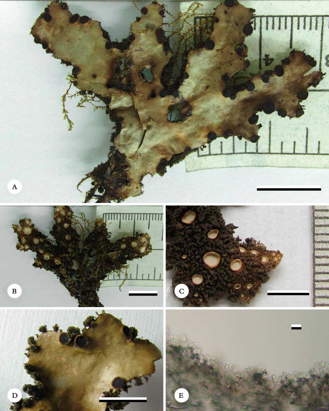
(Fig. 3)
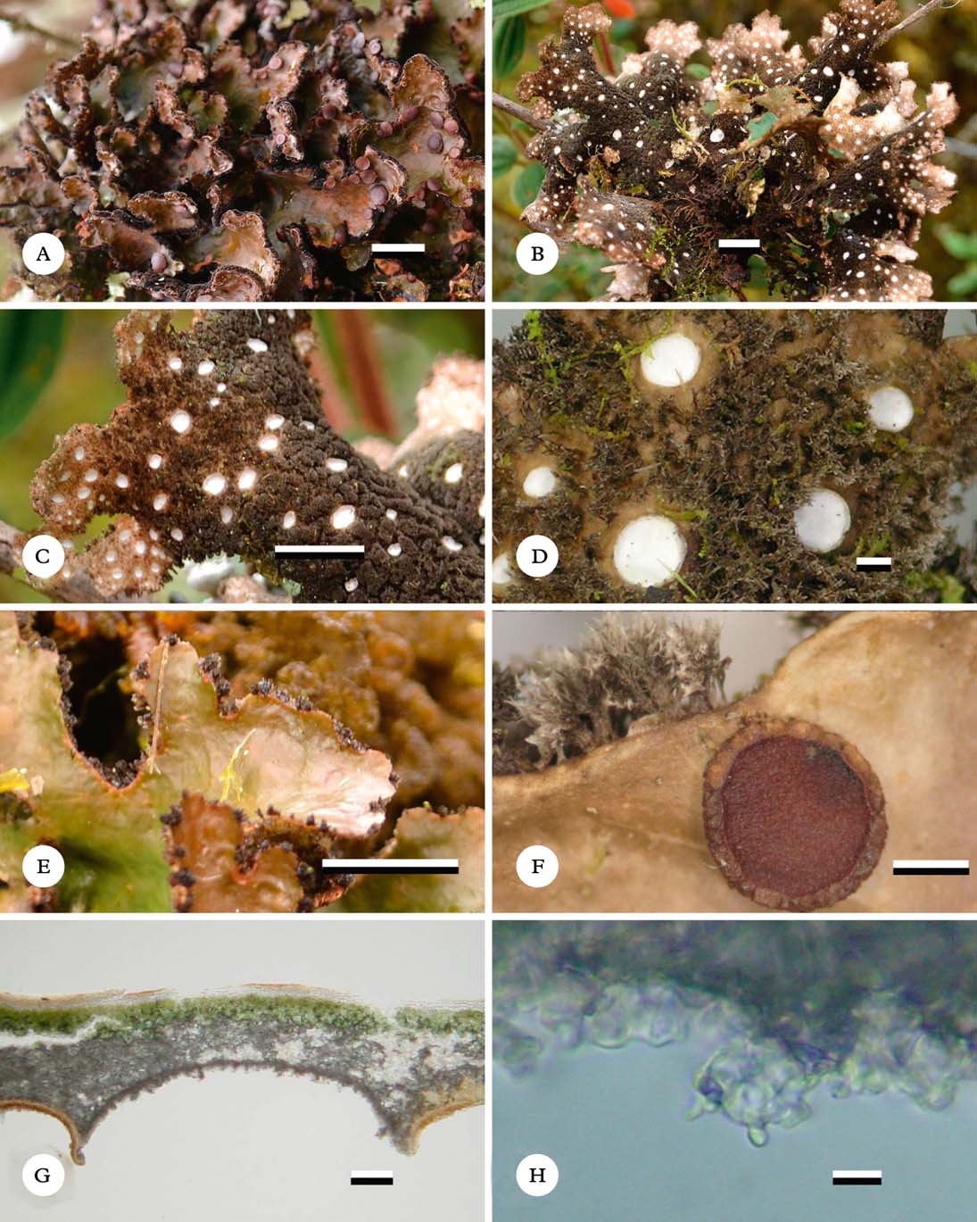
Sticta lumbschiana (A–E, Lücking 34051; F–H, Lücking 33364). A, thallus in situ; B–D, ventral view of thallus in situ showing detail of tomentum and cyphellae; E, lobe detail showing penicillate cilia; F, lobe in the herbarium, enlarged showing apothecium and marginal, penicillate cilia; G, section through cyphella; H, papillate cells of basal membrane showing single papilla per cell. Scales: A–C & E=10 mm; D & F=1 mm; G=100 µm; H=10 µm. In colour online.
Primary photobiont cyanobacterial (Nostoc). Basal stipe indistinct, up to 5 mm long. Thallus palmate to irregular, up to 15 cm diam., moderately branched, with 3–5 branches for every 5 cm of radius, branching pattern polytomous to anisotomous. Lobes laciniate to flabellate, ascendant to erect, adjacent to imbricate, involute to slightly canaliculate, with their tips rounded and involute and their margins entire to crenate, not thickened; lobe internodes (6–)8–13(–15) mm long, (5–)7–20(–25) mm wide, coriaceous, fragile. Upper surface smooth to scrobiculate towards the tips, dark olive when fresh, reddish brown and darker towards the tips in the herbarium, glossy, the marginal line concolourous to brown-black; surface glabrous, without papillae, without pruina, with irregular, abundant, cream-coloured maculae. True marginal cilia abundant, penicillate to agglutinate, dark greyish brown, up to 1·5 mm long. Vegetative propagules absent. Medulla compact, white to cream-coloured, K+ ochraceous-yellow, C−, KC−, P−. Lower surface smooth to slightly undulate, beige to brown-black towards the centre. Primary lower tomentum dense and thick but sparse and thinner towards the margin, spongy to penicillate, soft, greyish to reddish brown with their tips white. Secondary lower tomentum arachnoid, pale, sometimes indistinct. Rhizines absent. Cyphellae abundant, 1–10 per cm2 towards the thallus centre and 21–40 per cm2 towards the margin, dispersed, rounded, cupuliform to urceolate with wide pore, erumpent to sessile, below the level of the primary tomentum, with the margin raised to slightly involute, white to cream-coloured, without tomentum; pore (1·0–)1·25–2·50(–3·0) mm diam. towards the thallus centre; basal membrane pubescent, white, K+ yellow, C−, KC−, P−.
Upper cortex paraplectenchymatous, 35–45 µm thick, differentiated into two layers: upper layer formed of 1–2 cell layers, of 3–5 µm diam., cell walls 2·5 µm thick and cell lumina rounded to isodiametric, 1·25–2·50 µm diam.; lower layer formed of 3–4 cell layers; cells 5–11 µm diam., cell walls 0·60–1·25 µm thick and cell lumina rounded to isodiametric, 5–10 µm diam. Photobiont layer 75–100 µm thick, cells 10–25 µm diam. Medulla 110–200 µm thick, medullary hyphae 2·5 µm wide, without crystals. Lower cortex paraplectenchymatous, 20–25 µm thick, with 2–3 cell layers; cells 5–11 µm diam., cell walls 0·6–2·5 µm thick. Lower primary tomentum 160–650 µm long, formed of fascicles of more than 20 hyphae, hyphae simple, septate with their tips interwoven. Lower secondary tomentum 12–25 µm long, of single hyphae, hyphae branched, moniliform with their tips free. Cavity of the cyphellae 90–280 µm high; cells of basal membrane with one papilla per cell. Apothecia abundant, biatorine, up to 380 µm high, with indistinct stipe; principally submarginal, dispersed, subpedicellate, basal invagination pronounced, up to 3 mm diam.; disc reddish brown, glossy, K−; margin entire to tomentose, tomentum short-pubescent, with sparse larger trichomes, dark brown, evanescent in mature apothecia. E xcipulum up to 120 µm thick. Hymenium up to 125 µm high; epihymenium up to 2·5 µm high, orange-brown, K−. Ascospores hyaline, 30–40×6–8 µm, fusiform, with 1–3 septa.
Pycnidia absent.
Etymology
It is an honour for us to name this new species after our colleague Thorsten Lumbsch, outstanding lichenologist and mycologist, for his invaluable contributions to lichenology and his support of this project on the genus Sticta.
Ecology and distribution
Colombian Andes. The species is known from 2100 to 3750 m altitude in (sub-)andine forests and páramo vegetation, in (semi-)exposed microsites. It was found in the Cordillera Central and the eastern slopes of the Cordillera Oriental (Rangel-Ch. Reference Rangel-Ch.1995, Reference Rangel-Ch. and Rangel-Ch.2000; Rodríguez et al. Reference Rodríguez, Armenteras, Morales and Romero2006; Moncada et al. Reference Moncada, Aguirre-C. and Lücking2013c ). Sticta lumbschiana grows on bark, fallen trunks and occasionally on soil, generally associated with liverworts of the genera Metzgeria, Bazzania, Plagiochila and Radula.
Notes
This species, as well as the following one, belongs to the S. dilatata group, characterized by pedunculate, much branched thalli having rather narrow, distinctly ciliate lobes that produce very large and conspicuous cyphellae with papillae on the basal membranes (Moncada Reference Moncada2012). The latter character is very consistent and characterizes entire clades within Sticta (Moncada et al. Reference Moncada, Lücking and Suárez2013b ).
The name S. dilatata (Nyl.) Vain. is misapplied in the literature. Zahlbruckner (Reference Zahlbruckner1925) indicated that Vainio (Reference Vainio1913) based his combination on S. laciniata var. dilatata Nyl., a taxon described from Colombia that was later renamed S. granatensis Nyl. (Nylander Reference Nylander1874). However, Vainio (Reference Vainio1913) based his combination on a different, morphologically and phylogenetically unrelated lichen, S. tomentosa var. dilatata Nyl., also described from Colombia (Nylander Reference Nylander1860).
The most distinctive feature of S. lumbschii is the presence of one papilla per cell of the basal membrane of the cyphellae, whereas other species in this group have more than one papilla. It also has a shorter stipe and narrower lobes than S. dilatata, and the lobes are slightly scrobiculate; furthermore, its apothecia have a tomentose margin, at least when young (see also key below).
Additional specimens examined. Colombia: Antioquia: Mun. Medellín, Estación Forestal Piedras Blancas, 2100 m, 8 viii 1980, R. Bernal & G. Galeano s. n. (COL). Cundinamarca: Mun. Choachí, El Verjón. P. E. Matarredonda, 04°33′08·0″N, 73°59′55·0″O, 3220 m, 2011, B. Moncada 4587c (UDBC); Mun. Guasca, Páramo de Guasca, 3200 m, 2011, R. Lücking 33364b, 33364, 34038, 34051 (F, UDBC); Mun. La Calera, Zona de Amortiguación Parque Nacional Natural Chingaza, Vía del Ángulo al Rincón del Oso, 3600 m, 2011, B. Moncada 4994, 4995 (UDBC); ibid., R. Lücking 34038, 34051 (F, UDBC). Nariño: Mun. La Cruz, La Estancia, sector Loma larga, 3310 m, 2001, S. L. Díaz 1980b (CAUP, COL). Risaralda: Mun. Santa Rosa de Cabal, Costado Occidentral, Cordillera Central, 1 km NE de la Finca la Sierra, 3750 m, 1984, J. Aguirre & H. Sipman 5518, 5522 (B, COL).
Sticta macrocyphellata Moncada & Coca sp. nov.
MycoBank No.: MB804397
GenBank ITS barcoding sequence: KC732662
Differing from Sticta silverstonii in the smooth upper surface and the cells of the membrane of the cyphellae having 2–4 papillae per cell.
Type: Colombia, Risaralda, Mun. Santuario, Parque Nacional Natural Tatamá, Planes de San Rafael, towards Monte Zancudo, 2800 m, 14 January 2011, L. F. Coca et al. 1267 (FAUC—holotype).
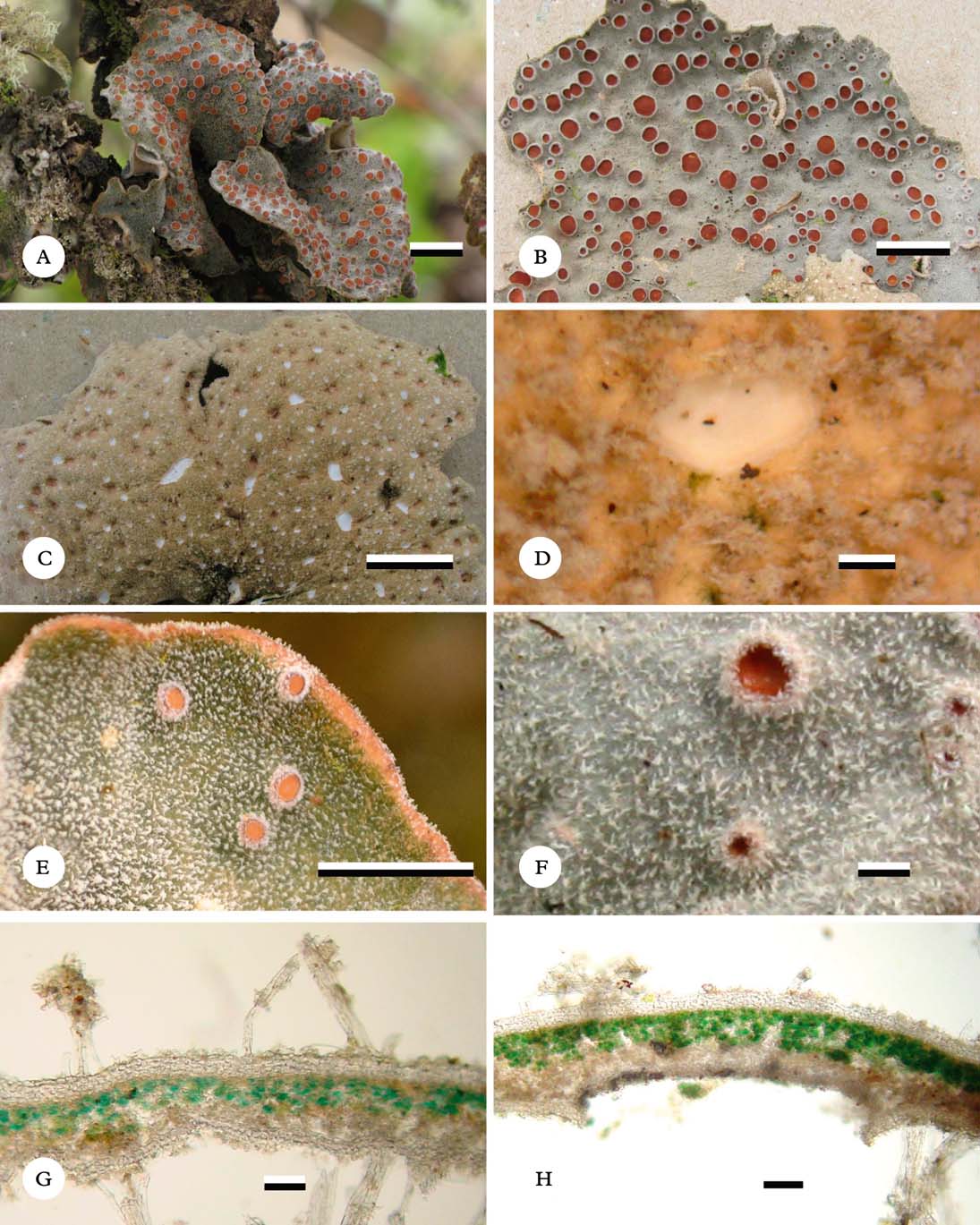
(Fig. 4)
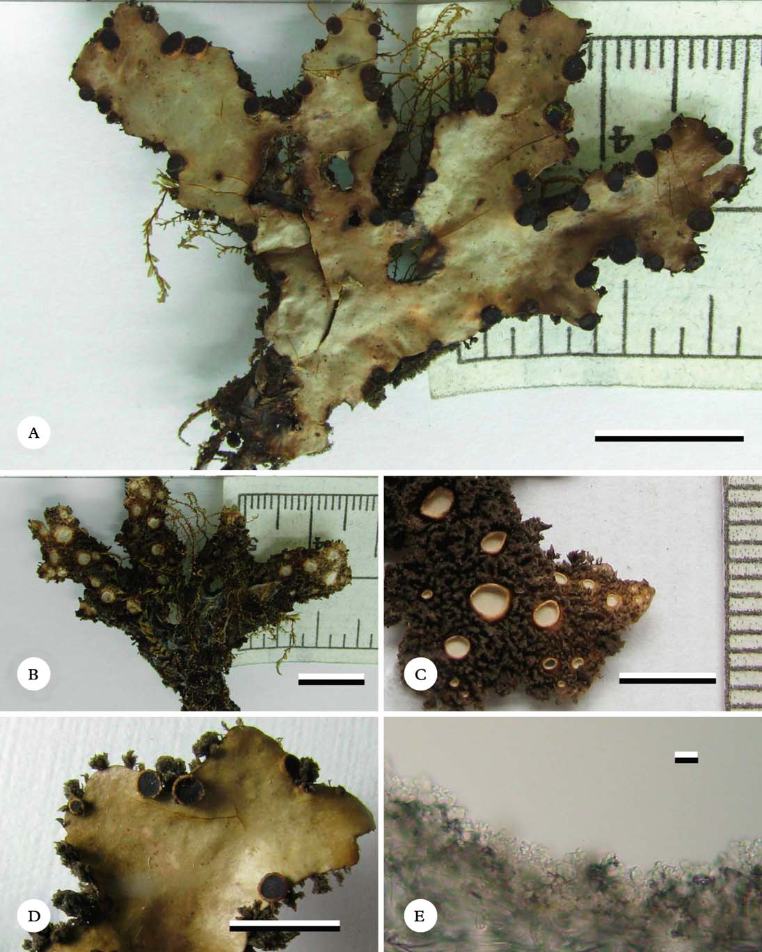
Sticta macrocyphellata (holotype). A, dorsal view of thallus; B, ventral view of thallus; C, detail showing lower tomentum and cyphellae; D, lobe detail showing apothecia and penicillate marginal cilia; E, papillate cells of basal membrane showing several papillae per cell. Scales: A & B=10 mm; C & D=5 mm; E=10 µm. In colour online.
Primary photobiont cyanobacterial (Nostoc). Basal stipe absent. Thallus irregular, up to 10 cm diam., much branched, with 6–10 branches for every 5 cm of radius, branching pleurotomous. Lobes laciniate to ligulate, horizontal to slightly ascendent, adjacent to slightly imbricate, plane to slightly canaliculate, with their tips rounded to truncate and plane to revolute and their margins entire to sinuose, not thickened; lobe internodes (3–)4–8(–12) mm long, (3–)5–9(–10) mm wide, subcoriaceous, fragile. Upper surface smooth to shallowly scrobiculate, dark olive when fresh, pale cinnamon brown and darker towards the tips in the herbarium, glossy, the marginal line brown; surface glabrous, without papillae, without pruina, with punctiform, abundant, cream-coloured maculae. True marginal cilia abundant, penicillate, golden brown to brown-black with their tips free, up to 2 mm long. Vegetative propagules absent. Medulla lax, cream-coloured, K+ ochraceous-yellow, C−, KC−, P−. Lower surface smooth, beige to dark brown, glossy. Primary lower tomentum sparse, thick, spongy to arachnoid, soft, dark brown with paler tips. Secondary lower tomentum absent. Rhizines absent. Cyphellae abundant, 1–10 per cm2 towards the thallus centre and 21–40 per cm2 towards the margin, dense, rounded, urceolate with wide pore to cupuliform, immersed to prominent, below the level of the primary tomentum, with the margin raised and involute to erect, cream-coloured to black, without tomentum; pore (0·7–)1·0–1·5(–2·5) mm diam. towards the thallus centre; basal membrane pruinose, cream-coloured to yellow, K− to K+ vine red in cyphellae with yellow colour, C−, KC−, P−.
Upper cortex paraplectenchymatous, 50–60 µm thick, differentiated into two layers: upper layer formed of 1–2 cell layers, 3–5 µm diam., cell walls 2·5 µm thick and cell lumina rounded to isodiametric, 1·25–2·50 µm diam.; lower layer formed of 2–3 cell layers; cells 6–16 µm diam., cell walls 1·25 µm thick and cell lumina rounded to isodiametric, 5–15 µm diam. Photobiont layer 25–37 µm thick, cells 10–15 µm diam. Medulla 65–140 µm thick, medullary hyphae 2·5 µm wide, with yellow crystals. Lower cortex paraplectenchymatous, 20–25 µm thick, with 2–3 cell layers; cells 6–13 µm diam., cell walls 1·25–2·50 µm thick. Lower primary tomentum 25–550 µm long, composed of fascicles formed of 12–20 hyphae, hyphae branched, septate with their tips interwoven. Cavity of the cyphellae 40–270 µm high; cells of basal membrane with 4–6 papillae each.
Apothecia abundant, principally laminal, dispersed, biatorine, up to 380 µm high, with indistinct stipe; subpedicellate, basal invagination pronounced, up to 2 mm diam.; disc reddish brown to brown-black when mature, glossy, K−; margin entire to ciliate, cream-coloured to dark brown when mature. Excipulum up to 130 µm thick. Hymenium up to 135 µm high; epihymenium up to 2·5 µm high, orange-brown, K−. Ascospores hyaline, 30–40×7–9 µm, fusiform, with 1–3 septa.
Pycnidia immersed.
Etymology
The epithet refers to the dense, large cyphellae prominent over much of the underside.
Ecology and distribution
Colombian Andes. Sticta macrocyphellata was found between 2200 and 3400 m in subandine to andine forests in semi-exposed microsites. It is known from the Cordillera Oriental, the eastern slopes of the Cordillera Occidental and the western slopes of the Cordillera Central (Rangel-Ch. Reference Rangel-Ch.1995, Reference Rangel-Ch. and Rangel-Ch.2000; Rodríguez et al. Reference Rodríguez, Armenteras, Morales and Romero2006; Moncada et al. Reference Moncada, Aguirre-C. and Lücking2013c ). The species grows on bark and rocks, often associated with bryophytes of the genera Plagiochila, Lophocolea, Bazzania, Omphalanthus and Macromitrium.
Notes
Like the preceding species, Sticta macrocyphellata belongs to the S. dilatata group, being characterized by rather narrow, distinctly ciliate lobes, large cyphellae, and cells of basal membrane of the cyphellae forming papillae (Moncada Reference Moncada2012; Moncada et al. Reference Moncada, Lücking and Suárez2013b ). It is most similar to S. silverstonii Moncada & Lücking, which differs in the distinctly ampullate upper surface (Moncada & Lücking Reference Moncada and Lücking2012). The five species currently known in the S. dilatata group are separated in the key below.
Additional specimens examined. Colombia: Cundinamarca: Mun. San Bernardo, La Soledad, W slope of Filo de Paquiló, 2900 m, 1981, J. Aguirre 2385 (COL). Magdalena: Mun. Santa Marta, Sierra Nevada de Santa Marta, Transecto Buritaca, 2700 m, 1977, G. van Reenen & O. Rangel 294 (B, COL, U). Meta: Mun. Cubarral, Parque Nacional Natural Sumapáz, Cerro Nevado del Sumapaz, Quebrada El Buque, 3400 m, 1973, A. M. Cleef 7788 (B, COL, U). Risaralda: Mun. Santa Rosa de Cabal, Vereda El Cedral., vía El Cedral, Laguna del Otún, 2200 m, 2003, B. Moncada & R. Dávila 1976 (UDBC).
Key to species of the Sticta dilatata group
-
1 Upper surface ampullate (caused by ‘imprint’ of cyphellae from lower surface); marginal cilia simple to fasciculate; cyphellae covering almost entire lower surface; apothecia principally marginal ... Sticta silverstonii
Upper surface plane; marginal cilia always penicillate to fasciculate; cyphellae separated by narrow areas of tomentum; apothecia laminal to submarginal ... 2
-
2(1) Basal membrane of cyphellae with only 1 papilla per cell; lobe surface scrobiculate towards the tips; cyphellae up to 3 mm wide ... Sticta lumbschiana
Basal membrane of cyphellae with 4–6 papillae per cell; lobe surface plane to shallowly scrobiculate towards the tips; cyphellae up to 1·5 mm wide, if wider (up to 2·5 mm), then lobe surface plane ... 3
-
3(2) Lobes up to 25(–50) mm wide, usually shallowly scrobiculate towards the tips; cyphellae up to 1·5 mm wide; apothecial margins verrucose to crenulate ... Sticta dilatata
Lobes up to 10(–15) mm wide, plane; cyphellae up to 2·5 mm wide; apothecial margin entire or ciliate ... 4
-
4(3) Cyphellae plane, up to 1·8 mm wide; apothecial margin entire ... Sticta papillata
Cyphellae urceolate with wide pore, up to 2·5 mm wide; apothecial margin often ciliate ... Sticta macrocyphellata
Sticta parahumboldtii Moncada & Lücking sp. nov.
MycoBank No.: MB804398
GenBank ITS barcoding sequence: KC732550
Differing from Sticta humboldtii in the more fragile thallus, the K+ pale ochraceous medulla, and the K+ pale salmon reaction of the cyphellae membrane.
Type: Colombia, Cundinamarca, Mun. Bogotá D.C., Alrededores Laguna de Chisacá, 04°17′21·7″N, 74° 12′26·9″O, 3724 m, 15 August 2010, B. Moncada et al. 4016 (UDBD—isotype; COL—isotype).
(Fig. 5)
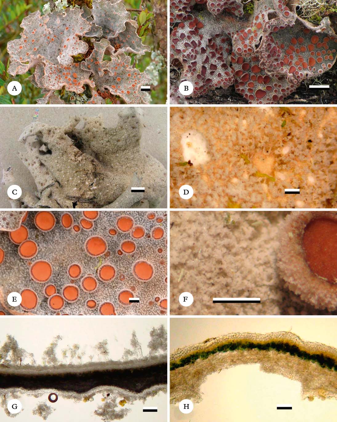
Sticta parahumboldtii (A & B, Lücking s.n., holotype; E & F, Lücking 34043; C, D, G & H, Betancourt 144f). A, thallus in situ; B, dorsal view of lobe in the herbarium; C, ventral view of thallus showing tomentum and immersed cyphellae; D, detail of lower tomentum and cyphella; E & F, detail of dorsal tomentum and apothecia; G, section through thallus showing upper and lower tomentum; H, section through cyphella. Scales: A–C & E=10 mm; D & F=1 mm; G & H=100 µm. In colour online.
Primary photobiont cyanobacterial (Nostoc). Basal stipe absent. Thallus orbicular to suborbicular, up to 10 cm diam., sparsely branched, with 0–2 branches for every 5 cm of radius, branching pattern polytomous to anisotomous. Lobes suborbicular, horizontal to subpendulous, imbricate, undulate, with their tips rounded and undulate, involute and their margins entire, not thickened; lobe internodes (5–)16–35(–45) mm long, (17–)18–50(–60) mm wide, subcoriaceous to coriaceous, fragile. Upper surface scrobiculate to rugose or undulate, emerald green when fresh, beige to pale yellow-brown or whitish in the herbarium, opaque, the marginal line concolourous; surface arachnoid to spongy, without papillae, without pruina, with irregular, sparse, cream-coloured maculae. True marginal cilia abundant, mingled with projections of the upper and lower tomentum, fasciculate to agglutinate, white to cream-coloured, up to 0·2 mm long. Vegetative propagules absent. Medulla compact, white to cream-coloured, K+ pale yellow-ochraceous, C−, KC−, P−. Lower surface undulate, cream-coloured. Primary lower tomentum dense up to the margin, thick but thinner towards the margin, spongy, soft, cream-coloured. Secondary lower tomentum arachnoid, pale. Rhizines absent. Cyphellae abundant, 21–40 per cm2 towards the thallus centre and 100–200 per cm2 towards the margin, dispersed, rounded to irregular, plane to urceolate with wide pore, immersed to erumpent, below the level of the primary tomentum, with the margin levelled to slightly involute to erect, cream-coloured, with tomentum; pore (0·1–)1·0–2·5(–5·0) mm diam. towards the thallus centre; basal membrane pubescent, white to cream-coloured, K+ yellow to pale salmon-coloured, C−, KC+ yellow-ochraceous, P−.
Upper cortex paraplectenchymatous, 25–35 µm thick, uniform, formed of 3–4 cell layers, cells 4–10 µm diam., cell walls 1·25–2·50 µm thick and cell lumina rounded to isodiametric, 2·5–7·5 µm diam. Photobiont layer 50–60 µm thick, cells 10–15 µm diam. Medulla 60–140 µm thick, medullary hyphae 2·5 µm wide, without crystals. Lower cortex paraplectenchymatous, 20–30 µm thick, with 1–2 cell layers; cells 4–12 µm diam., cell walls 1·25–2·50 µm thick. Primary upper tomentum 110–370 µm long, composed of fascicles formed of 6–12 hyphae, hyphae branched, apically moniliform with their tips free. Secondary upper tomentum 15–25 µm long, of single hairs, hyphae branched, moniliform with their tips free. Lower primary tomentum 120–400 µm long, composed of fascicles formed of 12–20 hyphae, hyphae branched, apically moniliform with their tips interwoven. Lower secondary tomentum 15–35 µm long, of single hairs, hyphae branched, moniliform with their tips free. Cavity of the cyphellae 80–100 µm high; cells of basal membrane without papillae.
Apothecia abundant, principally laminal, dispersed, pedicellate, biatorine, up to 400 µm high, with distinct stipe; basal invagination pronounced, up to 3 mm diam.; disc orange to reddish orange, becoming reddish brown in the herbarium, opaque, K−; margin tomentose to hirsute, tomentum white but proper margin cream-coloured. Excipulum up to 105 µm wide, with tomentum. Hymenium up to 95 µm high; epihymenium up to 2·5 µm high, orange, K−. Ascospores hyaline, 27–35×7·5–10·0 µm, fusiform, with 1–3 septa.
Pycnidia immersed.
Etymology
The epithet of this new species (Greek: ‘para’=next to, near) reflects the similarity to, and close relationship with, Sticta humboldtii.
Ecology and distribution
Colombian Andes. The species was found between 3000 and 4000 m in high-andine forests and páramo vegetation, in shaded to semi-exposed microsites. It is known from the Cordillera Central and Cordillera Occidental, and the western slopes of the Cordillera Oriental (Rangel-Ch. Reference Rangel-Ch.1995, Reference Rangel-Ch. and Rangel-Ch.2000; Rodríguez et al. Reference Rodríguez, Armenteras, Morales and Romero2006; Moncada et al. Reference Moncada, Aguirre-C. and Lücking2013c ), growing epiphytically and on mossy soil, generally associated with species of Metzgeria, Frullania and Leptogium.
Notes
Sticta parahumboldtii is one of only a few species in the genus with an upper tomentum, cyanobacterial photobiont, and lack of vegetative propagules. Two other species in Colombia have a very similar morphology: S. humboldtii Hook. (Hooker Reference Hooker, Kunth and Bonpland1822, Reference Hooker1829; Galloway Reference Galloway1995) and S. pseudohumboldtii (see below). Their taxonomy and phylogenetic relationships are discussed under the latter species.
Additional specimens examined. Colombia: Cauca: Mun. Puracé, Sur del Volcán del Puracé, Filo de la Cordillera San Francisco, 3400 m, 1943, J. Cuatrecasas 14585, 14597 (COL); Volcán del Puracé, cabeceras Río Vinagre, El Alfombrado, 3800 m, 1972, A. M. Cleef 637a (COL); Volcán del Puracé, 1–2 km W of Alto de San Rafael, 3300 m, 1972, A. M. Cleef 2644 (COL, U); Mun. Santa Rosa, Macizo Colombiano, páramo de las Papas, alrededores de Valencia, 3150 m, 1958, H. G. Barclay & P. Juajibioy 5881 (COL). Cundinamarca: Mun. Bogotá D.C., Parque Nacional Natural Sumapaz, Páramo de Chisacá, 3600 m, 22 iii 1983, M. Castro s. n. (UPTC); Corregimiento Nazareth, sector Andabobos, 3610 m, 2004, B. Moncada & R. Dávila 2278, 2304 (UDBC); Alrededores Laguna de Chisacá, 04°17′21·7″N, 74°12′26·9″W, 3730 m, 2008, B. Moncada & A. López 2647 (UDBC); ibid., I. Ardila & B. Gómez 191 (UDBC); ibid., 3724 m, 2010, B. Moncada et al. 4016, 4119 (UDBC); ibid., 3600 m, 2010, L. Betancourt 144f, 144g (UDBC); Mun. Choachí, Páramo del Verjón, 3500 m, 1940, J. Cuatrecasas 10450 (COL); ibid., 3050–3350 m, 2001, E. Duarte et al. 93 (UPTC); ibid., 3220 m, 2007, D. Hernández et al. 21 (UDBC); ibid., L. Castro et al. 16 (UDBC); Mun. Cogua, Páramo entre Cógua and San Cayetano, cercanías Laguna Seca, 3685 m, 1972, A. M. Cleef 6139b (COL); Mun. Fómeque, Parque Nacional Natural Chingaza, Sendero La Arboleda, quebrada Babilonia, 3020 m, 2003, B. Moncada & R. Dávila 1584 (UDBC); Mun. Guasca, Piedras Gordas, 3200 m, 1988, H. Sipman & J. Aguirre 27378, 27380 (B, COL); Mun. La Calera, Páramo de Palacio, Hacienda la Siberia, 3500 m, 1953, L. E. Mora 638 (COL); Páramo de Siberia, 3470 m, 1971, G. Guzmán 9110 (COL); Páramo de Palacio, Hacienda la Siberia, km 17·5 camino La Calera to La Mina, 3000 m, 1971, G. Guzmán 9117, 9122 (COL); ibid., 3450 m, 1971, G. Guzmán 9780 (COL); Mun. Villapinzón, Páramo de Villapinzón, 3300 m, 2002, M. Pinzón 1396 (COL). Meta: Mun. Cubarral, Parque Nacional Natural Sumpáz, Cerro Nevado del Sumapaz, Quebrada El Buque, 3450 m, 1973, A. M. Cleef 7882 (COL, U). Risaralda: Mun. Pereira, Parque Nacional Natural Los Nevados, Laguna de Otún, 3930 m, 1980, T. Boekhout 174 (COL). Tolima: Mun. Ibagué, Las Juntas, vía Nieves, 3500 m, 2002, D. Moreno & P. Hincapié 153 (COL); Las Juntas, Vía la Cueva, 4000 m, 2002, D. Moreno & P. Hincapié 183 (COL); Mun. Santa Isabel, Valle del Río Totarito, 3650 m, 1980, H. Valencia & T. Boekhout 131, 154b (COL). Valle del Cauca: Mun. Tuluá, Corregimiento de Barragán, hoya del Río Bugalagrande, Páramo de Bavaya, 3520 m, 1946, J. Cuatrecasas 20196 (COL).
Sticta pseudohumboldtii Moncada & Lücking sp. nov.
MycoBank No.: MB804399
GenBank ITS barcoding sequence: KC011071
Differing from Sticta humboldtii in the coriaceous thallus, the K− to K+ pale yellow medulla, and the urceolate, prominent to sessile cyphellae with K+ dark yellow reaction.
Type: Colombia, Cundinamarca, El Verjón, Parque Ecológico Matarredonda, 04°33′37·7″N, 74°00′02·2″O, 3220 m, 23 January 2011, B. Moncada 4614 (UDBC—holotype).
(Fig. 6)
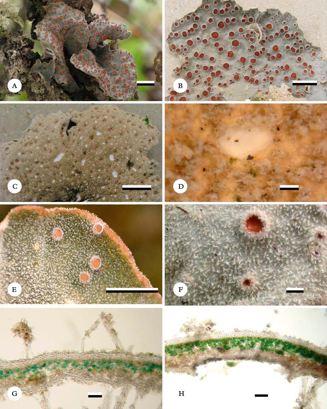
Sticta pseudohumboldtii (Moncada 4586). A & B, thallus in situ; C, ventral view of thallus; D, detail of lower tomentum and cyphellae; E & F, detail of upper tomentum and apothecia; G, section through thallus showing upper and lower tomentum; H, section through cyphella. Scales: A–C=10 mm; D–F=1 mm; G & H=100 µm. In colour online.
Primary photobiont cyanobacterial (Nostoc). Basal stipe absent. Thallus orbicular to irregular, up to 10 cm diam., moderately branched, with 3–5 branches for every 5 cm of radius, branching pattern anisotomous to polytomous. Lobes flabellate to suborbicular, horizontal to subpendulous, imbricate, undulate, with their tips rounded to truncate, involute and their margins entire to crenate, not thickened; lobe internodes (8–)10–20(–25) mm long, (3–)10–45(–55) mm wide, coriaceous. Upper surface undulate to scrobiculate, emerald green to green-brown when fresh, greenish grey to pale brown or whitish in the herbarium, opaque, the marginal line concolourous or brownish; surface arachnoid to spongy, with papillae towards the margin, without pruina, with irregular, sparse, cream-coloured maculae. True marginal cilia abundant, mingled with projections of the upper and lower tomentum, fasciculate to agglutinate, white to cream-coloured, up to 0·2 mm long. Vegetative propagules absent. Medulla compact, white to cream-coloured, K− or K+ pale yellow, C−, KC−, P−. Lower surface undulate, cream-coloured. Primary lower tomentum dense up to the margin, thick but thinner towards the margin, spongy to intertwined, soft to rough, cream-coloured to greyish brown. Secondary lower tomentum arachnoid, pale. Rhizines absent. Cyphellae abundant, 21–40 per cm2 towards the thallus centre and 100–200 per cm2 towards the margin, dispersed, rounded to angular, urceolate with wide pore, prominent to sessile, below the level of the primary tomentum, with the margin elevated and involute to erect, cream-coloured, with tomentum; pore (0·7–)1·2–2·5(–3·5) mm diam. towards the thallus centre; basal membrane pubescent, white to cream-coloured, K+ dark yellow, C−, KC+ pale yellow, P−.
Upper cortex paraplectenchymatous, 20–25 µm thick, uniform, formed of 3–4 cell layers, cells 4–10 µm diam., cell walls 1·25–2·50 µm thick and cell lumina rounded to isodiametric, 5–8 µm diam. Photobiont layer 25–40 µm thick, cells 10–15 µm diam. Medulla 55–75 µm thick, medullary hyphae 2·5 µm wide, without crystals. Lower cortex paraplectenchymatous, 12–18 µm thick, with 1–2 cell layers; cells 4–10 µm diam., cell walls 1·25–2·50 µm thick. Primary upper tomentum 110–370 µm long, composed of fascicles formed of 4–6 hyphae, hyphae branched, apically moniliform with their tips free. Secondary upper tomentum 20–25 µm long, of single hairs, hyphae branched, moniliform with their tips free. Lower primary tomentum 130–500 µm long, composed of fascicles formed of 12–20 hyphae, hyphae branched, apically moniliform with their tips interwoven. Lower secondary tomentum 20–25 µm long, of single hairs, hyphae branched, moniliform with their tips free. Cavity of the cyphellae 110–230 µm high; cells of basal membrane without papillae.
Apothecia abundant, principally laminal, dispersed, pedicellate, biatorine, up to 400 µm high, with distinct stipe, basal invagination pronounced, up to 5 mm diam.; disc orange to reddish brown, opaque, K−; margin tomentose to hirsute, tomentum white to brown. Excipulum up to 100 µm wide, with tomentum. Hymenium up to 130 µm high; epihymenium up to 2·5 µm high, orange, K−. Ascospores hyaline, 25–38×7·5–8·5 µm, fusiform, with 1–3 septa.
Pycnidia immersed.
Etymology
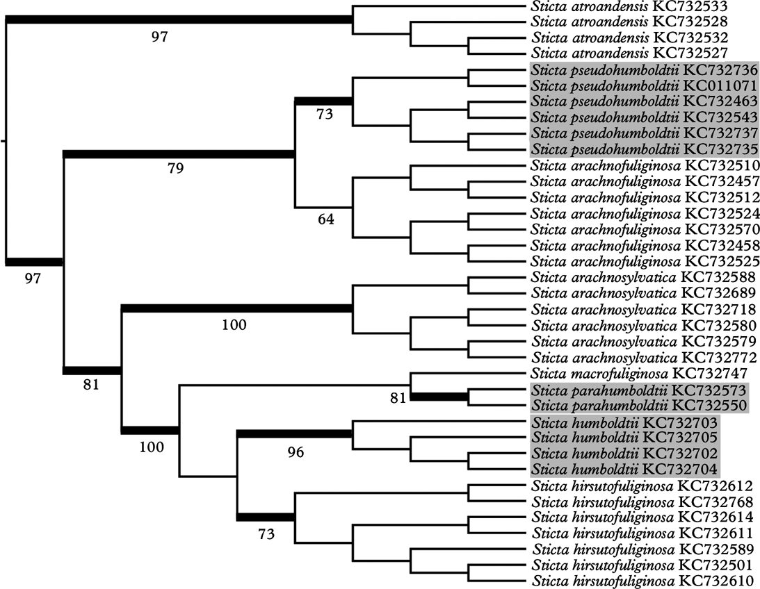
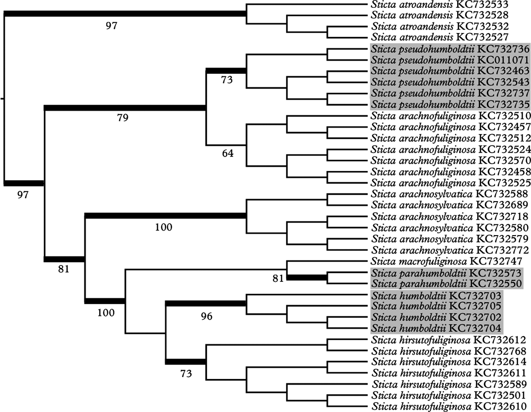
The epithet of this new species [Greek: ‘pseudo’=false] was chosen since the species resembles Sticta humboldtii but, contrary to S. parahumboldtii (see above), is not the closest relative of S. humboldtii but sister to the morphologically different S. arachnofuliginosa.
Ecology and distribution
Colombian Andes. The species was found between 1900 and 3975 m in subandine forests and páramo vegetation. It is known from all three major Cordilleras (Rangel-Ch. Reference Rangel-Ch.1995, Reference Rangel-Ch. and Rangel-Ch.2000; Rodríguez et al. Reference Rodríguez, Armenteras, Morales and Romero2006; Moncada et al. Reference Moncada, Aguirre-C. and Lücking2013c ), growing epiphytically, generally associated with Metzgeria spp., Heterodermia circinalis, Leptogium spp., and Oropogon bicolor.
Notes
Based on study of the type material of Sticta humboldtii (Hooker Reference Hooker, Kunth and Bonpland1822, Reference Hooker1829; Galloway Reference Galloway1995) and our field experience with the species in Costa Rica and the northern Andes, we initially considered this to be a rather well-defined species, characterized by large thalli with a dense tomentum on the upper surface and densely tomentose apothecial margins. Molecular phylogeny, however, demonstrated that three species were involved (Fig. 7), forming a clade with other dorsally tomentose species that produce isidia (Moncada et al. Reference Moncada, Lücking and Suárez2013b ). The isidiate species are rather well distinguished by their unique lobe morphologies, such as the deeply faveolate S. arachnofuliginosa Moncada & Lücking (Moncada & Lücking Reference Moncada and Lücking2012), which is sister to S. pseudohumboldtii (Fig. 7). The three non-isidiate species, S. humboldtii, S. parahumboldtii, and S. pseudohumboldtii, can be separated using the key below.
Partial ITS phylogeny of the Sticta humboldtii clade, highlighting the apotheciate species corresponding to the S. humboldtii morphodeme (adapted from Moncada et al. Reference Moncada, Lücking and Suárez2013b ). Thick branches indicate bootstrap support >70% and support values given.
Additional specimens examined. Colombia: Antioquia: Mun. Medellín, Estación Forestal Piedras Blancas, 2100 m, 1974, K. Dumont et al. 1592 (COL). Boyacá: Mun. Socha, Carretera Socha-Sácama, pasando el páramo de Pisba, 3050 m, 1982, J. Aguirre et al. 2854 (COL). Caldas: Mun. Manizales, Río Blanco, 2000 m, 18 iii 2008, F. Sarria & S. Valdes s.n. (CUVC); Mun. Villa María, Nevado del Ruíz, costado noroccidental, a 500 m de la estación de Televisión, 3900 m, 1979, H. Sipman & H. Valencia 10476 (B, COL). Cauca: Mun. Puracé, Páramo de San Rafael, cerca de la cascada del río San José, 3200–3350 m, 1961, J. Cuatrecasas y L. Willard 26289, 26290 (COL); Bosque en los alrededores de Pilimbalá, 3470 m, 1972, A. M. Cleef & A. Fernández-P. 493b (COL, U); Parque Nacional Natural Puracé, Pilimbalá-Laguna de San Rafael, 3300 m, 1984, J. Aguirre & H. Sipman 5978 (B, COL); Carretera La Plata-Puracé, cerca a la cabaña del Inderena, 3300 m, 1984, J. Aguirre & H. Sipman 6018 (B, COL); Termales de San Juan, 3000 m, 1985, J. Aguirre & H. Sipman 5919 (B, COL); Parque Nacional Natural Puracé, Pilimbalá-Laguna de San Rafael, 3280 m, 2000, B. Moncada & R. Dávila 523 (UDBC); Pilimbalá-Volcán Puracé, 3710 m, 2000, B. Moncada & R. Dávila 623 (UDBC); Mun. Santa Rosa, Macizo Colombiano, páramo de las Papas, entre El Boquerón y La Hoyola, 3200–3510 m, 1958, H. Bischler 711 (COL). Cundinamarca: Mun. Bogotá D.C., Parque Nacional Natural Sumapaz, Páramo de Chisacá, Santa Rosa, Bodegas de Sumapaz, Valle del río Colorados (o Santa Rosita), 3460 m, 1976, T. v. d. Hammen & R. Jaramillo-M. 4325 (COL); Alrededores de Laguna Grande, 3700 m, 1984, J. Aguirre & H. Sipman 5003 (B, COL); ibid., 3500 m, 1986, H. Sipman et al. 34499 (B, COL); Carretera a la Laguna Larga, 3500 m, 1986, N. Cardona de Hollaender et al. 38 (CUVC); Corregimiento Nazareth, Cerro al costado oriental de la Caseta Los Pinos, 3460 m, 2004, B. Moncada & R. Dávila 2208, 2243 (UDBC); Alrededores Laguna de Chisacá, 04°17′21·7″N, 74°12′26·9″W, 3730 m, 2008, B. Moncada & A. López 2658 (UDBC); ibid., 3734 m, 2008, D. Beltrán et al. 26 (UDBC); ibid., I. Ardila & B. Gómez 186, 188, 209 (UDBC); ibid., 3724 m, 2010, B. Moncada et al. 4022b, 4104, 4114, 4116, 4117, 4120, 4122, 4123 (UDBC); Mun. Choachí, Páramo de Cruz Verde, 3150 m, 1938, J. Cuatrecasas 450b, 450d (US); Páramo de Cruz Verde, Vertiente hacia Choachí, 3200 m, 1961, J. Idrobo & Dumont 4536 (COL); Páramo del Verjón, 3220 m, 2007, D. Hernández et al. 19 (UDBC); ibid., K. Malaver et al. 70 (UDBC); ibid., L. Castro et al. 40 (UDBC); ibid., Y. Carreño et al. 21, 28 (UDBC); Sendero el Silencio., 3220 m, 2007, A. López et al. 28 (UDBC); El Verjón, Parque Ecológico Matarredonda, 04°33′37·7″N, 74°00′02·2″W, 2900–3220 m, 2010, B. Moncada 3276, 3285 (UDBC); ibid., 3220 m, 2011, B. Moncada 4586 (UDBC); Mun. Fómeque, Parque Nacional Natural Chingaza, Río La Playa-Chuza, 3183 m, 1999, C. Cuellar et al. 94 (UDBC); ibid., 3154 m, 1999, C. Gaitán et al. 99 (UDBC); Río la Playa, Valle de los Frailejones, 3154 m, 1999, B. Moncada & R. Dávila 277, 308 (UDBC); Sendero La Arboleda, quebrada Babilonia, 3060 m, 2003, B. Moncada & R. Dávila 1491b, 1513, 1529, 1591b (UDBC); Parque Nacional Natural Chingaza, 04°44′19″N, 73°50′36″W, 3460 m, 2011, D. Fonseca & F. Martínez 31 (UDBC); Mun. Guasca, Páramo de Guasca, carretera Guasca-Gachetá, 3300 m, 1979, H. Sipman et al. 10611 (B, COL); ibid., 3100 m, 1983, L. B. Cruz 499 (COL); Carretera a Bogotá, 3380 m, 2001, D. Ros 178 (COL); Páramo de Guasca, 3350 m, 2011, B. Moncada 4768b, 4769a (UDBC); ibid., R. Lücking 33385, 34024, 34036, 34042 (UDBC, F); Mun. La Calera, Páramo de Palacio, Hacienda la Siberia, 3500 m, 1960, L. E. Mora 909 (COL); Bosque Alto Andino, abajo del páramo de Palacio, 3100 m, 1960, L. E. Mora 974 (COL); Hacienda La Siberia, km 17·5 camino La Calera a la Mina, 3400 m, 1971, G. Guzmán 9114, 9773 (COL); ibid., 3450 m, 1983, L. B. Cruz 329a (COL); Parque Nacional Natural Chingaza, Monterrendondo, 04°44′11·6″N, 73°50′31·3″W, 3430 m, 2011, B. Moncada 4620, 4635a (UDBC); Laguna de Siecha, 04°44′18·8″N, 73°50′36″W, 3456 m, 2011, Y. Orozco & R. Soto 83 (UDBC); ibid., 2011, Y. Orozco & R. Soto 104 (UDBC); Zona de Amortiguación Parque Nacional Natural Chingaza, Vía del ángulo al Rincón del Oso, 3600 m, 2011, B. Moncada 4921, 4928, 4964, 4980 (UDBC); Mun. Villapinzón, Páramo de Villapinzón, 3300 m, 2002, M. Pinzón 1409, 1472, 1483, 1507, 1522, 3001 (COL). Huila: Mun. La Plata, Vereda La Candelaria, cascada del Río La Candelaria, 2300 m, 1984, J. Aguirre & H. Sipman 6143, 6145c (COL). Risaralda: Mun. Pereira, Parque Nacional Natural Los Nevados, Laguna de Otún, margen occidental, 3975 m, 1980, T. Boekhout 8b (COL); Mun. Santa Rosa de Cabal, Alrededores Finca La Sierra, 3740 m, 1980, J. Aguirre 1371 (COL); Costado Occidental, Cordillera Central, Finca La Sierra, 3750 m, 1984, J. Aguirre & H. Sipman 5458 (B, COL).
Key to species of the Sticta humboldtii group
-
1 Cyphellae urceolate (with their margin distinctly raised and involute and the cavity more than 100 µm high), prominent to sessile (basal membrane at or above lower cortex level), mostly rounded to slightly angular; upper tomentum dense, hiding the surface beneath ... Sticta pseudohumboldtii
Cyphellae plane (with their margin levelled or slightly raised and erect and the cavity less than 100 µm high), immersed to erumpent (basal membrane below lower cortex level), usually angular; upper tomentum not dense, at least partially exposing the surface beneath ... 2
-
2(1) Lobes with pale marginal line; medulla K+ ochraceous; cyphellae K+ pale yellow to pale salmon ... Sticta parahumboldtii
Lobes with dark marginal line; medulla K+ pale yellow; cyphellae K+ yellow ... Sticta humboldtii
This study was supported by a grant from the National Science Foundation: Neotropical Epiphytic Microlichens – An Innovative Inventory of a Highly Diverse yet Little Known Group of Symbiotic Organisms (DEB 715660 to The Field Museum; PI R. Lücking). The Caterpillar® company and Field Museum donor and board member Robert H. Gordon provided additional funds for the molecular studies. The Universidad Distrital Francisco José de Caldas is thanked for the support to the lichen herbarium and the curatorial work of the UDBC collections, and we especially acknowledge the invaluable help of laboratory assistant Alejandra Suárez. The curators of the herbaria cited in this study, in particular Carlos Parra (COL), were extremely helpful in providing access to relevant collections and data. We are indebted to David Galloway and an anonymous reviewer for comments on an earlier version of the manuscript that helped to improve it considerably.